
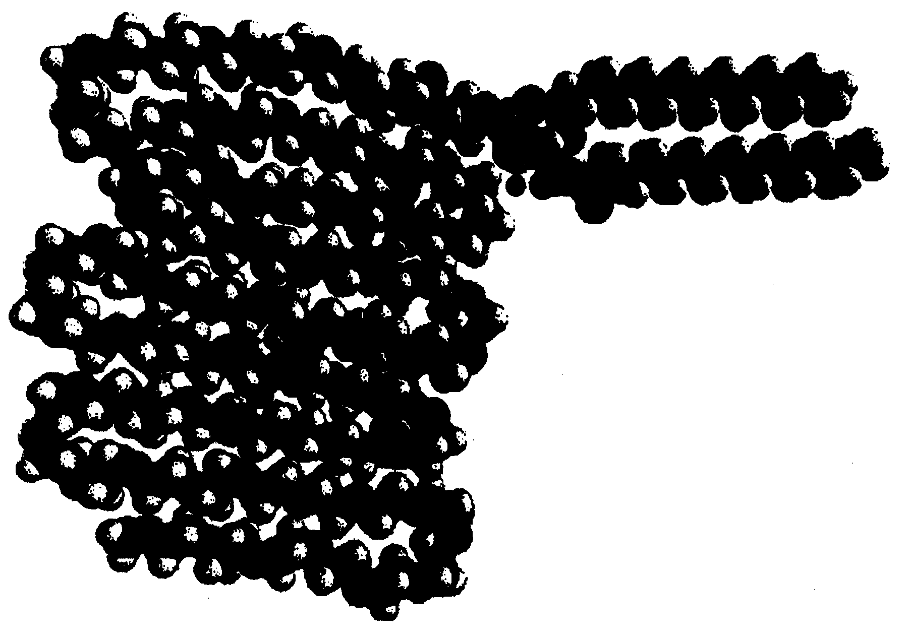
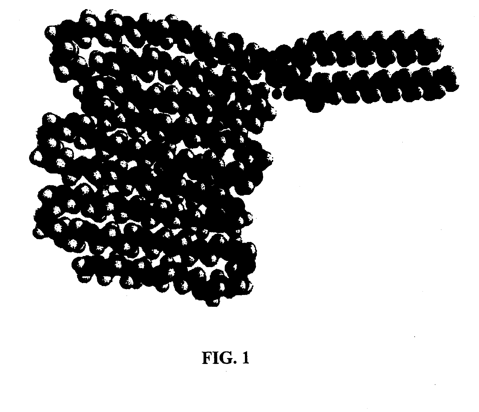
Rgd peptide attached to bioabsorbable stents

a bioabsorbable device and peptide technology, applied in the direction of peptide/protein ingredients, prosthesis, blood vessels, etc., can solve the problems of occluded blood conduits, intimal flaps or torn arterial linings which can collapse, and still pose significant problems, so as to enhance the growth rate of endothelium and enhance the regeneration rate of endothelium on the surface.

Inactive Publication Date: 2008-06-19
ABBOTT CARDIOVASCULAR
View PDF7 Cites 0 Cited by
  • Summary
  • Abstract
  • Description
  • Claims
  • Application Information

AI Technical Summary

Benefits of technology

[0012]Provided herein is a method of forming a medical device, an example of which is a stent such as a bioabsorbable stent, comprising a chemo-attractant attached to a surface layer of polymer on the medical device including a chemo-attractant for endothelial progenitor cells (EPCs). The method comprises providing a spacer compound comprising a hydrophobic moiety and a hydrophilic moiety, grafting or embedding the spacer compound to the surface layer of the polymer to cause the hydrophobic moiety to be grafted to or embedded within the surface layer of polymer, and attaching a chemo-attractant to the hydrophilic moiety. The chemo-attractant is chemically bonded to the bioabsorbable stent via a spacer compound. In some embodiments, the spacer compound comprises a hydrophobic moiety and a hydrophilic moiety. The spacer compound can be grafted to the surface of the bioabsorbable stent. The hydrophobic moiety can be embedded in the stent, and the chemo-attractant can be attached to the hydrophilic moiety. Upon implantation, the hydrophilic moiety can be projected from the surface of the medical device toward a physiologic environment. The chemo-attractant can then recruit EPCs so as to enhance the growth rate of endothelium on the surface of the device. It is important to note that a linker of this construct allows for more freedom for EPCs to access to the chemo-attractant so that the regeneration rate of endothelium on the surface can be enhanced.

Problems solved by technology

Problems associated with the above procedure include formation of intimal flaps or torn arterial linings which can collapse and occlude the blood conduit after the balloon is deflated.
However, ISR still poses a significant problem given the large volume of coronary interventions and their expanding use.
However, this study fails to take into account the accessibility of the RGD to the endothelial progenitor cells (EPCs) so that the endothelial cell growth enhancing effect described therein is limited.

Method used


Image

  • Rgd peptide attached to bioabsorbable stents
  • Rgd peptide attached to bioabsorbable stents
  • Rgd peptide attached to bioabsorbable stents

Examples


Embodiment Construction

[0016]Provided herein is a bioabsorbable stent including a chemo-attractant for endothelial progenitor cells (EPCs). The chemo-attractant is chemically bonded to the bioabsorbable stent via a spacer compound. In some embodiments, the spacer compound comprises a hydrophobic moiety and a hydrophilic moiety. The spacer compound can be grafted to the surface of the bioabsorbable stent. The hydrophobic moiety can be embedded in the stent, and the chemo-attractant can be attached to the hydrophilic moiety. Upon implantation, the hydrophilic moiety can be projected from the surface of the medical device toward a physiologic environment. The chemo-attractant can then recruit EPCs so as to enhance the growth rate of endothelium on the surface of the device. It is important to note that a spacer compound of the present invention allows for more freedom for EPCs to access to the chemo-attractant so that the regeneration rate of endothelium on the surface can be enhanced.

[0017]In some embodimen...


PUM

PropertyMeasurementUnit
molecular weightaaaaaaaaaa

Abstract

Provided herein is a method of forming medical device that includes RGD attached to the device via a spacer compound. The method comprises providing a spacer compound comprising a hydrophobic moiety and a hydrophilic moiety, grafting or embedding the spacer compound to the surface layer of the polymer to cause the hydrophobic moiety to be grafted to or embedded within the surface layer of polymer, and attaching a chemo-attractant to the hydrophilic moiety.

Description

CROSS REFERENCE TO RELATED APPLICATIONS[0001]The present application is a divisional of U.S. Ser. No. 11 / 453,679 filed Jun. 14, 2006. The aforementioned patent application is incorporated herein by reference in its entirety.BACKGROUND[0002]1. Field of the Invention[0003]This invention is generally related to bioabsorbable devices having RGD attached thereto.[0004]2. Description of the State of the Art[0005]Percutaneous coronary intervention (PCI) is a procedure for treating heart disease. A catheter assembly having a balloon portion is introduced percutaneously into the cardiovascular system of a patient via the brachial or femoral artery. The catheter assembly is advanced through the coronary vasculature until the balloon portion is positioned across the occlusive lesion. Once in position across the lesion, the balloon is inflated to a predetermined size to radially compress the atherosclerotic plaque of the lesion to remodel the lumen wall. The balloon is then deflated to a smalle...

Claims


Application Information

Patent Timeline
Patent Type & Authority Applications(United States)
IPC IPC(8): A61F2/82A61K31/337A61K31/565A61K38/44A61K31/436A61K31/573A61K31/56A61K31/506
CPCA61L31/047A61L31/10C08L71/02
Inventor GALE, DAVID C.HUANG, BINKLAUS, KLEINE
Owner ABBOTT CARDIOVASCULAR
Learn More