Particle Based Binding Assay

a technology of particle binding and assay, applied in the field of particle binding assay, can solve the problems of colouring forming substrates having to be added to the membrane, increasing signal to noise ratio, and more complete separation of unbound materials, etc., and achieves the effect of overcompensating sensitivity deficiencies, high background label count, and small but still optimal amount of ligands or anti-ligands

Active Publication Date: 2008-07-03
ORION DIAGNOSTICA
View PDF5 Cites 13 Cited by
  • Summary
  • Abstract
  • Description
  • Claims
  • Application Information

AI Technical Summary

Benefits of technology

[0029]The present invention overcomes the sensitivity deficiencies of prior art methods by exploiting unlabelled and labelled particles of different size and a porous surface. A common problem with the label used in prior art methods is that it has an inappropriate signal intensity and signal to noise ratio. The size of the capture particles in relation to label particles in the present invention is optimised to consume a smaller but still optimal amount of ligand or anti-ligand when compared to small particles. According to the present invention, high background label counts, due to the high unspecific label binding, can be decreased by using a porous surface, which possess low unspecific label binding, for the separation of the immunocomplex from the unbound material. Moreover, the sensitivity of the assay can be increased by using a binding substance affixed to label particles, thus increasing the specific activity of the labelled binding substance and the assay sensitivity.

Problems solved by technology

In solid phase immunoassays, the sensitivity of the assay can be increased by washing the solid phase bound immunocomplex, which results in more complete separation of unbound material and thus in increase of signal to noise ratio.
When an enzyme label is used a disadvantage is that a colouring forming substrate must be added to the membrane.
This results in the loss of sensitivity of the assay.
Said application does not disclose any separation of unbound material from agglutinate using a filtration process.
However, the use of small particles is disadvantageous since the consumption of ligand or anti-ligand on the particle is higher the smaller the particles are.
However, there is often unspecific binding of labelled binding substance due to the porosity of the membrane.
Hence, three dimensional membranes require considerable washing to reduce unspecific binding.
The methods disclosed in the above prior art documents rely mainly on visual detection of analyte and are, therefore, not sensitive enough to measure low concentrations of analyte.
Moreover, the level of signal created by the label is not, in all circumstances, intensive enough.
However, the above electrical methods did not meet the needs of a simple, fast, sensitive, inexpensive and easy-to-use method to perform an immunodiagnostic assay.

Method used


Image

  • Particle Based Binding Assay
  • Particle Based Binding Assay
  • Particle Based Binding Assay

Examples


example 1

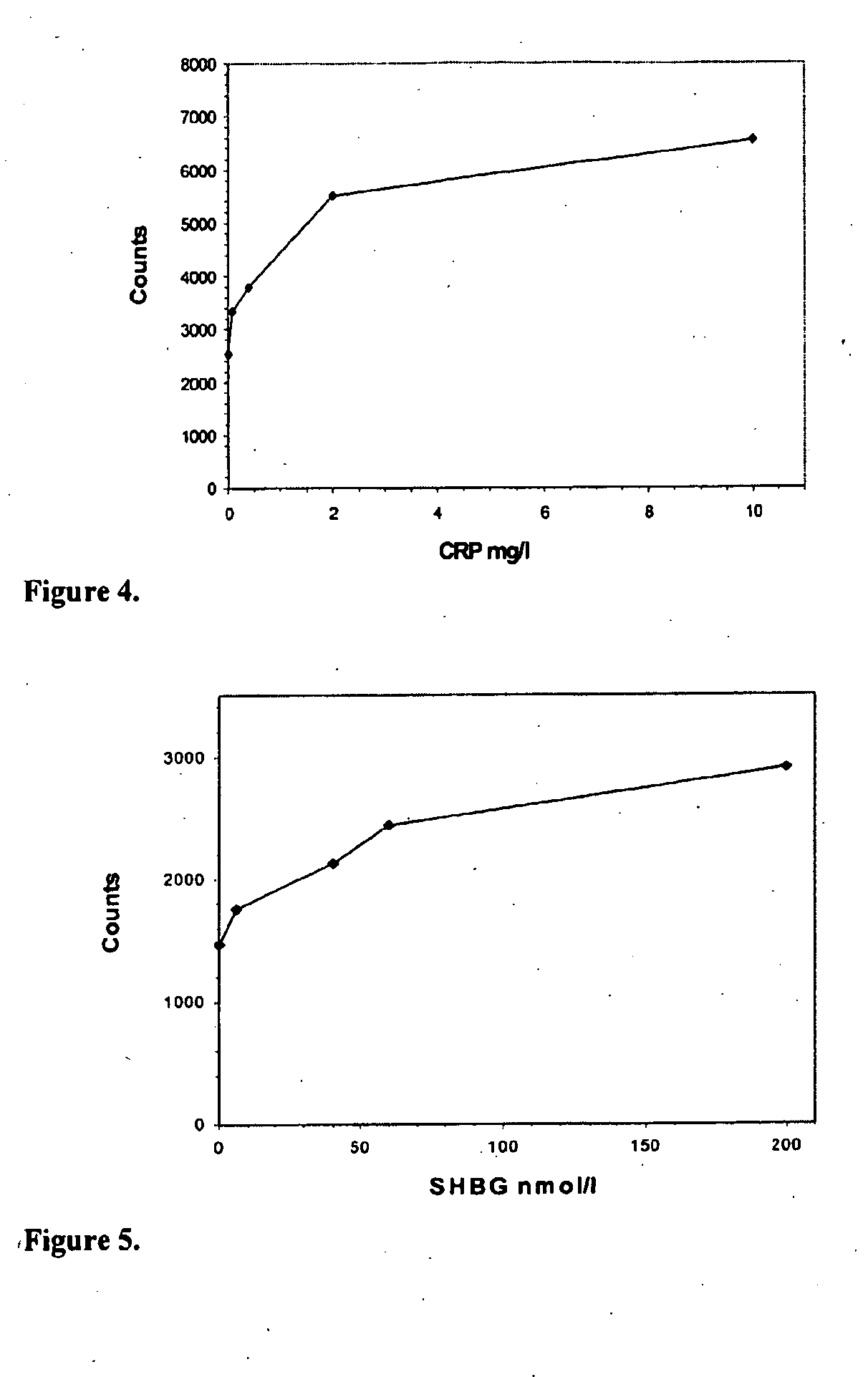
[0126]Procedure for sequential assay of hCRP with first and second particle bound binding substance exploiting polyclonal F(ab′)2-fragments and measuring immunocomplex formation on the grid used for separation of said complex (FIG. 4).

[0127]Human C-reactive protein (hCRP) assays are used as an indicator of inflammation. In the present example hCRP was determined in a calibrator sample. First, a sample containing hCRP at a concentration between 0-10 mg / l and 0.1 ml of the buffer (Tris based) are added to small centrifugal tubes. Thereafter, 10 μl of 0.25% latex particles (diameter 10 μm) coated with anti-CRP F(ab′)2 fragment are added and the tubes are incubated for 5 min with mixing. After incubation the tubes are centrifuged for 0.5-3 min (about 16000×g) and the supernatants are discarded. Latex particles with bound CRP are then washed to remove unbound CRP by means of. centrifugation and aspiration. The particles are resuspended to 0.1 ml of buffer and 10 μl of label containing bi...

example 2

[0128]Procedure for sequential assay of hSHBG with first and second particle bound binding substance exploiting monoclonal antibodies and measuring immunocomplex formation on the grid used for separation of said complex (FIG. 5).

[0129]Human sex hormone binding globulin (hSHBG) assays are used as one indicator for hormone status. In the present example recombinant hSHBG was determined in a sample of cell culture medium. First, a sample containing hSHBG at a concentration between 0-200 nmol / l and 0.1 ml of the buffer are added to small centrifugal tubes. Thereafter, 10 μl of 0.25% latex particles (diameter 10 μm) coated with monoclonal anti-SHBG antibody are added and the tubes incubated for 5 min with mixing. After incubation the tubes are centrifuged for 0.5-3 min (about 16000×g) and the supernatants are discarded. Latex particles with bound SHBG are then washed to remove unbound SHBG by means of centrifugation and aspiration. The particles are resuspended to 0.1 ml of buffer and 10...


PUM

PropertyMeasurementUnit
diameteraaaaaaaaaa
diameteraaaaaaaaaa
diameteraaaaaaaaaa

Abstract

The present invention provides a method for detecting the presence or amount of an analyte in a sample, said method comprising: a porous surface to which particles having attached thereto a binding substance, analyte and binding substance coated label particles are added. If said analyte is present in the sample an immuno- or chemical reaction occurs in the liquid phase. The separation of bound complex from unbound material is achieved by using said surface where the separation occurs mainly two dimensionally on the surface of the porous surface (e.g. grid). Interestingly, the disclosed surface enables a separation where said complexes are distributed two dimensionally on the porous surface (e.g. grid), whereas unbound materials are distributed three dimensionally. Accordingly, said surface enables both a two and a three dimensional separation.

Description

FIELD OF THE INVENTION[0001]The present invention relates to separation of formed immunocomplex or any biomolecular complex from unbound material on a porous surface (e.g. grid) comprising holes / openings mutually compatible with used particles. The invention discloses a heterogeneous immunoassay performed in two-particle assay format where the size and different characteristics of the particles are exploited, for detection and quantification of an analyte in a sample. In the method, large particles are used to catch analyte from a sample, separate and distinguish said analyte from body fluids, whereas small particles are used as labelling means.BACKGROUND OF THE INVENTION[0002]Several methods use immunoassay techniques for detection and quantification of antigen in a specimen. Two types of immunoassay systems are currently used. In a homogeneous system, the assay is performed in a single phase. In a heterogeneous system, the immunoassay is performed in two steps. The additional step...

Claims


Application Information

Patent Timeline
Patent Type & Authority Applications(United States)
IPC IPC(8): G01N33/53G01N33/543G01N33/545G01N33/553G01N33/552G01N33/538
CPCG01N33/538Y10S435/971Y10S436/824Y10S435/975G01N33/537
Inventor LUOTOLA, JUHANINIKULA, HANNUMURTOVUORI, MIRA
Owner ORION DIAGNOSTICA
Why Eureka
Try Eureka