Optical biopsy method for precancerous lesion diagnosis and an endoscope apparatus thereof

a biopsy method and precancerous lesion technology, applied in the field of precancerous lesion diagnosis, can solve the problems of increasing the difficulty of cure, morphologic diagnosis will not be able to differentiate early stage and moderate to severe atypical hyperplasia, and the incidence of cancer will be declined, the probability of change is reduced, and the sensitivity and specificity of precancerous lesion detection is improved.

Inactive Publication Date: 2008-07-03
SHANGHAI SHENGBIAO SCI & TECH CO LTD
View PDF4 Cites 13 Cited by
  • Summary
  • Abstract
  • Description
  • Claims
  • Application Information

AI Technical Summary

Benefits of technology

[0018]When using the method and apparatus of this invention to diagnose the lesion of the tested tissue, the doctors can selectively observe the white light images, the intrinsic fluorescence images and the intrinsic fluorescence spectrum curves of the tested site according to their needs. The computer processes these images and spectrums curves so ifs possible to identify the location of the precancerous lesion and the nature of the precancerous lesion clearly and rapidly in multiple ways, which improves the sensitivity and specificity of detection of the pr

Problems solved by technology

The cause of this is that there to no great breakthrough in the diagnosis of the tumors, especially in the diagnosis of mucous membrane tumors.
However, these can only confirm if there is a space-occupying lesion, but cant toll the lesion is benign or malignant.
Morphologic diagnosis won't be able to differentiate early stage and moderate to severe atypical hyperplasia because the origin of the lesion is only minor biochemical changes in the mucous membrane which are not detectable by the ultrasound, endoscopy, CT or MRI.
It's learned from the developmental way of gastric cancer that the occurrence of metastasis through blood and lymph system from invasive cancer has taken place

Method used


Image

  • Optical biopsy method for precancerous lesion diagnosis and an endoscope apparatus thereof
  • Optical biopsy method for precancerous lesion diagnosis and an endoscope apparatus thereof
  • Optical biopsy method for precancerous lesion diagnosis and an endoscope apparatus thereof

Examples


Embodiment Construction

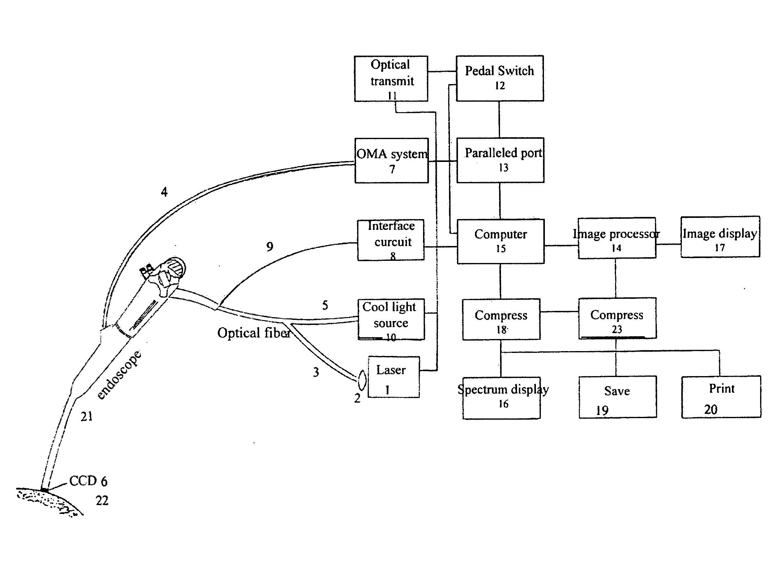
[0029]We will describe the structure and the usage of this invention in detail by reference to the attached figures of an embodiment as follows:

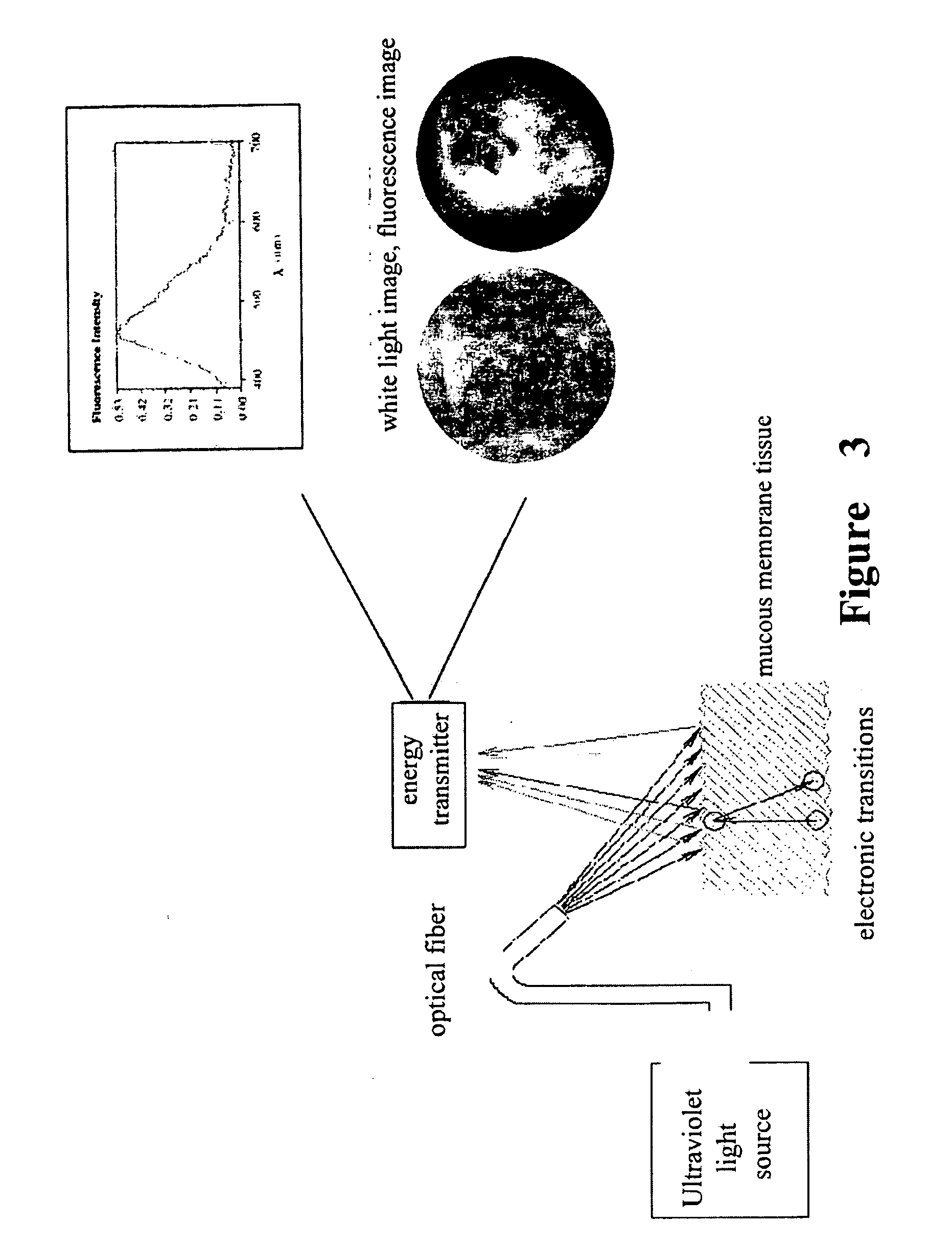
[0030]This invention is based on biochemistry and applies the spectrum technology to the spectrum detection of human body tissues. A diagnostic apparatus that uses optical biopsy (LIF, laser induced fluorescence), a worldwide-recognized precancerous detection technology, is invented. The diagnostic method and diagnostic criteria of LIF is known and approved internationally.

[0031]Principle of Related Optical Biopsy Technology:

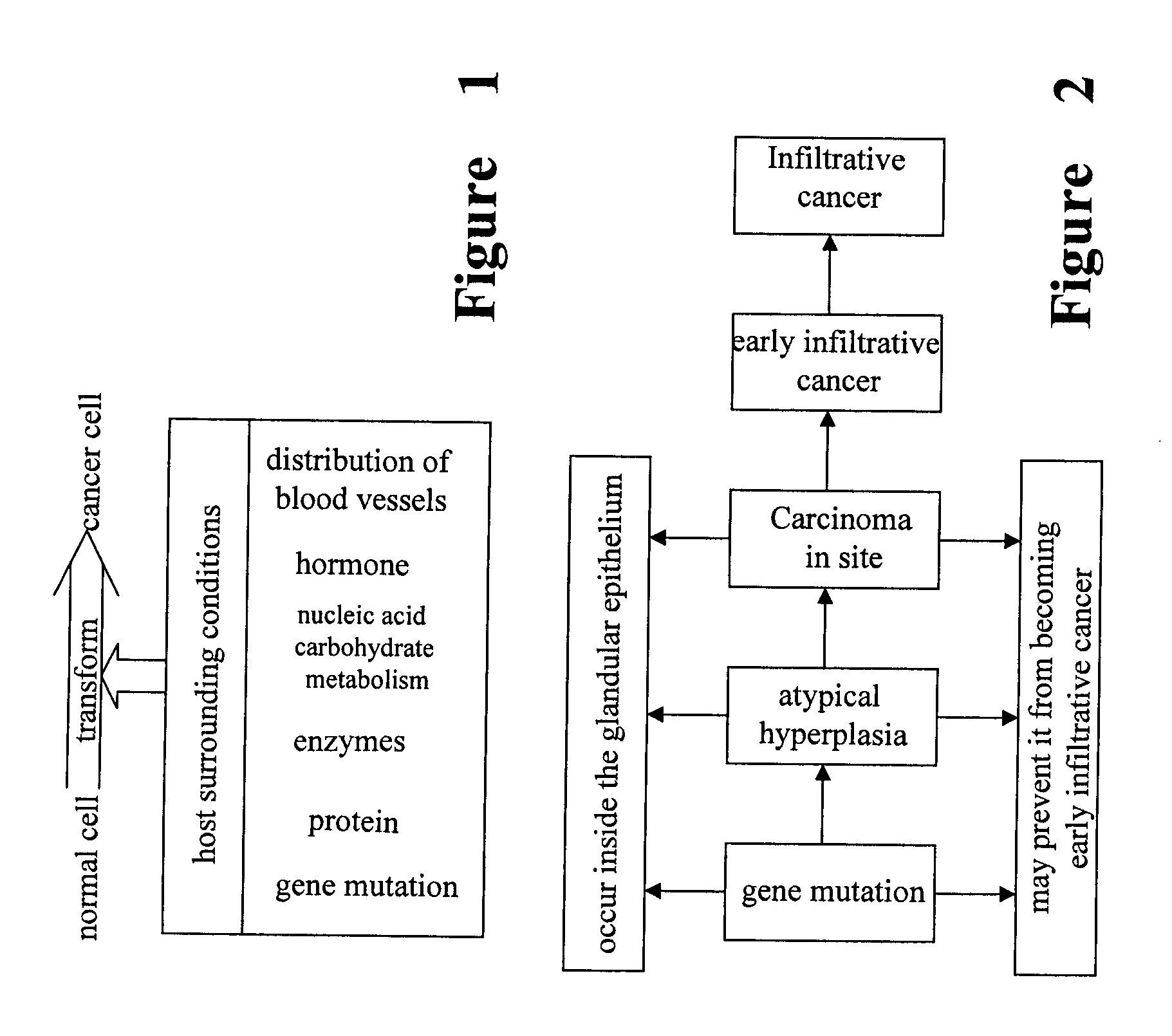
[0032]Firstly, see FIG. 1, the basic biological character of cancerous cells is malignant proliferation, poor differentiation, infiltration and metastasis. These are well known morphologic changes. The viewpoint of biochemical change that normal cells transform cancerous cells is that, this transformation starts from the mutations of genes induced by carcinogenic factors, and then these gene mutations may result in intra...


PUM


Abstract

A optical Biopsy method and an apparatus are used in the diagnosis of precancerous lesion for locating the place and determining the level of malignant tumor. The apparatus comprises light source (1, 10) a light channel system, an endoscope (21) and a circuit system. The light sources include an excited light (1) and a cold light source (10). The cold light source and the excited light in the light channel system go through the end of the light guide of the endoscope via optical fiber bundle and irradiate the tested living tissue (22). The white light image signal and the intrinsic fluorescence image signal reflected from the tested living tissue (22) are received by a weak fluorescence CCD (6) that tightly connects to the end of the endoscope (21) and then transmit to the circuit system via a signal wire (9) to produce the image in the display (17). The weak fluorescence signal reflected from the tested living tissue (22) is transmitted to the circuit system via the weak fluorescence fiber bundle (4) protruding from the forceps hole of the endoscope to produce the spectrum image (16).

Description

[0001]A optical Biopsy method for precancerous lesion diagnosis and an endoscope apparatus thereof.[0002]This application is a continuation application of Ser. No. 11 / 692,843, filed on Mar. 28, 2007, which is a continuation of application Ser. No. 10 / 533,623, filed Apr. 29, 2005, which is a 371 of PCT / CN2003 / 000917, filed Oct. 29, 2003FIELD OF INVENTION[0003]This invention involves a method and its apparatus for the diagnosis of precancerous lesion. In primarily, this invent provides a optical Biopsy method for precancerous lesion diagnosis and a medical endoscope apparatus thereof.BACKGROUND OF INVENTION[0004]At present the incidence and mortality of malignant tumors are very high. The cause of this is that there to no great breakthrough in the diagnosis of the tumors, especially in the diagnosis of mucous membrane tumors. The diagnosis of these tumors still depends on the principle and method of morphology, i.e. the inspection of doctors, ultrasound, endoscopy, CT, MRI. However, t...

Claims


Application Information

Patent Timeline
IPC IPC(8): A61B1/06A61B6/00A61B5/00
CPCA61B5/0084A61B5/0071
Inventor ZENG, KUNYU, ZHENFENHUANG, HAOZHU, CAILIN
Owner SHANGHAI SHENGBIAO SCI & TECH CO LTD
Why Eureka
Try Eureka