Diagnostic Method for Brain Damage-Related Disorders

a brain damage and diagnostic method technology, applied in the direction of biological material analysis, peptides, drug compositions, etc., can solve the problems of affecting the diagnostic value of brain damage biomarkers, unable to accurately predict clinical status and functional outcome, and no biological marker is currently available for routine brain diagnosis

Active Publication Date: 2008-09-11
UNIVERSITY OF GENEVA
View PDF3 Cites 52 Cited by
  • Summary
  • Abstract
  • Description
  • Claims
  • Application Information

AI Technical Summary

Benefits of technology

[0053]The term “control” refers to a normal subject (human or non-human mammal), i.e. one not suffering from a brain damage-related disorder (also called a “healthy donor”), and also to a sample taken from the same subject that provided the diagnostic sample, but at an earlier time.
[0054]References to an increased or decreased concentration compared with a sample of a control do not imply that a step of comparing is actually undertaken, since in many cases it will be obvious to the skilled practitioner that the concentration is abnormally high or low. Further, when the stages of a brain damage-related disorder are being monitored progressively, the comparison made can be with the concentration previously seen in the same subject in earlier progression of the disorder.

Problems solved by technology

No biological marker is currently available for the routine diagnosis of brain damage-related disorders including cerebrovascular, dementia and neurodegenerative diseases.
Most of them have not proved useful indicators of the extent of brain damage and accurate predictors of clinical status and functional outcome.
In fact, the diagnostic value of biomarkers for brain damage-related disorders has been hampered by their late appearance and a delayed peak after the damage event, their poor sensitivity and specificity, and the limited understanding of the mechanisms governing the release of these molecules into the CSF and ultimately in the blood.
As a result of these limitations, the use of brain damage-related disorder biomarkers is currently limited to research settings and none has been recommended for routine assessment [8].

Method used


Image

  • Diagnostic Method for Brain Damage-Related Disorders
  • Diagnostic Method for Brain Damage-Related Disorders
  • Diagnostic Method for Brain Damage-Related Disorders

Examples


example 1

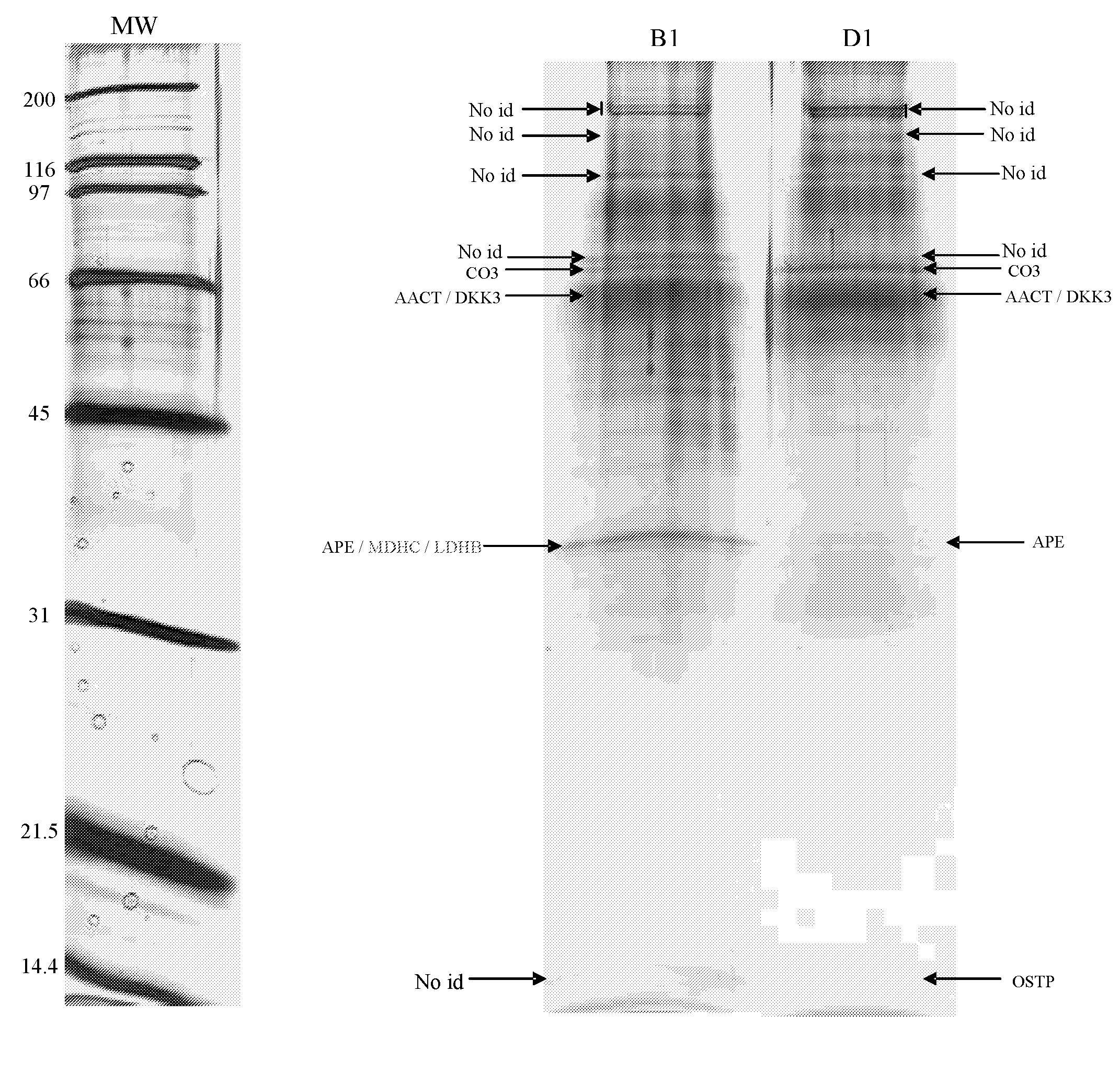
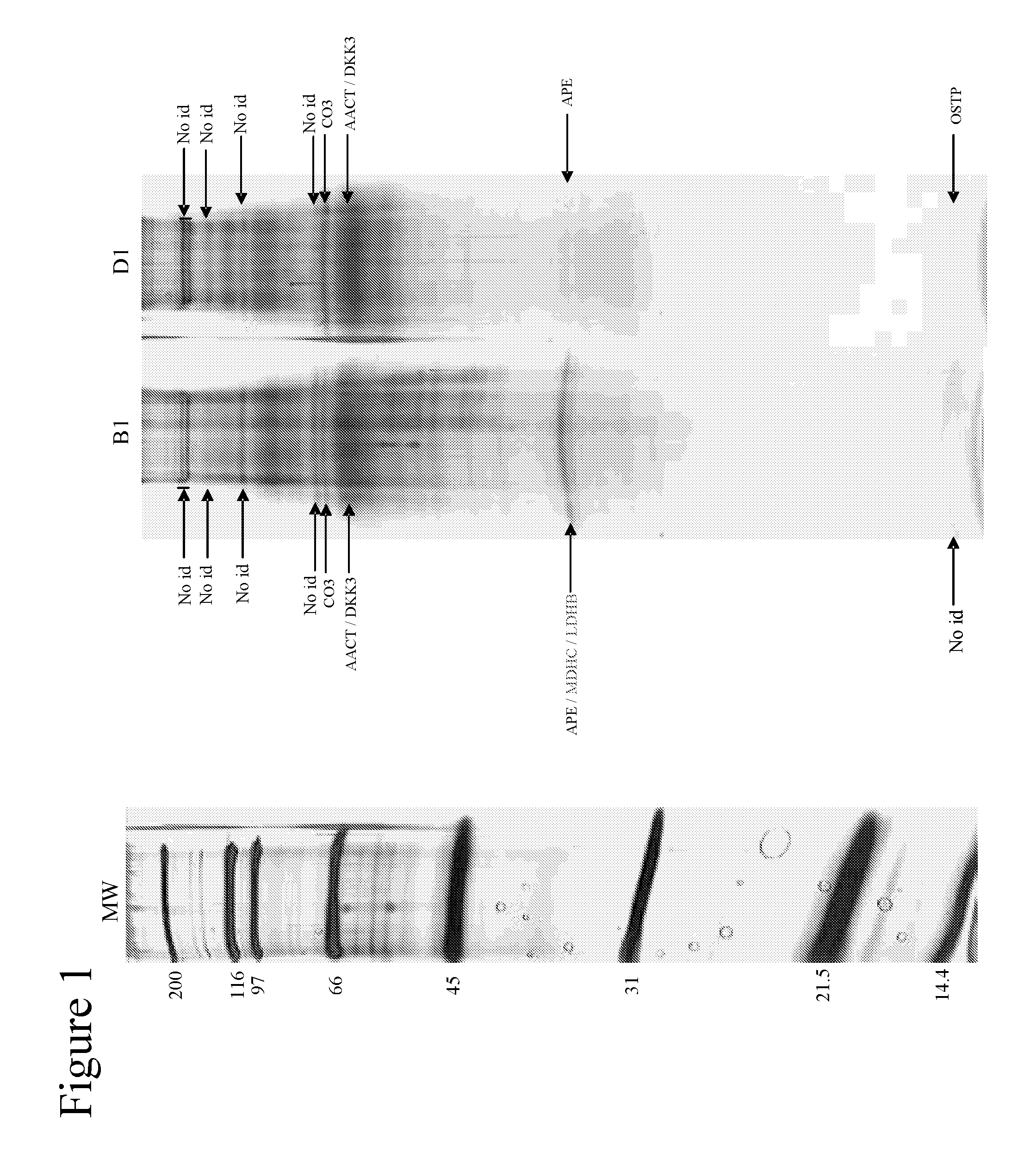
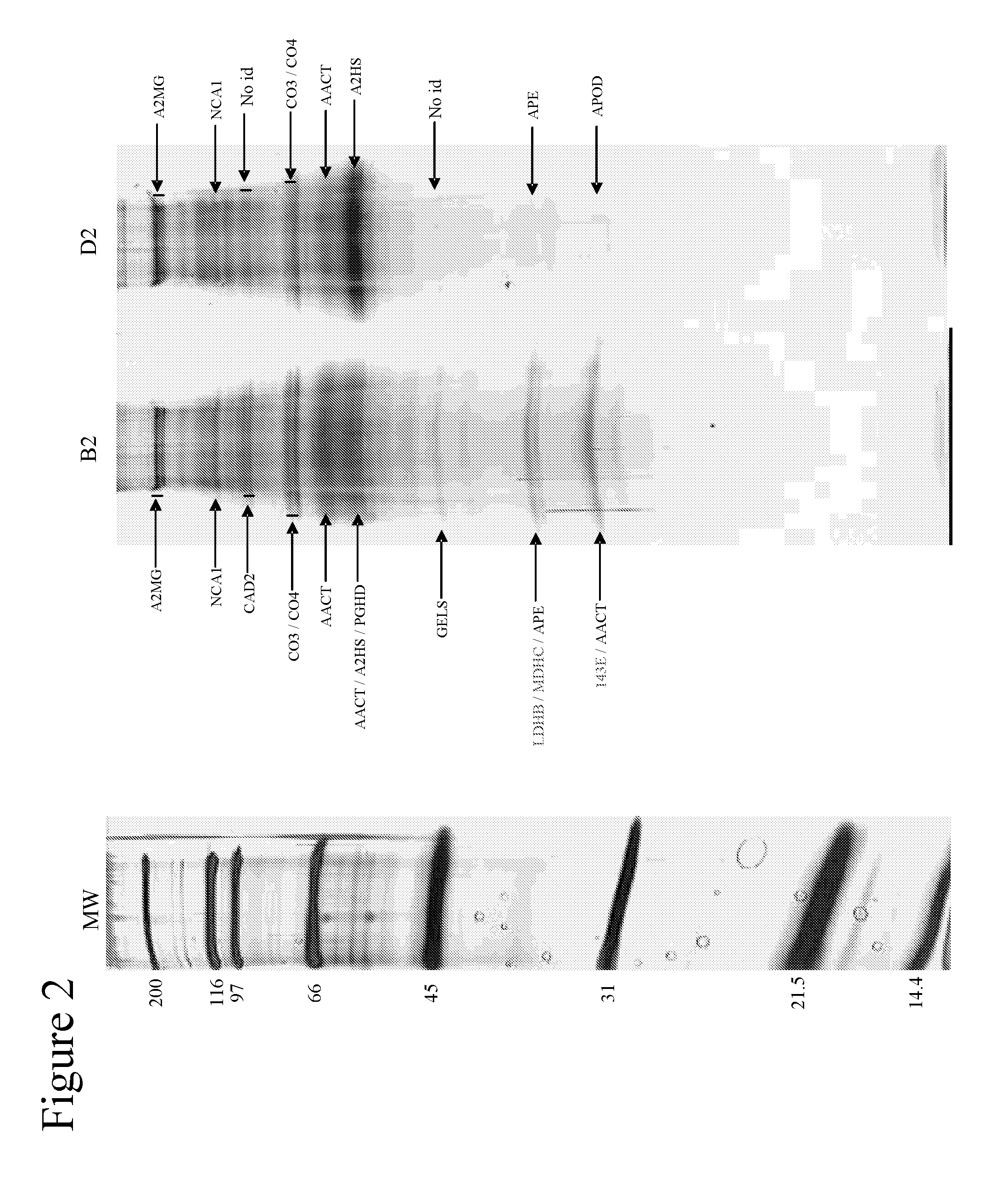
[0087]Using one-dimensional gel electrophoresis (1-DE) separation of cerebrospinal fluid (CSF) proteins and mass spectrometry techniques, 58 polypeptides named in Table 1 were found elevated or decreased in the CSF of deceased patients, used as a model of massive brain damage.

Study Population and Sample Handling

[0088]Twenty CSF samples were used for the proteomics-based approach aiming at discovering brain damage-related disorder markers. Five of these samples were obtained at autopsy from deceased patients 6 hours after death with no pathology of the central nervous system. Fifteen others were collected by lumbar puncture from living patients who had a neurological workup for benign conditions unrelated to brain damage (atypical headache and idiopathic peripheral facial nerve palsy). CSF samples were centrifuged immediately after collection, aliquoted, frozen at −80° C. and stored until analysis.

CSF Depletion Fractionation

[0089]Immunodepletion of human serum albumin, transferrin, h...

example 2

Introduction

[0092]One of the proteins identified as being upregulated in deceased CSF was evaluated as a potential biomarker of cerebrovascular disease, an example of a brain damage-related disorder. A survey of stroke patients was carried out and the results are shown in FIGS. 5 to 7. An ELISA intensity signal was obtained for Ubiquitin fusion degradation protein 1 homolog (UFD1) in plasma samples of the patients and of negative control patients. Plasma samples were taken from patients between 0-24 hours and / or after 72 hours of arrival at emergency hospital, and were matched for age / sex with samples from control patients.

[0093]ELISA was performed using 96-well Reacti-Bind™ NeutrAvidin™ coated Black Plates (Pierce, Rockford, Ill.). Plates were first rinsed in Borate Buffer Saline pH 8.4 (BBS) (100 mM H3BO3, 25 mM Na2B4O7 (Sigma, St Louis, Mo., USA), 75 mM NaCl (Merck, Darmastadt, Germany)) on a NOVAPATH™ washer (Bio-Rad, Hercules, Calif.). Then, 50 μl of biotin-conjugated antibody ...

example 3

[0097]This Example provides additional data showing plasma levels of UFDP1 in stroke and control patients. Additional data has been obtained from two cohorts of patients and controls, the smaller from Geneva, and a more comprehensive panel from the US.

[0098]ELISA was performed using 96-well Reacti-Bind™ NeutrAvidin™ coated Black Plates (Pierce, Rockford, Ill.). Plates were first rinsed in Borate Buffer Saline pH 8.4 (BBS) (100 mM H3BO3, 25 mM Na2B407 (Sigma, St Louis, Mo., USA), 75 mM NaCl (Merck, Darmastadt, Germany)) on a NOVAPATH™ washer (Bio-Rad, Hercules, Calif.). Then, 50 μl of relevant biomarker specific biotin-conjugated antibody (2 μg / mL) prepared in the dilution buffer A at pH 7 was added and incubated for one hour at 37° C. Plates were then washed 3 times in BBS in the plate washer. 50 μl of antigen or plasma was then added and incubated for one hour at 37° C. Recombinant protein antigens were diluted at 100, 50, 25, 12.5, 6.25, 3.125, 1.56 ng / ml in dilution buffer A to g...


PUM

PropertyMeasurementUnit
concentrationsaaaaaaaaaa
temperatureaaaaaaaaaa

Abstract

A brain damage-related disorder is diagnosed in a subject by detecting at least one polypeptide, or a variant or mutant thereof, in a sample of body fluid taken from the subject, wherein the polypeptide is one for which the level is either increased or decreased in cerebrospinal fluid from deceased patients compared to cerebrospinal fluid from healthy donors.

Description

BACKGROUND OF THE INVENTION[0001]1. Field of the Invention[0002]This invention relates to a diagnostic method for brain damage-related disorders.[0003]No biological marker is currently available for the routine diagnosis of brain damage-related disorders including cerebrovascular, dementia and neurodegenerative diseases. This invention relates to the use of cerebrospinal fluid from deceased patients as a model for the discovery of brain damage-related disorder markers, and to the use of such markers in diagnosis of human and animal brain damage-related disorders.[0004]2. Description of the Related Art[0005]Over the last two decades, a number of biological markers (biomarkers) have been studied in the cerebrospinal fluid (CSF) and serum of patients with brain damage-related disorders, including creatine kinase-BB [1], lactate dehydrogenase [2], myelin basic protein [3], S100 protein [4], neuron-specific enolase (NSE) [5], glial fibrillary acidic protein [6] and tau [7]. Most of them ...

Claims


Application Information

Patent Timeline
Patent Type & Authority Applications(United States)
IPC IPC(8): G01N33/68C12Q1/26C12Q1/32C12Q1/48C12Q1/40C12Q1/00C12Q1/44G01N33/573A61K38/44A61K38/45A61K38/46A61K39/00A61K38/02A61K38/16A61K38/17C40B40/10A61P25/00
CPCG01N33/6893G01N2800/30G01N2800/28G01N33/6896A61P25/00C07K14/4702C07K14/475C07K14/705C12N9/10C12N9/14G01N33/53
Inventor HOCHSTRASSER, DENISSANCHEZ, JEAN-CHARLES
Owner UNIVERSITY OF GENEVA
Why Eureka
Try Eureka