Nanoparticle delivery systems for membrane-integrating peptides

a technology of membrane integration and nanoparticles, applied in the direction of peptide/protein ingredients, drug compositions, antibacterial agents, etc., can solve the problems of peptides presenting particular problems, cell death, and general inapplicability, and achieve the effect of increasing ldh release and reducing cell proliferation

Inactive Publication Date: 2008-11-06
WASHINGTON UNIV IN SAINT LOUIS
View PDF6 Cites 3 Cited by
  • Summary
  • Abstract
  • Description
  • Claims
  • Application Information

AI Technical Summary

Benefits of technology

[0094]The role of cholesterol in modulating the melittin activity on the cell membrane was assessed by depleting cells of cholesterol before the cell proliferation assay and flow cytometry (as described above). C-32 Melanoma cells were treated with either 0.25 mM or 0.5 mM methyl beta cyclodextrin (Sigma, St. Louis, Mo.) for 15 min at 37° C. to deplete cholesterol. To quantify the amount of cholesterol depleted by methyl beta cyclodextrin, total lipids were extracted from melanoma cells by standard procedure. Briefly, 106 C-32 melanoma cells grown in 6-well tissue culture plates were scraped by a rubber policeman and the pellet obtained post-centrifugation was treated with 200 μl chloroform-methanol (2:1). After spinning top speed in a microcentrifuge, the organic phase was collected and vacuum dried. The dried lipids were then dissolved in 20 μl of 2-propanol containing 10% triton-X 100 as assay samples. The total cholesterol content in the cells was determined by using the Amplex Red Cholesterol Assay kit (Sigma, St. Louis, Mo.). Cholesterol standard was used for calibration as per manufacturer's recommendations.
[0095]The effect of cholesterol depletion on cell death was studied by both the MTT assay and annexin V-FITC / 7-AAD staining by flow-cytometry. At low concentrations, methyl beta cyclodextrin (0.25 mM and 0.5 mM) cause a 7% and 12% depletion of cholesterol respectively, but no change in cell proliferation. When such cholesterol-depleted cells were treated with αvβ3-targeted melittin nanoparticles, a significant difference (p<0.05) was found in the cell viability of normal and cholesterol depleted cells for both the concentrations of melittin-nanoparticles (5 and 10 μM). The dramatic reduction in cell proliferation after 12% cholesterol depletion and treatment with melittin-nanoparticles (100% cell death for both 5 and 10 μM melittin-nanoparticles) was analyzed with flow cytometry of annexin-V FITC and 7-AAD staining. Under these conditions, cell debris was evident consistent with necrotic cell death in this case.
[0096]Cytochrome c is a water-soluble 15 kDa protein residing in the mitochondrial intermembrane space and is released early during apoptosis. To determine if melittin-loaded nanodroplets trigger release of cytochrome c, C-32 melanoma cells were treated with varying concentrations of melittin-nanoparticles for 1 hour at 37° C. following which the mitochondria were isolated by using the Mitochondrial Isolation Kit (Pierce Biotechnology, Rockford, Ill.). The cytochrome c present in the mitochondrial and cytosol fractions thus obtained was assayed by using the cytochrome c ELISA kit (Invitrogen, Carlsbad, Calif.).
[0097]The cytochrome c release assay indicates that both free melittin and αvβ3-targeted melittin-nanoparticles cause a dose-dependent release of cytochrome c from the mitochondria of melanoma cells.
[0098]Lactate dehydrogenase (LDH) is a stable enzyme that is rapidly released from the cells upon plasma membrane damage. The lactate dehydrogenase release assay kit was obtained from BioVision (Mountain View, Calif.) and the assay performed according to manufacturers instructions. Briefly, C-32 melanoma cells were either treated with free melittin or melittin-nanoparticles at various concentrations for 1 hour at 37° C. and the amount of lactate dehydrogenase released quantified. Cells treated with 0.1% triton-X 100 were taken as 100% release.
[0099]There was no significant difference in LDH release from control cells and cells treated with melittin-loaded nanoparticles indicating that cell membrane integrity was maintained. However, free melittin caused a dose-dependent increase in LDH release. At a concentration of 1 μM, 19.17 (±4.2) percent LDH release was observed while 68.14 (±7.1) percent release was observed at 5 mM. Taken together, the LDH release and cytochrome c release data therefore suggest that free melittin at the lower concentration causes release of both LDH and cytochrome c, but at the higher concentrations causes a greater release of LDH (68.14±4.3%) than cytochrome c (55.48±6.1%). For αvβ3-targeted melittin-nanoparticles, there was a dose dependent increase in cytochrome c release, but no LDH release up to a concentration of 25 μM.

Problems solved by technology

None of the drugs contemplated by Lanza, et al., are membrane-integrating peptides that may undergo endocytosis or that may form pores in the lipid bilayer cellular membrane which can result in cell death due, for example, to non-specific control of substances that enter and exit the cell.
These peptides present particular problems and are generally not practical as, for example, antitumor agents, due to their nonspecificity and frequently their overall cytotoxicity in vivo.

Method used


Image

  • Nanoparticle delivery systems for membrane-integrating peptides
  • Nanoparticle delivery systems for membrane-integrating peptides
  • Nanoparticle delivery systems for membrane-integrating peptides

Examples


example 1

Preparation of Perfluorocarbon Nanoparticles

[0056]Perfluorocarbon nanoparticles were synthesized as described by Winter, P. M., et al., Arterioscler. Thromb. Vasc. Biol. (2006) 26:2103-2109. Briefly, a lipid surfactant co-mixture of egg lecithin (98 mol %) and dipalmitoyl-phosphatidylethanolamine (DPPE) 2 mol % (Avanti Polar Lipids, Piscataway, N.J.) was dissolved in chloroform, evaporated under reduced pressure, dried in a 50° C. vacuum oven and dispersed into water by sonication. The suspension was combined with either perfluoro-octylbromide (PFOB), or perfluoro-15-crown ether (CE) (Gateway Specialty Chemicals, St. Peters, Mo.), and distilled deionized water and continuously processed at 20,000 lbf / in2 for 4 min with an S110 Microfluidics emulsifier (Microfluidics, Newton, Mass.). αvβ3-integrin targeted nanoparticles were made by incorporating 0.1 mole % peptidomimetic vitronectin antagonist conjugated to polyethylene glycol (PEG)2000-phosphatidylethanolamine (Avanti Polar Lipids,...

example 2

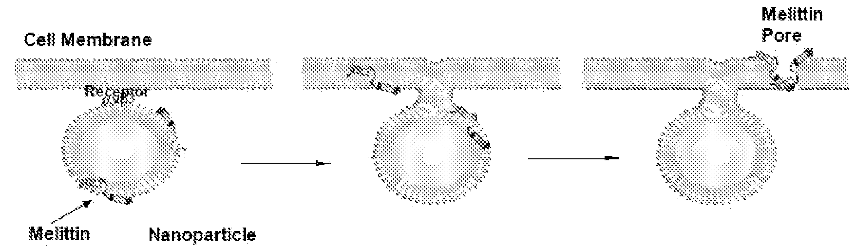
Incorporation of Melittin onto Nanoparticles

[0058]Melittin-loaded nanoparticles were formulated by mixing known amounts of melittin to perfluorocarbon nanoparticles. Pure melittin peptide material produced by solid-state peptide synthesis was obtained from Dr. Robert Mecham at Washington University Medical School, Department of Cell Biology and Physiology. The melittin was dissolved in 100 mM KCl (pH 7, mM HEPES) at 0.1 mM and 2-20 mL was added to 50 μl of nanoparticle suspension with mixing. After incubation at room temperature for 10 min, the nanoparticles were washed twice by centrifugation (100 g, 10 min) to remove the unbound melittin. The melittin in the supernatant was quantified by measuring the tryptophan fluorescence (described below). Depending on the amount of melittin added, the melittin-loaded nanoparticles yielded molar lipid / melittin ratios ranging from 1,000 to 40.

example 3

Characterization of Melittin Nano-Emulsions

A. Size Distribution and Zeta Potential

[0059]The size distribution of melittin-loaded nanoparticles was determined by photon correlation spectroscopy (PCS) on a Malvern Zetasizer 3000HS (Malvern Instruments, Malvern, UK). Zeta potential (ζ) values for the nanoparticles were determined with a Brookhaven Instruments PALS Zeta Potential Analyzer (Brookhaven Instruments). Data were acquired in the phase-analysis light scattering (PALS) mode following solution equilibration at 25° C. The Smoluchowski approximation was employed to calculate 4 from the measured nanoparticle electrophoretic mobility (μ):

μ=∈·ζ·(1.5) / η

where ∈ and η are the dielectric constant and the absolute viscosity of the medium, respectively.

[0060]The melittin-carrying nanoparticles exhibited a mean diameter of 358.8 nm (polydispersity index 0.011) when measured at an angle of 90 degrees using the Malvern Particle Size Analyzer. The zeta potential (ζ) was −31.5 mV. Measurements ...


PUM

PropertyMeasurementUnit
temperatureaaaaaaaaaa
pHaaaaaaaaaa
hydrophobicaaaaaaaaaa

Abstract

Compositions which comprise emulsions of nanoparticles for delivery of membrane-integrating peptides are described. The nanoparticles comprise a liquid hydrophobic core coated with a lipid/surfactant layer which contains the membrane-integrating peptides. Methods to use such compositions are also described.

Description

CROSS-REFERENCE TO RELATED APPLICATIONS[0001]This application claims priority from U.S. Provisional Application 60 / 905,227 filed 5 Mar. 2007, and from U.S. Provisional Application 60 / 991,654 filed 30 Nov. 2007. The contents of these documents are incorporated herein by reference.STATEMENT OF RIGHTS TO INVENTIONS MADE UNDER FEDERALLY SPONSORED RESEARCH[0002]This work was supported in part by grants from the U.S. government. The U.S. government has certain rights in this invention. The work was supported in part by U.S. National Institutes of Health grants U54-CA-119342 and HL-073646.TECHNICAL FIELD[0003]The invention relates to delivery of membrane-integrating peptides especially those that would otherwise be non-selectively cytotoxic in vivo. More specifically, the invention relates to delivery of such peptides using self-assembling emulsions of nanoparticles wherein a hydrophobic core is coated with a lipid / surfactant layer.BACKGROUND ART[0004]U.S. Pat. No. 6,676,963 to Lanza, et a...

Claims


Application Information

Patent Timeline
Patent Type & Authority Applications(United States)
IPC IPC(8): A61K49/00A61K9/14A61P43/00A61K38/00
CPCA61K9/1075A61K9/127A61K38/1767A61K47/48815A61K47/48238A61K47/488A61K47/48069A61K47/546A61K47/62A61K47/6907A61K47/6911A61P31/04A61P31/12A61P35/00A61P43/00
Inventor SCHLESINGER, PAULSOMAN, NEELESHLANZA, GREGORYWICKLINE, SAMUEL A.
Owner WASHINGTON UNIV IN SAINT LOUIS
Why Eureka
Try Eureka