
Ultrasound Measurement System and Method

a measurement system and ultrasound technology, applied in tomography, applications, instruments, etc., can solve the problems of large cumbersome devices, inability to readily adapt to handheld ultrasound systems, and large size of static mode scanners, etc., to achieve the effect of simple and convenien

Inactive Publication Date: 2008-11-13
SIGNOSTICS LTD
View PDF21 Cites 122 Cited by
  • Summary
  • Abstract
  • Description
  • Claims
  • Application Information

AI Technical Summary

Benefits of technology

[0015]Preferred embodiments broadly disclose novel systems in which ultrasonic measurement and imaging can be conveniently performed with less complexity and cost than previously available devices. The preferred embodiment devices possess a range of novel characteristics whereby the cost of medical ultrasound scanning is significantly and advantageously reduced and which also enhances the ease of use and convenience of their operation to the level at which they are operable by a primary care physician.
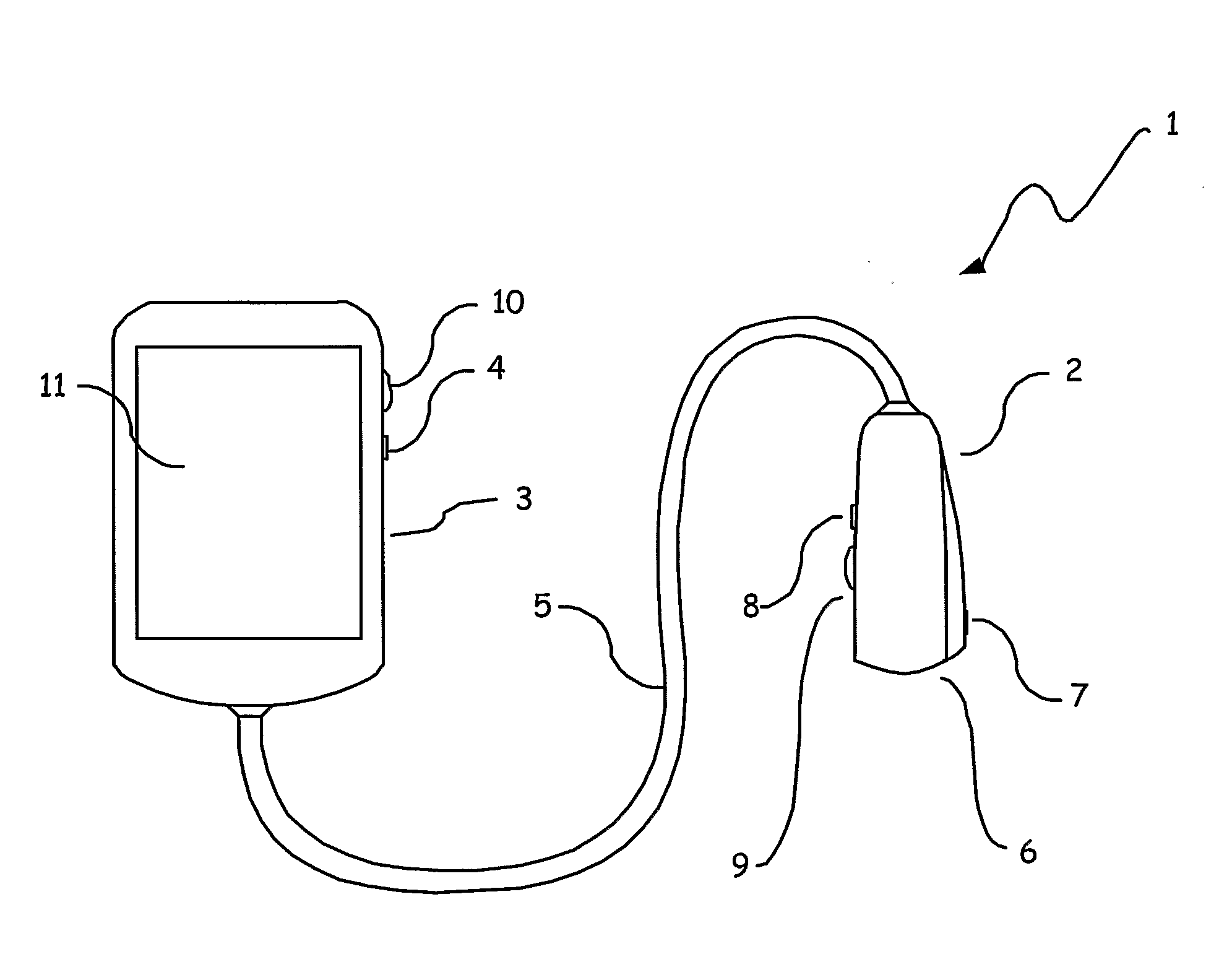
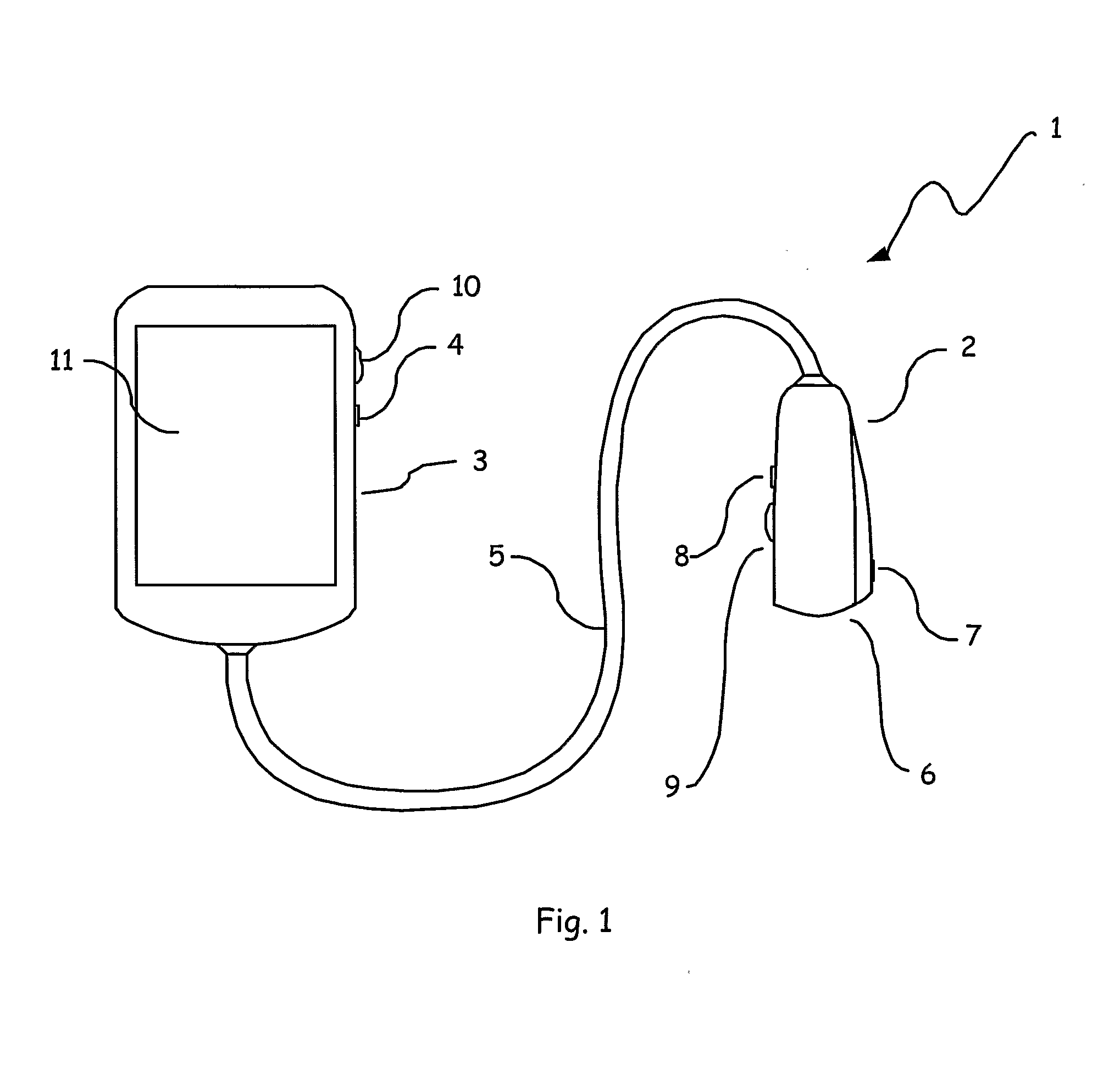
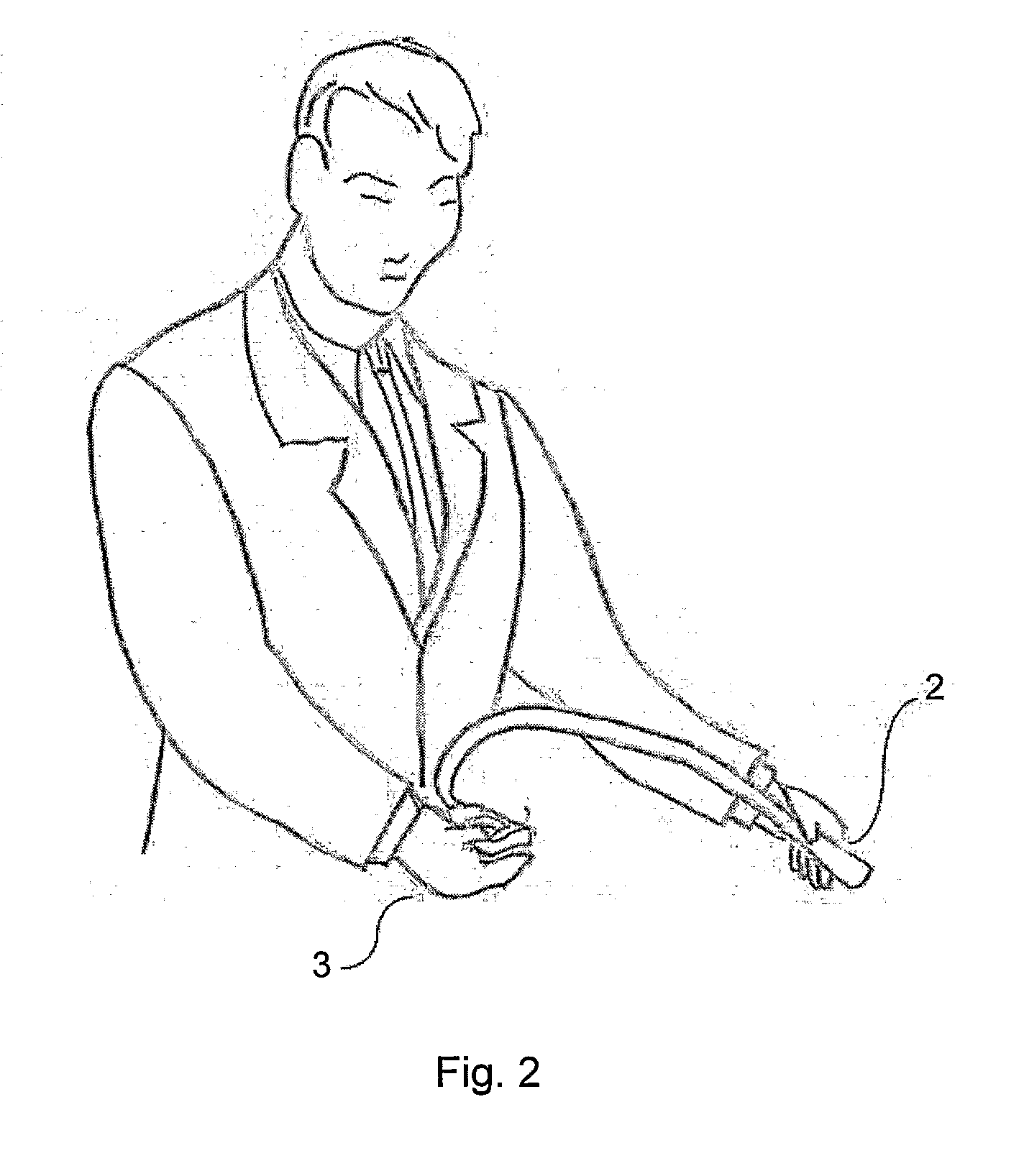
[0016]Preferred embodiments of the invention include a handheld display and user input host system connected to an ultrasound transducer via a cable. The handheld display system and the ultrasound transducer system are manufactured to be of similar volume and mass, facilitating a balanced load when the system is carried around a user's neck or over a user shoulder. The systems and cable are also of a size to be conveniently folded and placed in a user's pocket.
[0017]The ultrasound transducer system consists of one or more elements for transmitting and receiving ultrasonic waves with associated transmission circuitry and receiver amplifiers. The receiver circuitry includes analog to digital converters for converting the electrical representations of the received ultrasonic energy to digital data. The ultrasound transducer system also contains a controller for communicating with the host system, controlling operation of the ultrasound apparatus, and accepting user inputs from local mechanical or electrical switches and user input means. A preferred embodiment also contains circuitry for measuring the orientation and / or position of the transducer relative to a starting point or external reference, a temperature sensor, and a means to store local calibration data. The position / orientation measurement data is processed with the calibration data according to the temperature and input, and combined with the ultrasound data before being transmitted over the cable to the host system, enabling a position measurement system of high accuracy without the host system being aware of the means of position measurement. The position and orientation measurement allows an ultrasound transducer where the transmission pulse is transmitted in a fixed relative position to the ultrasound transducer, but moved in space by the user moving the probe.
[0018]The ultrasound transducer system can include an ultrasound gel storage and dispensing system, removing the requirement to carry a bottle of ultrasound gel, and a camera, for recording scan locations.
[0020]The host processing and display system could advantageously contain communications components such as those enabling wireless network communications, and software enabling the interfacing to host computers or servers containing medical records databases, providing a simple and convenient means for transferring patient data to an electronic records system.

Problems solved by technology

The static mode scanners were large cumbersome devices, and the techniques used are not readily suited to a handheld ultrasound system.
The nature of these devices, as well as the motor driving circuitry, adds size, power consumption, and cost to the device.
Additionally, the motor itself and associated moving parts reduces the reliability of the device.
Electronic beam steering removes the need for a motor to produce real time images, but the cost of producing transducers with arrays of crystals is high.
The probe cost is not an important factor in state-of-the-art ultrasound diagnostic systems, as the overall equipment cost is several times the probe cost.
The power consumption for electronic systems is also high, and is generally proportional to the number of channels being simultaneously operational.
The cost and power consumption of the Sonosite systems is far less than the large cart based systems, but still too expensive for most primary care physicians.
However, both products still consist of expensive and power hungry multi-element transducer arrays resulting in a costly imaging system.
The result is an increase in the cost of hand-carried systems, rather than a decrease.
The requirement for the tethered transducer to have a separate stationary electromagnetic transmitter is well suited to cart or desk based systems, where the host processing unit does not move, but is not suitable for handheld systems where the host processing unit is moving.
The requirement for a wireless communications system in the probe increases cost and power consumption, requiring additional components for the wireless communications system and a separate battery for the ultrasound probe.
The system also requires a separate battery supply for the ultrasound processor and transducer, and incurs the overhead of the wireless communications scheme in power consumption limiting the battery life and utility of the device.

Method used


Image

  • Ultrasound Measurement System and Method
  • Ultrasound Measurement System and Method
  • Ultrasound Measurement System and Method

Examples


Embodiment Construction

[0030]The background art provides several devices possessing unwieldy modes of operation. There is a need to integrate more fully the processing, recording, communication, display and control of ultrasound equipment and to reduce its cost and operational complexity such that it can be used by primary care physicians.

[0031]The preferred embodiment broadly disclose novel systems in which ultrasonic measurement and imaging can be conveniently performed with less complexity and cost than previously available devices. The preferred embodiment devices possess a range of novel characteristics whereby the cost of medical and veterinary ultrasound scanning is significantly and advantageously reduced and which also enhances the ease of use and convenience of their operation to the level at which they are operable by a primary care physician.

[0032]According to the invention there is provided an ultrasonic measurement and imaging system. An example embodiment is illustrated in FIG. 1. The syste...


PUM


Abstract

An ultrasound measurement system including a handheld display and processing means, an ultrasound transducer, a processing means of a substantially similar weight to the handheld display and processing means, and a transmission cable interconnecting the handheld display and processing means with the ultrasound transducer and processing means, the cable being of sufficient length to provide a means to mechanically locate the system around the neck of a user.

Description

FIELD OF THE INVENTION[0001]The present invention relates to a low cost and efficient medical ultrasound imaging, measurement and recording system with a configurable interface that supports a variety of medical ultrasound probes.BACKGROUND OF THE INVENTION[0002]Ultrasound was first investigated as a medical diagnostic imaging tool in the 1940's. George Ludwig was the first scientist to use amplitude mode (A-mode) ultrasound to detect foreign bodies in tissue. This is described in the report by Ludwig et al., “Considerations underlying the use of Ultrasound to detect Gallstones and Foreign Bodies in Tissue”, Naval Medical Research Institute Reports, Project #004 001, Report No. 4, June 1949. In the early 1950's Wild and Reid constructed a B-mode scanning system using a mechanically mounted rotating transducer, described in Wild, J. J. and Reid, J. M. (1952) “Application of echo-ranging techniques to the determination of structure of biological tissues”. Science 115:226-230 (1952). U...

Claims


Application Information

Patent Timeline
IPC IPC(8): A61B8/00
CPCA61B8/4281A61B8/4455A61B8/462A61B8/56A61B2560/0431G01S7/003G01S15/8906A61B8/4427
Inventor BARTLETT, STEWART GAVINCOSTELLO, ROGER MICHAEL
Owner SIGNOSTICS LTD
Learn More