Targeted bone marrow protection agents

a bone marrow and protection agent technology, applied in the direction of drug compositions, phosphorous compound active ingredients, peptide/protein ingredients, etc., can solve the problems of affecting the survival rate of p53 gene, so as to inhibit cell death and inhibit cell death.

Inactive Publication Date: 2009-01-08
SEMAFORE PHARMA INC
View PDF9 Cites 3 Cited by
  • Summary
  • Abstract
  • Description
  • Claims
  • Application Information

AI Technical Summary

Problems solved by technology

The p53 gene is, however, susceptible to damage.
Treatment of such cancers, however, is impeded by active p53 protein in normal tissue.
Such sensitivity to chemical or radiation-based therapeutics causes damage to normal tissue, while the therapeutics act on malignant, target tissue.
For example, p53-mediated damage to the lymphoid system, the hematopoietic system, intestinal epithelium, and hair follicles contribute to collateral damage associated with cancer therapies, which often limits the maximum tolerated doses of drugs in treatment regimens.
However, preventing the body's natural response to genotoxic stress may, in turn, permit abnormal cell growth.

Method used


Image

  • Targeted bone marrow protection agents
  • Targeted bone marrow protection agents
  • Targeted bone marrow protection agents

Examples


example 1

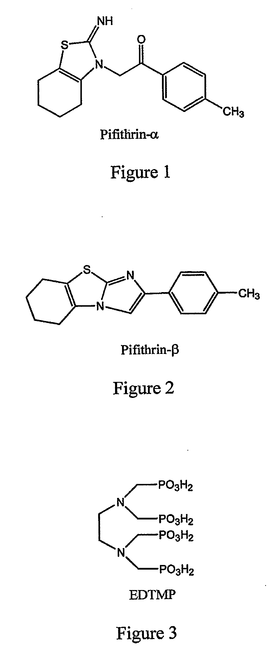
[0104]This example demonstrates a method of preparing pifithrin-α.

[0105]A multigram sample of pifithrin-α (the chemical structure of which is set forth in FIG. 1) suitable for the derivatization experiments, can be prepared according to literature methods as shown in FIG. 10 (see, for example, International Patent Application WO 00 / 44364; Tasaka et al., J. Heterocyclic Chem., 34, 1763 (1997); and Andreani et al., J. Med. Chem., 38, 1090 (1995)). Referring to FIG. 10, the 2-aminothiazole (C) is prepared from the reacting cyclohexanone (A) with thiourea (C). Then the solvent is removed, and the solid is recrystallized from hexane. This sample (C) is then dissolved with a slight excess of commercially available p-methylphenacyl bromide in toluene and then stirred for 48 hours at room temperature, at which time PFT-α precipitates out of solution as the HBr salt. PFT-α can be converted into pifithrin-β simply by refluxing in methanol for 6 hours.

example 2

[0106]This example demonstrates a method of making chemical modifications to pifithrin-α which allow for reversible covalent attachments.

[0107]With the sample prepared in accordance with Example 1, a number of different chemistries can be utilized for reaction with this particular substituted beta-ketothiazole. These different methods are shown in FIG. 11. Referring to FIG. 11, the preparation of the enol ether derivative (E) (or enol acetate, R2=acyl group) will be performed under basic conditions in the presence of an alkylating agent (or acylating agent) by known methods (Eroclspm et al., J. Org. Chem., 30, 1050 (1965)). The enol ether (and enol acetate) will then be evaluated for stability under relevant conditions. Relevant conditions for all of these conversions will be at either pH=7.4 (to simulate approximate physiological pH conditions) or pH=5 (to simulate bone resorption pH) in 50 mM sodium phosphate buffer (to simulate physiological conditions) at 37 degrees centigrade. ...

example 3

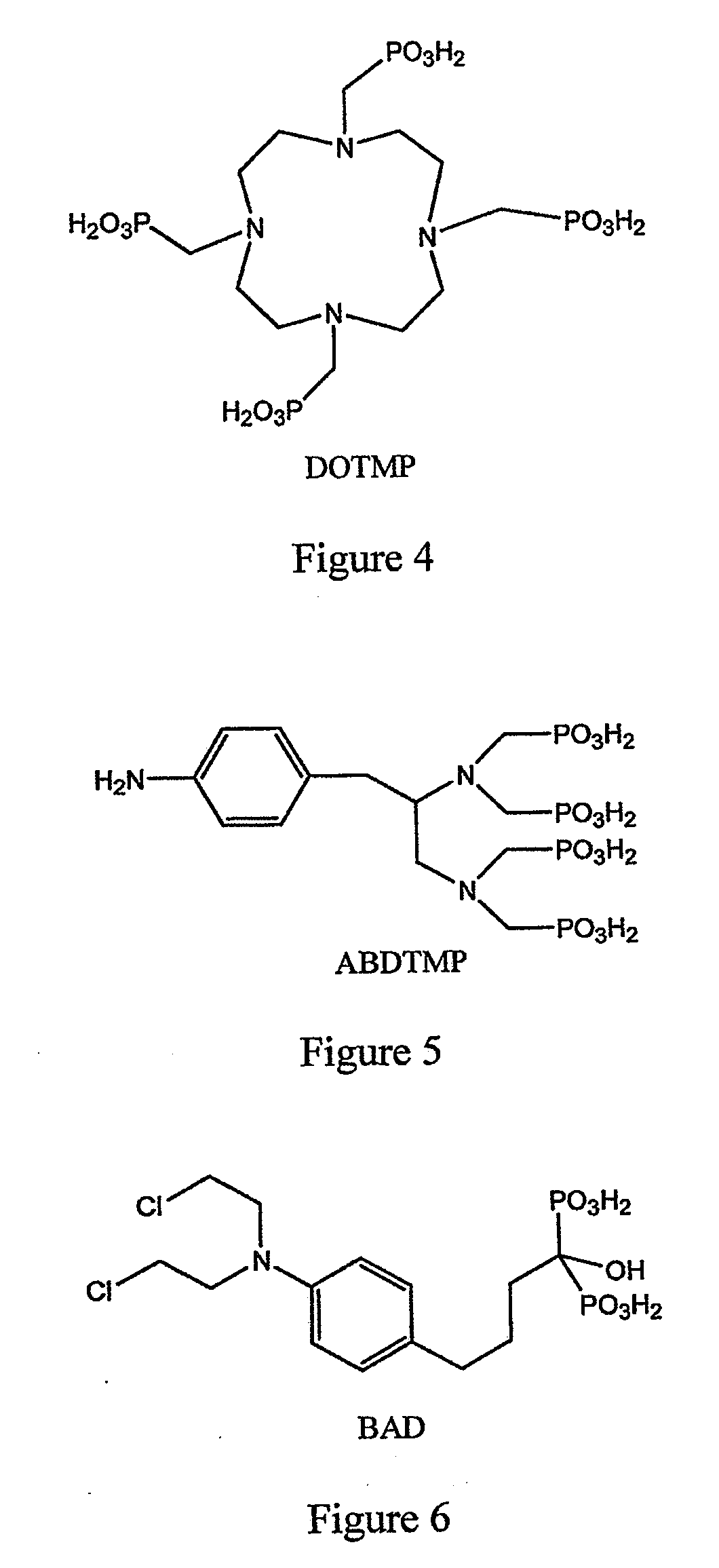
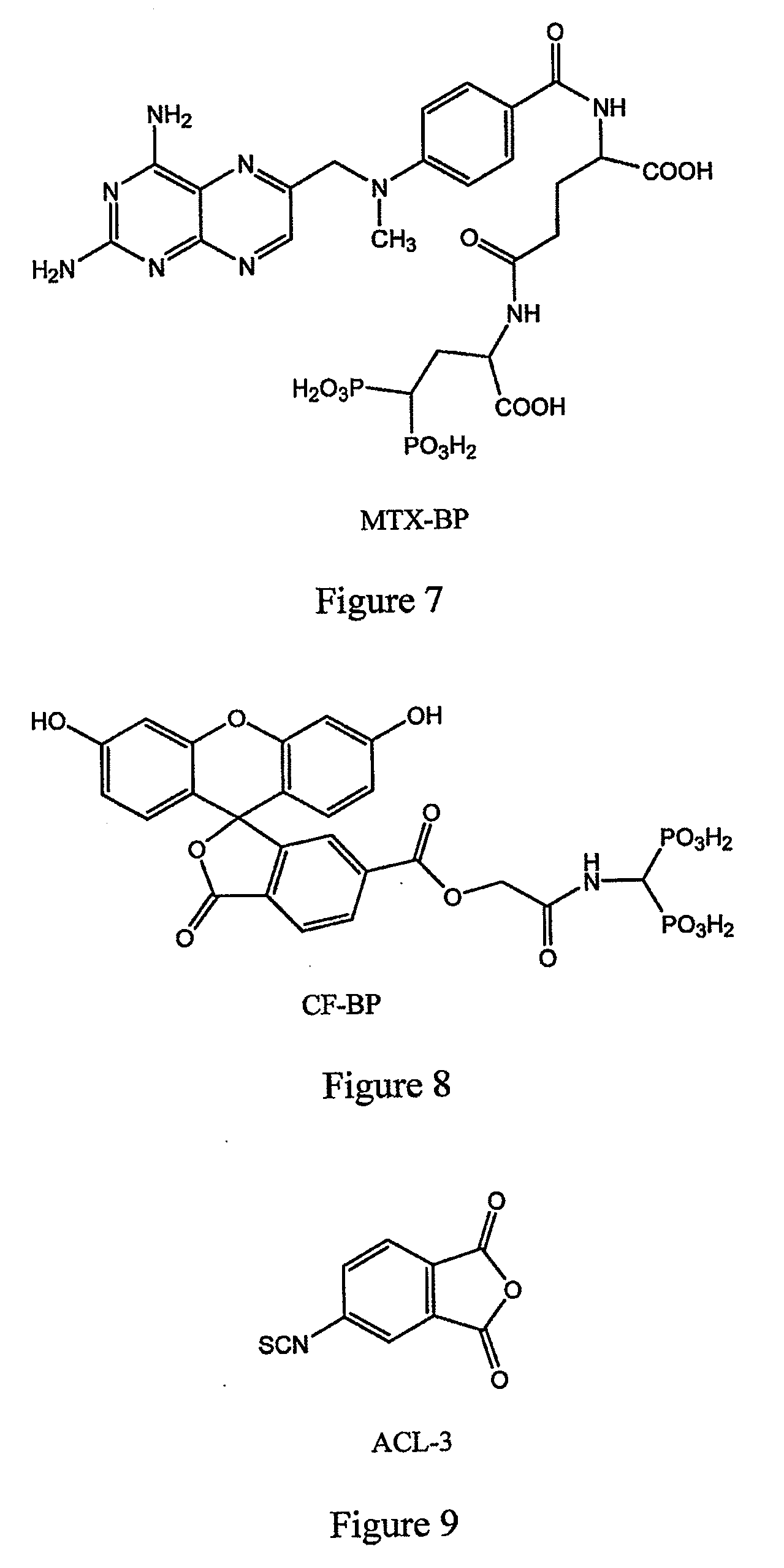
[0117]This example illustrates attachment of a cell protection factor, PFT-α, to a bone targeting agent via an acid-cleavable linker.

[0118]Preferably, the cell protection factor of the inventive compound is released from a bone targeting agent via an acid-enhanced cleavage mechanism. The acid sensitive linker ACL-3 (FIG. 6) is expected to acylate pifithrin-α (FIG. 1) on the imine NH moiety leaving the isothiocyanato group available to react with nucleophiles present on a bone-seeking moiety as shown in FIG. 15. This attachment through amines has been shown to be quite acid-sensitive (International Patent Application WO 94 / 00145). Given the facile hydrolysis of acylated imines, it can be expected that the acylated imine using ACL-3 would be even faster. This facile cleavage using the ACL-3 system is believed to occur because under acidic conditions the neighboring group effect of the ortho-carboxylate group facilitates cleavage. This chemistry is illustrated in FIG. 15. Reaction of P...


PUM

PropertyMeasurementUnit
pHaaaaaaaaaa
pHaaaaaaaaaa
pHaaaaaaaaaa

Abstract

The invention provides a method of inhibiting cell death in a mammal. The method comprises administering to a mammal an effective amount of a composition comprising a cell protection factor covalently linked to a bone targeting agent via a linkage that is cleaved under physiological conditions, whereby the cell protection factor is released from the bone targeting agent in vivo to inhibit cell death. The invention further provides a compound comprising a cell protection factor covalently attached to a bone targeting agent via a linker that is cleaved under physiological conditions.

Description

CROSS-REFERENCE TO RELATED PATENT APPLICATIONS[0001]This is a continuation of co-pending U.S. patent application Ser. No. 10 / 817,622, filed Apr. 2, 2004, claiming the benefit of U.S. Provisional Patent Application No. 60 / 460,289, filed Apr. 3, 2003. The disclosures of the '622 application and the '289 application are incorporated by reference.STATEMENT REGARDING FEDERALLY SPONSORED RESEARCH AND DEVELOPMENT[0002]This invention was made in part with Government support under Grant Number 1R43CA96259-01 awarded by the National Cancer Institute. The Government may have certain rights in this invention.FIELD OF THE INVENTION[0003]This invention pertains to materials and methods for inhibiting cell death in a mammal.BACKGROUND OF THE INVENTION[0004]Programmed cell death, known as apoptosis, is a tightly regulated cellular process which serves, in part, to prevent proliferation of abnormal or damaged cells. A subset of genes responsible for initiating cellular processes leading to apoptosis...

Claims


Application Information

Patent Timeline
Patent Type & Authority Applications(United States)
IPC IPC(8): A61K31/663A61K31/428A61K31/429A61K38/02C07D513/04C07F9/6539C07D277/38C07D277/82A61P35/00A61K31/662A61K41/00A61P43/00C07D277/40
CPCA61K31/428A61K31/429A61K31/662C07D513/04A61K41/00C07D277/40A61K31/663A61P35/00A61P43/00
Inventor GARLICH, JOSEPH R.DURDEN, DONALD L.SMITH, TIM C.
Owner SEMAFORE PHARMA INC
Why Eureka
Try Eureka