Use of Anti-integrin antibodies for reducing scar tissue formation

a technology of anti-integrin and scar tissue, applied in the field of biochemistry and physiology, can solve the problems of scarring and tissue detachment from the underlying membrane, and the problems are particularly acute, and achieve the effect of reducing deleterious granulation and minimizing tissue damage collateral

Inactive Publication Date: 2009-02-12
FACET BIOTECH CORP
View PDF25 Cites 7 Cited by
  • Summary
  • Abstract
  • Description
  • Claims
  • Application Information

AI Technical Summary

Benefits of technology

[0005]The present invention provides methods for controlling granulation in the region of injured tissue. In this manner, methods of the invention aid in minimizing tissue damage collateral to an initial injury. Accordingly, the present invention provides methods of reducing deleterious granulation that involve applying a granulation inhibitor to an injured or diseased tissue. The diseased or injured tissue may be part of an eye, joint or associated with a bursae. Some methods are useful for treating injured or diseased tissue produced by a condition such as keloid formation, burns or scleroderma. Other methods provide treatment for injured or diseased tissue associated with a disease causing tissue inflammation. Exemplary diseases of this type include f rheumatoid Arthritis, Wegener's Granulomatosis, Churg-Strauss-allergic granulomatosis, eosinophilic granulomata, midline granuloma, desmoid, sarcoidosis, macular degeneration, proliferative vitreoretinopathy, proliferative diabetic retinopathy, uterine fibroids, arteritis temporalis and Takayasu's arteritis. Diseases involving fibrosis resulting from inflammation also respond to the treatments described herein, for example, Crohn's disease, idiopathic pulmonary fibrosis, and allergic pulmonary fibrosis. Granulation inhibitors useful as medicaments in treating diseases such as those described above include antibodies, small organic molecules, and nucleic acid, protein and peptide antagonists.

Problems solved by technology

A particular problem in wound healing is the scarring and tissue detachment from underlying membranes caused by fluid accumulation resulting from excessive granulation in and around the wound site.
These problems are particularly acute in wounds to the eye and other tissues, such as joint cartilage.

Method used


Image

  • Use of Anti-integrin antibodies for reducing scar tissue formation
  • Use of Anti-integrin antibodies for reducing scar tissue formation
  • Use of Anti-integrin antibodies for reducing scar tissue formation

Examples


example 1

Construction of M200 Chimera from Murine IIa1 Anti-α5β1 Integrin

[0159]This example describes construction of the chimeric antibody M200.

[0160]A. Starting DNA Sequences of IIa1 and 200-4 VH and VL Domains

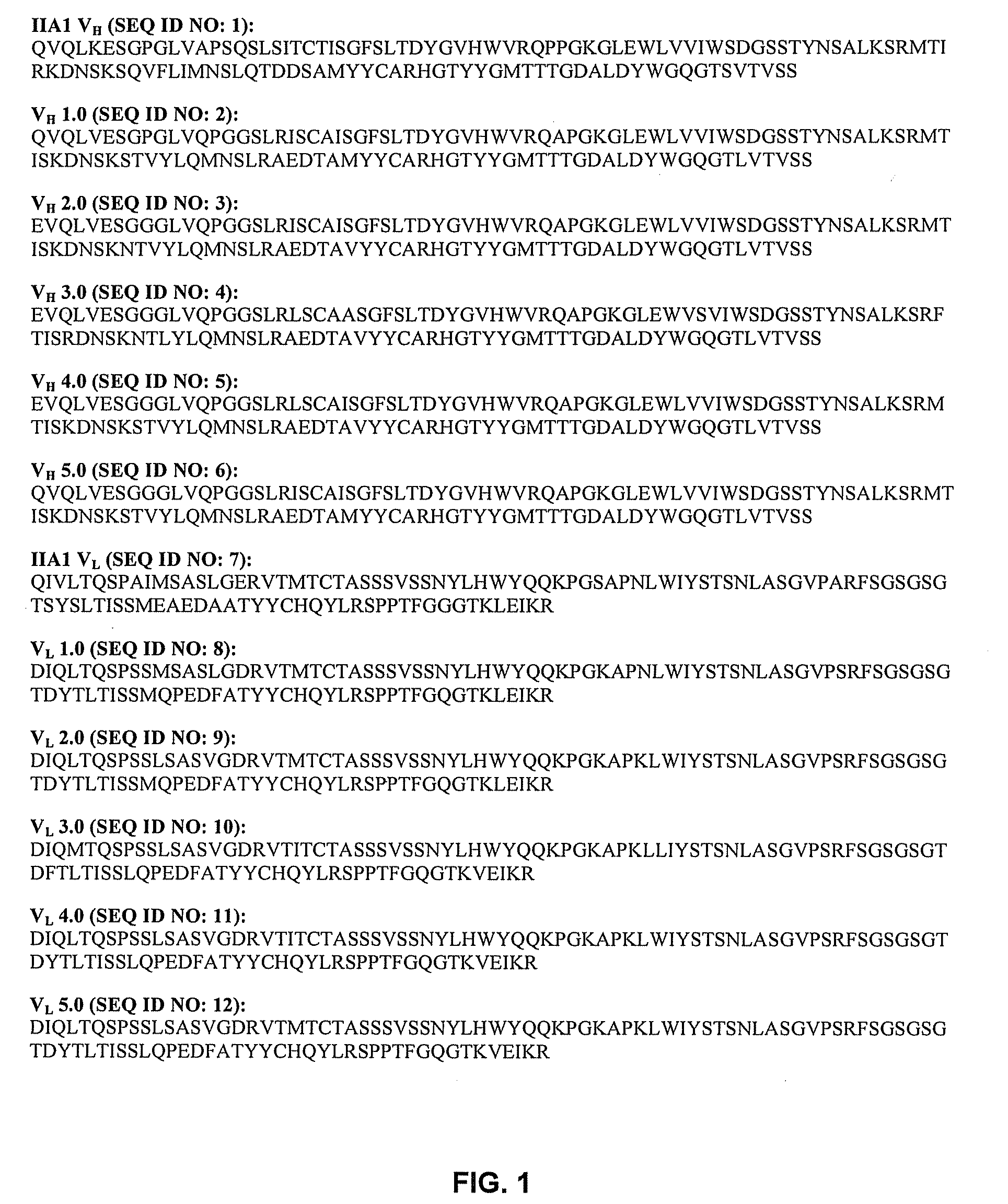
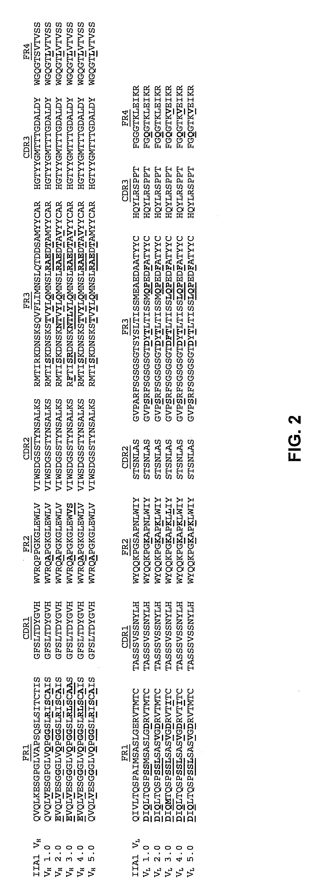
[0161]The variable heavy (VH) and light (VL) domains of the mouse anti-human α5β1 integrin antibody, IIA1 (Pharmingen, San Diego Calif.) were cloned from the IIA1 hybridoma cDNA and sequenced as part of the initial construction of the 200-4 antibody. FIG. 3 shows the cDNA sequences of the IIA1 VH (SEQ ID NO: 13) and VL (SEQ ID NO: 14) domains. During the construction of the 200-4 mouse / human chimeric IgG4 antibody from IIA 1, silent XhoI restriction sites (CTCGAG) (SEQ ID NO: 29) were introduced into the framework 4 regions of both IIA1 VH and VL. The 200-4 VH (SEQ ID NO: 15) and VL (SEQ ID NO: 17) DNA sequences containing these silent XhoI sites, as found in expression constructs DEF38 IIA 1 / human G4 chimera and NEF5 IIA1 / K chimera, are shown in FIG. 4. These 200-4 VH and VL sequenc...

example 2

Generation of Fab Fragment F200 from M200

[0172]This example describes making Fab fragment F200.

[0173]Fab fragments are generated from M200 IgG starting material by enzymatic digest. The starting IgG is buffer exchanged into 20 mM sodium phosphate, 20 mM N-acetyl cysteine pH 7.0. Soluble papain enzyme is added, and the mixture is rotated at 37° C. for 4 hours. After digestion the mixture is passed over a protein A column to remove Fc fragments and undigested IgG are removed. Sodium tetrathionate is added to 10 mM and incubated for 30 minutes at room temperature. Finally, this preparation is buffer exchanged into 20 mM sodium phosphate, 100 mM sodium chloride, pH 7.4, to yield the F200 solution.

[0174]Because it is a Fab fragment, the F200 light chain DNA and amino acid sequences are the same as the M200 light chain. The complete F200 heavy chain DNA (SEQ ID NO: 27) and amino acid (SEQ ID NO: 28) sequences are shown in FIG. 11.

example 3

Maintenance of Granulation Inhibitor Serum Levels after Systemic Administration

[0175]This example shows that granulation inhibitor serum levels can be maintained through a regular dosing regime.

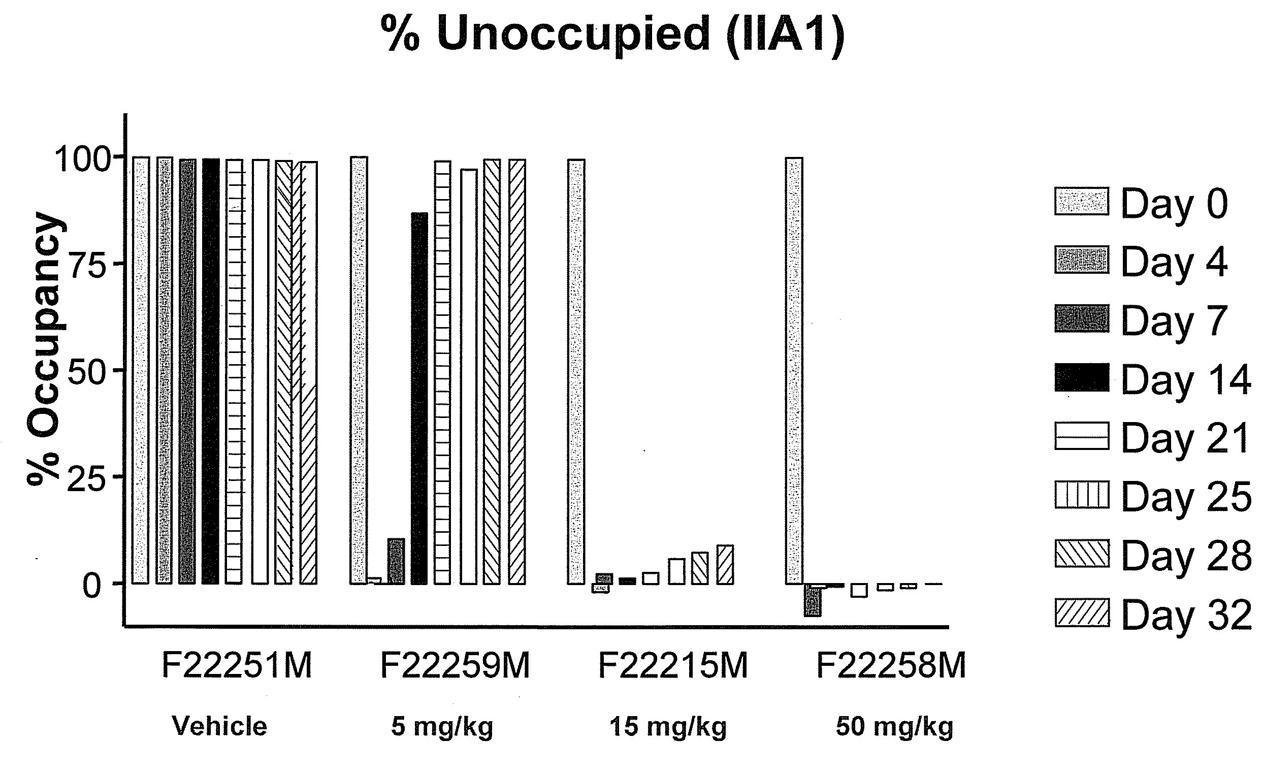
[0176]The dosing of each subject was through systemic delivery by intravenous infusion in the cephalic or saphenous vein. The dose volume for each animal was based on the most recent body weight measurement and was 50, 15 or 5 mg / kg. Intravenous infusion was conducted while the animals were restrained in primate chairs, using syringe infusion pumps. The animals were not sedated for dose administration. The dose schedule was once weekly for 4 weeks beginning on the day of laser injury.

TABLE 1Route ofGroup# of animalsadministrationPretreatmentTreatmentDoseDosing13IVlaseredVehicleNA4 doses, weekly21IVlaseredM200 5 mg / kg4 doses, weekly31IVlaseredM20015 mg / kg4 doses, weekly43IVlaseredM20050 mg / kg4 doses, weekly

[0177]The degree of saturation of α5β1 sites on CD14+ monocytes following intra venous a...


PUM

PropertyMeasurementUnit
diameteraaaaaaaaaa
temperatureaaaaaaaaaa

Abstract

The present invention provides methods that enable the user to identify inhibitors of tissue granulation in and around a wound site, thereby limiting excessive scar formation as the wounded tissue heals. The some granulation inhibitors identified using the methods of the invention inhibit granulation in and around a wound site up to five fold, with a corresponding decrease in the formation of scar tissue when tested on retinal injuries. Granulation inhibitors that can be identified using the methods of the present invention include antibodies, peptides, nucleic acids (aptamers), and non-peptide small molecules.

Description

CROSS REFERENCE TO RELATED APPLICATIONS[0001]This application is a continuation of U.S. application Ser. No. 10 / 818,068, filed Apr. 2, 2004, which claims priority from U.S. Provisional Application No. 60 / 460,642 filed Apr. 3, 2003, each of which is hereby incorporated by reference herein in its entirety.FIELD OF THE INVENTION[0002]The present invention relates to the field of biochemistry, and physiology, particularly to methods of enhancing wound healing. The methods provided enable the user to identify inhibitors useful as therapeutic agents to treat tissue granulation in and around a wound site, thereby limiting excessive scar formation as the wounded tissue heals. The granulation inhibitors identified using the methods of the invention inhibit granulation in and around a wound site up to five fold, with a corresponding decrease in the formation of scar tissue when tested on retinal injuries. In addition, these inhibitors inhibit macrophage behavior associated with lesions in RPE...

Claims


Application Information

Patent Timeline
Patent Type & Authority Applications(United States)
IPC IPC(8): A61K39/395A61P27/00A61P27/02C07K16/28
CPCA61K2039/505C07K16/2842C07K2317/56C07K2317/55C07K2317/24A61P17/00A61P17/02A61P19/02A61P27/00A61P27/02A61P29/00A61P35/00A61P43/00
Inventor JOHNSON, DALE E.JEFFRY, URSULA
Owner FACET BIOTECH CORP
Why Eureka
Try Eureka