
Method, compositions and classification for tumor diagnostics and treatment

a tumor and composition technology, applied in the field of tumor diagnostics and treatment, can solve the problems of not necessarily identifying benign or irregular tissues, increasing the incidence of either false positives or operator errors, and achieving the effects of enhancing nuclear medical imaging, reducing overall healthcare costs, and enhancing survival

Inactive Publication Date: 2009-03-26
ZUCKERMAN MATHEW MARK
View PDF2 Cites 15 Cited by
  • Summary
  • Abstract
  • Description
  • Claims
  • Application Information

AI Technical Summary

Benefits of technology

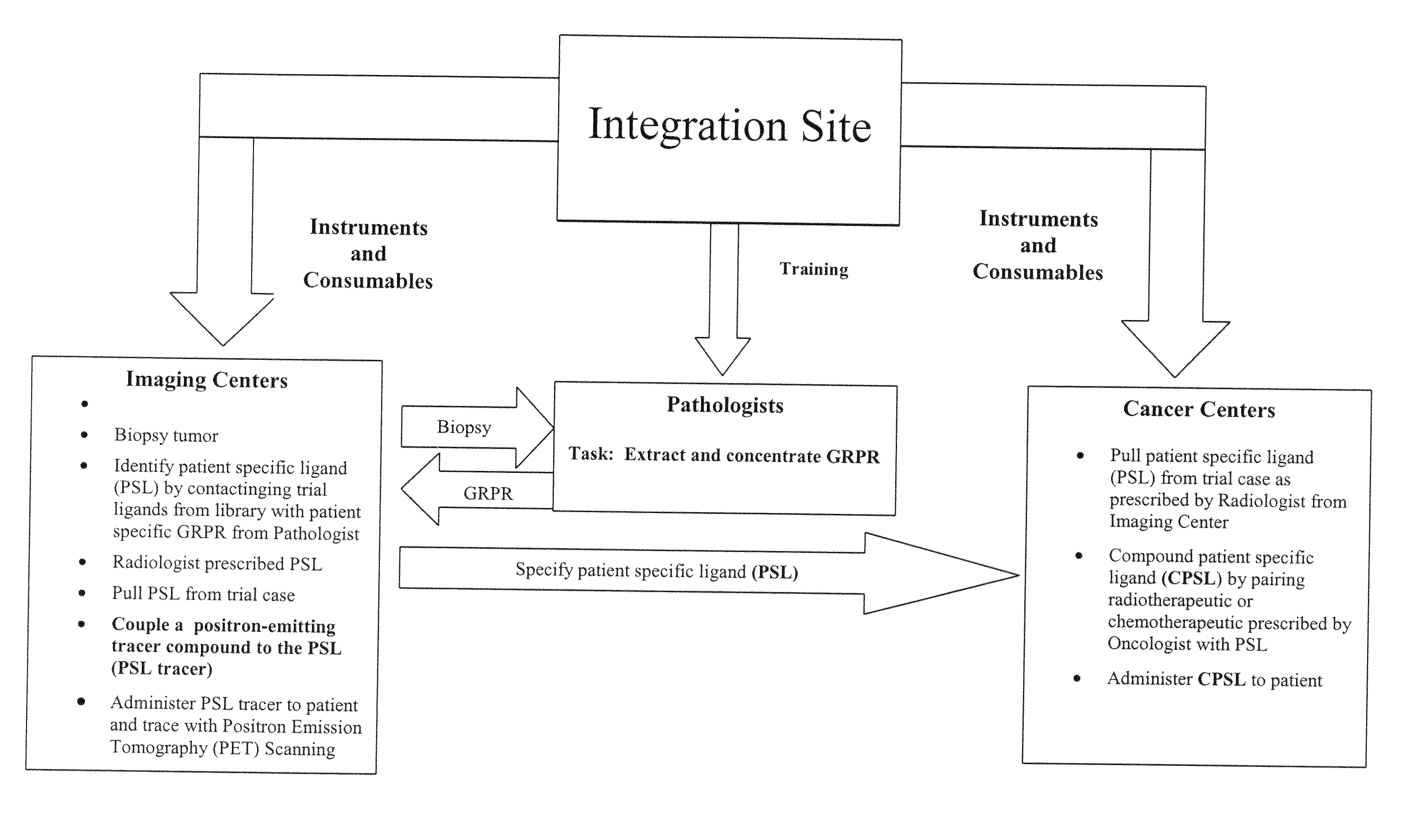
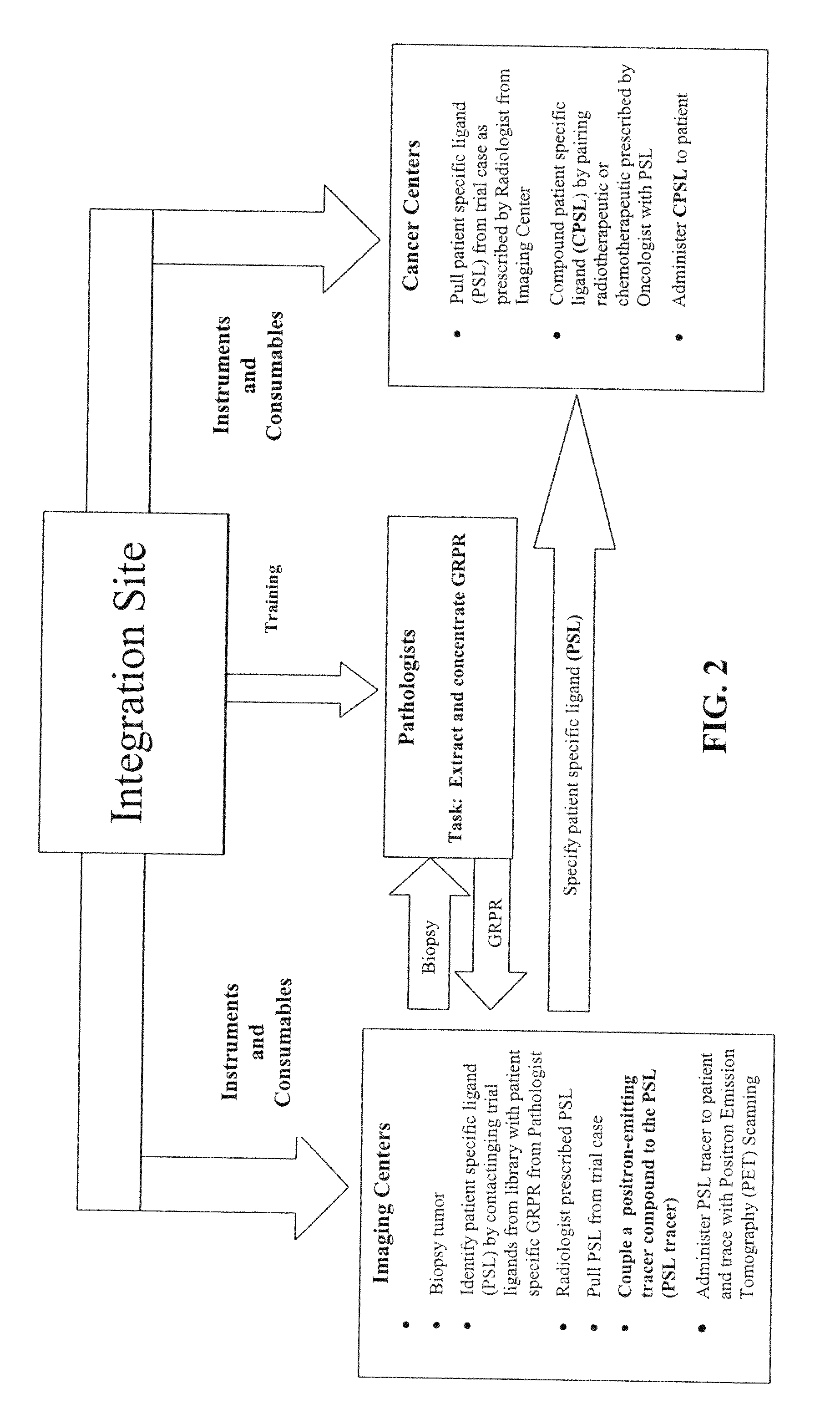
[0011]Earlier and more accurate detection of cancer has the potential to increase survival and reduce overall healthcare cost affiliated with cancer diagnosis. The present invention provides several diagnostic tools that allow earlier and more accurate detection of cancer. These tools utilize a group of tumor targeted patient specific ligands (PSL), along with radiopharmaceutical agents, to provide enhanced nuclear medical imaging. In the form of a diagnostic kit, or “Receptor TRAP”, these diagnostic tools will provide physicians with an easy and quick determination of the presence of certain cancers from a simple tissue, plasma, whole blood, or urine test. An exemplary, albeit non-limiting example is a technetium based oncology tracer for single photon emission computed tomography (SPECT). Data gathered from use of the SPECT tracer will enablement development of tracers for positron emission tomography (PET). As an oncology specific tracer, the combination of the PSL and PET tracer will enable specificity of PET imaging not heretofore possible. Because many diseases, particularly cancer, “target” the body's healthy cells resulting in abnormal cell behavior such as rapid growth or cell splitting, a cell-specific and, more importantly, patient-specific approach to cancer diagnosis and treatment will provide a crucial new and improved methodology for cancer diagnostics and therapeutics.
[0018]It is a further objective of the instant invention to provide diagnostic and therapeutic regimens that take advantage of the common features among tumors diverse in origins, locations, current status, or patients, in order to provide an economical and expedient way to diagnose and treat tumors.
[0019]It is yet another objective of the instant invention to provide methods and compositions that exploit the tumor receptor biomarker similarities to aid classification across tumors of diverse origins, by optimizing the isolation of ligands that bind to such receptor subtypes.
[0040]SPECT and PET differ from anatomically-based imaging modalities, such as MRI and X-rays, in that they assess the level of metabolic activity and perfusion in various organ systems. The process produces biologic images based on the detection of gamma rays that are emitted by a low dose radioactive substance such as the radioactive sugar FDG, which is the most common tracer used in conjunction with PET scans. The radioactive sugar can help in locating a tumor, because the faster growing cancer cells absorb sugar faster than other “normal” tissues in the body. FDG-PET has been shown to be effective in the staging of Hodgkin's and non-Hodgkin's lymphoma, and for staging of lung cancer and other tumors including cervical cancers. Improved disease staging can translate into better treatment decisions and overall prognosis.

Problems solved by technology

Positron emission tomography (PET) using radiolabeled 2-deoxyglucose to detect variations in a general location of specific cellular activity is a more recent addition; but, while this latter can putatively distinguish tumorous tissue, this is also a non-specific indicator of presumptive cancerous tissue, i.e. it cannot necessarily identify benign or irregular tissues.
The broader or more diffuse the potential location(s) for the tumorous tissues, the more time and effort must be spent locating and then studying and identifying the tumor—and the greater the incidence of either false positives or operator errors.

Method used


Image

  • Method, compositions and classification for tumor diagnostics and treatment
  • Method, compositions and classification for tumor diagnostics and treatment
  • Method, compositions and classification for tumor diagnostics and treatment

Examples


example 1

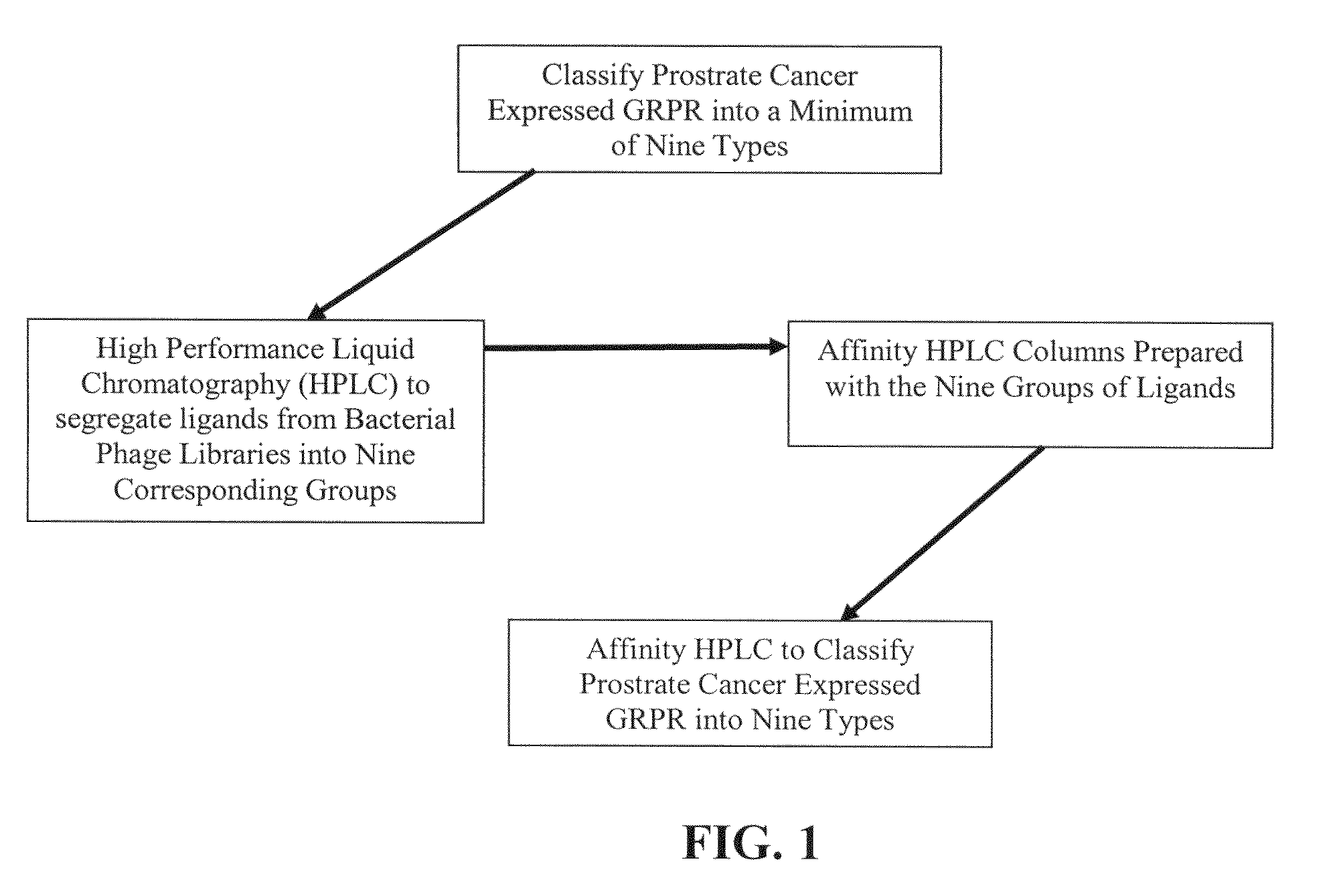
[0147]In the present invention a population 27 ligands plus the ligand in claim 1 is defined using combinations of the three active amino acids with first-order Chiral activity (Proline, Alanine and Arginine) that occur in six of the 27 positions. This population is reduced to a total population of nine by use of affinity HPLC analysis as taught herein. In the analysis extraction of the patient's GRP-r from prostate cancer tissue is performed by the steps of composite sampling, separation, enrichments, re-suspension and mining the active fragment. Tests are performed on 50 prostate cancer tumors from archival or biopsy specimens of 50 patients. Each sample is one milliliter of clear solid free liquid at a concentration of an estimated 300 micrograms per milliliter of GRP-r and is obtained from tissue samples of 1 grams wet weight of prostrate tissue. The steps in the preparation of the GRP-r are described in the following paragraphs.

Composite Tissue Sample:

[0148]Aliquots of surgical...

example 2

[0161]In the present invention an extract of the patient's GRP-r is captured in a trap containing suitable affinity chromatography media (the Trap) and the Level One ligand is: chemically tagged with fluorescence dye, contacted with the Trap and the presence of same measure in the discharge from the Trap. A reduction in the fluorescence in the discharge from the Trap greater than the expected dilution shows that GRP-r is present in the Trap and bound to some quantity of the ligand and presumes that the patient has cancerous cells. This presumption of cancer is to be confirmed by other tests not within the scope of this invention. The equipment used and the methods of analysis are described in the following paragraphs.

[0162]The equipment and major chemicals used are:

[0163]Fluorometer—Turner PicoFluor model number 80003 with mini cell adapter and cells model 8000-931; Fluorescent Dye—Molecular Probes AlexaFluor 488 a TFP ester model number A30005; Affinity Trap—Amershamphanmaciabiotec...

example 3

[0165]In the present invention radioactive tagging to form radiopharmaceuticals is added at the PET scan location or point of use by organometallic chemistry without the need for a cyclotron. The method used is described in the following paragraph.

[0166]The organometallic aquaion [99mTc(H2O)3(CO)3]+ is used as a radiosynthon for the labeling of the bioactive ligand molecule for use in PET scans. The NH2 terminus of the 27 mer ligand is functionalized to achieve radiolabeling by forming a high specific activity radiocomplex while maintaining the biological activity of the ligand. The aquaion is stable over a wide range of pH values and is characterized by excellent labeling efficiency. The labeling efficiency is associated with the presence on the three water molecules coordinated to the fac-M(CO)3 which is characteristic of the aquaion not only the amine donor group in the present invention but also with thiols, phosphines and thioesters as donor groups.

[0167]Any enumerated listing ...


PUM

PropertyMeasurementUnit
diameteraaaaaaaaaa
areaaaaaaaaaaa
affinityaaaaaaaaaa

Abstract

The present invention is directed towards classifying tumor biomarkers, particularly membrane receptors, and more particularly the gastrin-releasing peptide (GPR) receptors, identified in patient samples, then linking therapeutic agents (chemical, radiological, or biological) to patient-specific ligands that bind to such receptors, clinicians can produce diagnostic and treatment compositions and implement treatment regimens which, by using the classified and identified biomarkers, and due to their improved accuracy, increase success and decrease undesired side effects from such treatments.

Description

REFERENCE TO RELATED APPLICATIONS[0001]This application is a continuation of U.S. patent application Ser. No. 11 / 334,845, filed Jan. 19, 2006, which claims the benefit of U.S. Patent Application 60 / 645,077, filed on Jan. 19, 2005, all of which the contents are herein incorporated by reference in their entirety.FIELD OF THE INVENTION[0002]This invention relates to a method that enables identification and separation of ligands (proteins) that are specific to a cancerous cell and to at least a particular patient. It particularly relates to diagnostic and / or therapeutic applications, wherein the ligands can, after separation, be replicated and tagged with either a radioactive agent for the SPECT and / or PET tracer or a fluorescence (dye) for a Receptor TRAP. The invention most particularly relates to a technology platform which is based on gastrin releasing peptide receptors (GRP-R), which are useful as biomarkers for a number of cancer forms including renal cancer, prostate cancer and l...

Claims


Application Information

Patent Timeline
Patent Type & Authority Applications(United States)
IPC IPC(8): C07K14/00G01N33/53
CPCA61K51/0476C07K14/475C07K14/52C07K14/71G01N2500/00G01N33/57488G01N2333/475G01N2333/485C07K14/715
Inventor ZUCKERMAN, MATHEW MARK
Owner ZUCKERMAN MATHEW MARK
Learn More