Portable Pulmonary Injury diagnostic Devices And Methods

a pulmonary injury and portable technology, applied in the field of emergency medicine, can solve the problems of lungs collapse, rapid death from pulmonary injuries, injuries and deaths, etc., and achieve the effect of improving survival chances and minimal training and experien

Inactive Publication Date: 2009-06-11
VIRGINIA COMMONWEALTH UNIV
View PDF14 Cites 44 Cited by
  • Summary
  • Abstract
  • Description
  • Claims
  • Application Information

AI Technical Summary

Benefits of technology

[0044]The present inventors have invented, inter alia, a handheld device based on the frequency range 20 kHz to 50 kHz which may be used, e.g., by corpsmen to diagnose lung injury (such as, e.g., pneumothoraces) at the point of wounding in conscious and unconscious combat casualties. An exemplary inventive device uses low frequency ultrasound or sound waves, is automated, and includes an easy-to-use interface and readout, making the device non-operator dependent and requiring minimal training and experience. Early use of such an inventive device on an injured patient may improve chances of survival, such as, e.g., in the combat setting. The inventive technology also can be used in civilian trauma care including use by paramedics in the field and emergency physicians and surgeons in the trauma centers and community emergency departments.

Problems solved by technology

Injuries to the thorax can lead to a number of injuries and death.
Rapid death from these injuries usually takes the form of severe hypoxemia, hemorrhagic shock, or obstructive shock.
This causes the lungs to collapse and can be life threatening.
When “hemothorax” occurs, fluid enters the pleural space, putting pressure on the lungs and making breathing more difficult.
Hemothorax is as dangerous as pneumothorax, especially since the fluid that enters the pleural space is generally blood.
Additionally, there can exist combinations of pneumothorax and hemothorax termed hemopneumothorax which can be life-threatening; this condition usually occurs after trauma.
The dilemma with pneumothorax is that it sometimes is difficult to diagnose.
In the field, paramedics tap on the chest, using their fingers, to listen for a hollow sound, which is not an accurate method and can be misinterpreted, especially in high ambient noise.
In some cases, the chest tube has been inserted on the wrong side of the pneumothorax, which fails to cure pneumothorax and makes the problem even more life-threatening.
The most accurate conventional method to determine if pneumothorax is present is to use either an x-ray or CT scan, which takes time, which tends to reduce patient survival.
Although body armor has certainly reduced the absolute degree of penetrating injuries to the chest, this will last only until new munitions are developed that overcome the ability of the armor to prevent penetration.
In addition, new battlefield threats such as those involving improvised explosive devices have may be resulting in an increase in behind body armor trauma via blunt force mechanisms.
Although treatment of pneumothoraces ranging from tension to hemothorax is straightforward and a skill readily teachable to advanced field medics, the diagnosis of the injury based on physical exam is not.
Except for visualization of gross violation of the chest cavity, palpation of chest wall crepitance (from pleural air entering the chest subcutaneous tissue), palpation of flail chest wall segments, or visualization of tracheal deviation, the physical exam is extraordinarily limited in its ability to diagnose most pneumothoraces.
Auscultation of the chest to identify absent or diminished breath sounds and / or percussion of the chest to detect differences in conduction of mechanical energy are notoriously inaccurate even at sophisticated civilian trauma centers.
Although not as well studied, traditional percussion in this environment is unlikely to greatly improve the potential to make a correct diagnosis (Winter 1999).
Again, environmental issues (such as noise, access to the entire chest wall, etc.) will likely prevent this technique from being very useful.
Also, it is not uncommon for the injured to have bilateral pneumothoraces including hemothorax and hemopneumothorax, making the use of an uninjured lung and chest wall for comparison to the injured side not possible.
This technique is noninvasive and provides rich information, but is not practical as a combat medic or a civilian medic tool.
Even though high frequency ultrasound (MHz range) is portable and capable of detecting pneumothoraces, it is a highly operator dependent technique requiring significantly clinical training and expertise to perform (Chung, 2005, Jaffer 2005) Furthermore, high frequency ultrasound seems not to be meeting portability and ruggedness requirements needed for a field device nor to be satisfying cost concerns, so that a high-frequency ultrasound has not been viewed as a practical option for routine combat medic deployment.
Liberal performance of needle or tube thoracostomy when in doubt of the diagnosis, although occasionally life saving, and many times harmless, has its dangers including actually creating a pneumothorax (including tension pneumothorax), causing significant hemorrhage (if intercostal or lung vessels are injured), lung damage, and chest and systemic infection.
However, a portable device (such as a hand held device) which can function in high ambient noise, has proven to be a challenge to design and construct and before this invention apparently has not been provided.
A significant problem has been that typically large transducers and related amplifiers etc. are needed to propagate frequencies in the infrasound range (<20 Hz) needed to drive internal organs into resonance.
While these methods are generally reliable, they are not always available to an injured patient.
Conventional X-rays, CT scans, and ultrasound imaging require large scale equipment and are not always dependable.
When this occurs, the patient cannot breath sufficiently and can have lowered cardiac output.
If certain pneumothoraces are not diagnosed and treated, they may result in death.
Conventionally, x-rays or CT scans are used to identify pneumothorax, which requires large scale equipment and can be time consuming.
However, conventional thinking before this invention, relying on certain specific resonant frequencies from a diagnostic perspective, imposes limitations.

Method used


Image

  • Portable Pulmonary Injury diagnostic Devices And Methods
  • Portable Pulmonary Injury diagnostic Devices And Methods
  • Portable Pulmonary Injury diagnostic Devices And Methods

Examples


example 1

[0099]This Example uses a demodulating technique based on propagating an amplitude modulated (AM) signal in a non linear system as human soft tissue. Non parametric demodulation will result in the low frequency modulator and the carrier in the tissue. In this Example, a small transducer may be easily incorporated into a hand held architecture. The carrier in this Example is low frequency ultrasonic tones or low frequency ultrasonic noise. Frequencies between 25 and 50 kHz differentiate various induced fluid conditions in pig lung. The low frequency ultrasonic tones or low frequency ultrasonic noise (i.e., the stimulation) may be applied anywhere over the thorax. Preferably, the tracheal area is stimulated. Lung sounds may be recorded.

Low Frequency (>20 kHz) Ultrasound

[0100]Acoustic energy may be transformed into several other forms of energy, which may exist at the same time as heat, sonic and ultrasonic frequencies. When ultrasound is absorbed by tissue it produces alternate areas ...

example 1a

[0115]Assessment of Propagation Patterns: Fourier analysis was used to determine the spectra of signals with various transducer placements in a water / air simulated environment using a waveguide or tank.

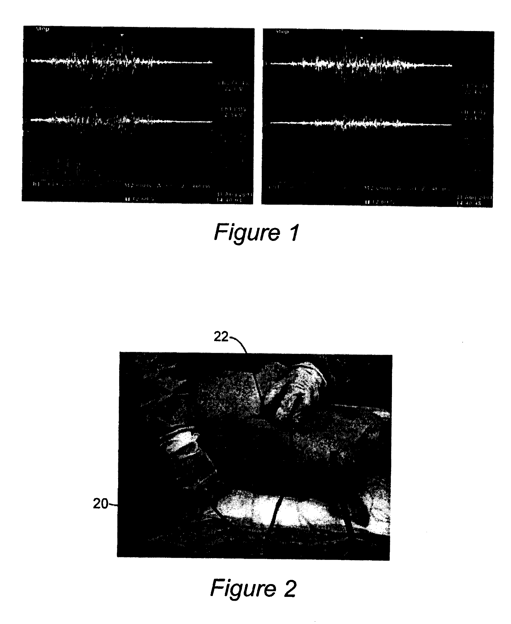
[0116]Two female adult Hampshire pigs weighing about 50 kg were used in a pilot study of a lab version of the novel hand held battle field interthoracic device. The animal study was approved by the local vertebrate animal review board. Each pig was sedated with ketimine and pentobarbital. The stimulating sensor 20 and recording sensor 22 are depicted in FIG. 2. Vibratory probes (transmitter and receiver pair) were positioned around the chest to measure the acoustic properties of each normal chest / lung. Care was taken to avoid placing the ribs in the acoustic beam. Data was acquired using a real time spectral analyzer in the surgical suite and processed off line via MatLab program developed for quantification. Immediate spectra plots were available in the lab. Two other experimental co...

example 2

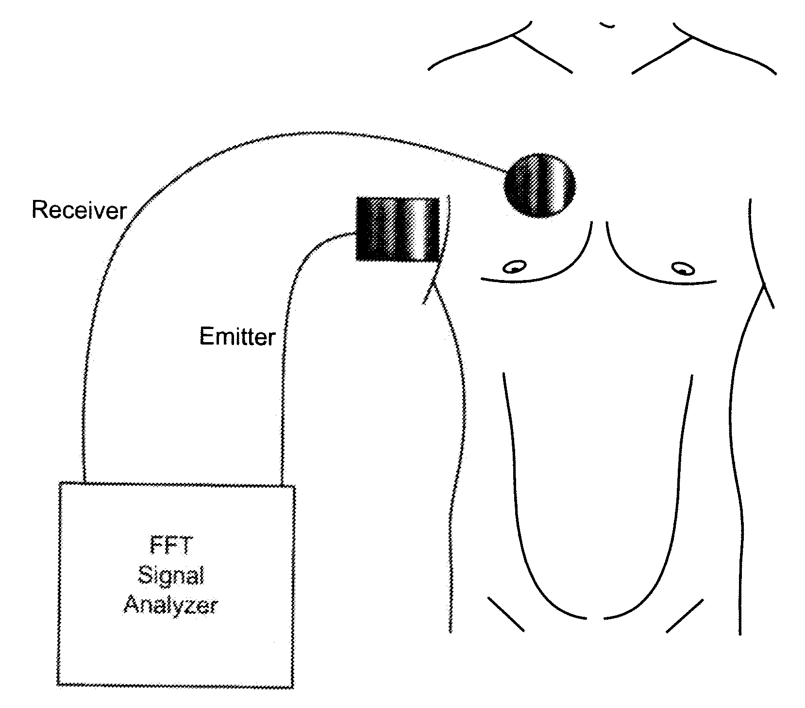
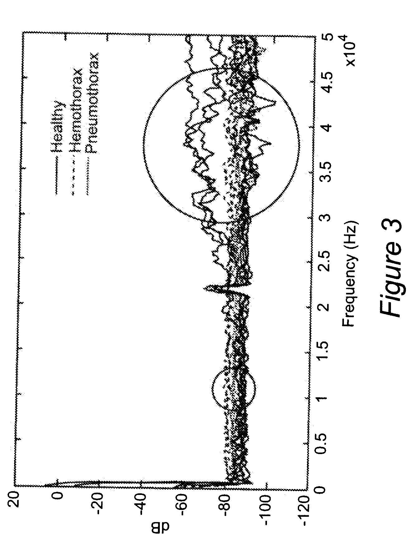
[0128]In this inventive example, a fast method and device capable of diagnosing pneumothorax uses high-frequency white noise sent through the chest cavity. The innovative portable pneumothorax detecting device of this example uses a transducer that emits a 10 Hz-50 kHz continuous band white noise sound. (FIG. 9) The pneumothorax detecting device generates vibro-acoustic wave frequencies, then transmits them from an emitter transducer to a receiver transducer located on the opposite side of the thorax. The receiving transducer, placed on the opposite side of the chest, senses the signal and sends the signal back to the real-time spectral analyzer for analysis. The spectral analyzer then computer the Fast Fourier Transform (FFT) on the signal and the result is displayed. The result indicates if pneumothorax or hemothorax is present. The velocity of the signal changes, due to attenuation, through different tissues inside the chest. When either air or fluid is present in the chest cavit...


PUM


Abstract

Lung injury such as pneumothorax now can be diagnosed reliably, portably and quickly. Vibro-acoustic waves are sent through the chest and the resulting wave is measured. By analyzing attenuation characteristics determined by the geometry of the chest structures, a determination can be made of whether the patient's pleural space is healthy, contains air (pneumothorax) or contains fluid (hemothorax).

Description

FIELD OF THE INVENTION[0001]The invention generally relates to emergency medicine, and especially relates to diagnosis of pulmonary injury including pneumothorax, hemothorax and other pulmonary injury.BACKGROUND OF THE INVENTION[0002]Injuries to the thorax can lead to a number of injuries and death. These injuries include but are not limited to: chest wall contusions, rib fractures, pneumothorax, tension pneumothorax, hemothorax, hemopneumothorax, pulmonary contusions, cardiac contusion, penetrating injuries to the heart, and great vessels of the thorax and esophageal injury. These injuries may occur secondary to both blunt and penetrating mechanisms. Rapid death from these injuries usually takes the form of severe hypoxemia, hemorrhagic shock, or obstructive shock. The vast majority of chest injuries leading to these modes of death whether in the civilian trauma or combat setting involve pneumothorax (including tension pneumothorax), hemopneumothorax, and hemothorax which can be ea...

Claims


Application Information

Patent Timeline
Patent Type & Authority Applications(United States)
IPC IPC(8): A61B5/08A61B8/00
CPCA61B5/4869A61B8/08A61B5/7257A61B5/0051
Inventor LENHARDT, MARTINWARD, KEVIN
Owner VIRGINIA COMMONWEALTH UNIV
Why Eureka
Try Eureka