
Cardiomyocytes and methods of producing and purifying cardiomyocytes

a technology of cardiomyocytes and myocytes, applied in the field of cardiomyocytes and methods of producing and purifying cardiomyocytes, can solve the problems of insufficient nutrient, oxygen, carbon dioxide, etc., and achieve the effect of promoting cardiomyocyte formation

Inactive Publication Date: 2009-06-18
NEOSTEM ONCOLOGY
View PDF3 Cites 5 Cited by
  • Summary
  • Abstract
  • Description
  • Claims
  • Application Information

AI Technical Summary

Benefits of technology

[0004]Growth Factors and Pathways Involved in Cardiac DevelopmentBone morphogenic protein 4 and 2 (BMP4, BMP2) induces cardiac differentiation in the cranial paraxial mesoderm, and blocks the differentiation of skeletal muscle precursors in these cells. Bone morphogenic proteins belong to the transforming growth factor beta super family (TGF-beta). Ligand binding to its receptor induces the formation of a complex in which the Type II BMP receptor phosphorylates and activates the Type I BMP receptor. The Type I BMP receptor then propagates the signal by phosphorylating a family of signal transducers, the Smad proteins. Currently, eight Smad proteins have been cloned (Smad 1-7 and Smad 9). Upon phosphorylation by the BMP Type I receptor, Smad1 can interact with either Smad4 or Smad6. The Smad1-Smad6 complex is inactive; however, the Smad1-Smad4 complex triggers the expression of BMP responsive genes. The ratio between Smad4 and Smad6 in the cell can modulate the strength of the signal transduced by BMP. BMP signaling is regulated at different molecular levels, most important—Noggin and other cystine knot-containing BMP antagonists bind with BMP-2, 4 and 7 and block BMP signaling. If activated, Smad6 binds type I BMP receptor and prevents Smad 1, 5 and 8 to be activated. The BMP family comprises: BMP1, BMP2, BMP3, BMP4, BMP5, BMP6, BMP7, BMP8a, BMP8b, BMP10, BMP15. The most important factors involved in the cardiac development are BMP2, BMP4, BMP7 and BMP10.

Problems solved by technology

As the embryo is growing thicker than 300-400 microns, the diffusion of nutrients, oxygen, and carbon dioxide is inadequate.
Failure to activate Cripto signaling results in conversion of ES cells into a neural fate.

Method used


Image

  • Cardiomyocytes and methods of producing and purifying cardiomyocytes
  • Cardiomyocytes and methods of producing and purifying cardiomyocytes
  • Cardiomyocytes and methods of producing and purifying cardiomyocytes

Examples


example 1

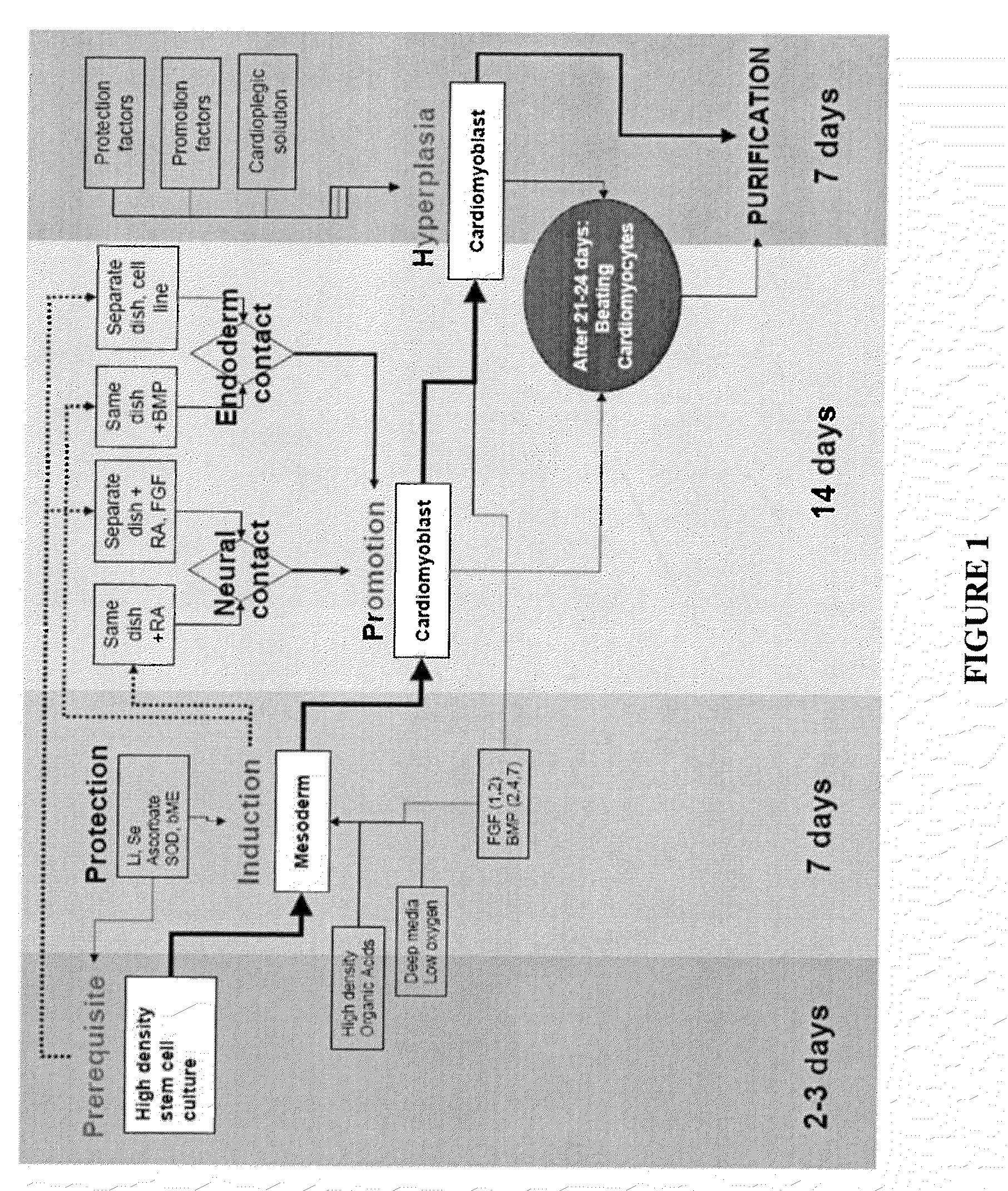
[0087]This example is of an exemplary protocol for obtaining a mixed cardiomyocyte population.[0088]1 One passage before differentiation, stem cells are plated in deep dishes which can carry 1-3 cm of supernatant. Stem cells are fed according to hES cells protocols.[0089]2. Incubator set to 37-37.5° C., 2-5% O2 5-6% CO2 and 98% H2O. Stem cells are overgrown in the original plated dishes: deep dishes (5-10 mm of supernatant) for increasing media volume will ensure hypoxia and enough nutriments for cellular overgrowth. The culture will stabilize after 3-5 days of overgrowth.[0090]3. Day 1-3 addition of retinoic acid (RA) 5 mM in DMSO to final concentration of 5 μM[0091]4. Day 1 to the end of the protocol. BMP4 10 ng / ml[0092]5. Day 1 to the end of the protocol FGF −10 ng / ml[0093]6. Replace the media daily up to day 10 and than every other day[0094]7. At day 20-25 first colonies with beating clumps and typical morphology[0095]8. Day 28. The cells are incubated for 1-2 hour in a 50% card...

example 2

[0100]This example is of an exemplary protocol for obtaining early purified cardiomyocyte population.[0101]1. One passage before differentiation the stem cells are plated in deep dishes which can carry 2-3 cm of supernatant. Stem cells are fed according to hES cells protocols.[0102]2. Stem cells are overgrown in the original plated dishes until layering of the colonies is observed (by the yellowish color on the surface) and the media is increased in volume to ensure hypoxia and nutriments. The culture will stabilize after 3-5 days of overgrowth[0103]3. In a separate dish a pre-differentiated culture consisting of pax6 / nestin positive active dividing cells of 50,000 cells / cm2, will incubate overnight the CardioMedia. Next day the media is collected and filtered. The culture can be obtained from primary culture of neural stem cells or by differentiating human embryonic stem cells for 5-7 days in a serum free media which contains 10 uM of retinoic acid and 5 ng / ml FGF2 added daily at f...

example 3

[0111]This example includes a description of media compositions used in examples 1 and 2. Cardio Media includes DMEM:F12 LO, B27 supplement, MEM-NE Aminoacids 1×, Glutamax 1×, Ascorbic acid 20 μg / ml, thyroid hormones T3 / 4 20 ng / ml and insulin 10 ug / ml Cardioplegic solution includes THAM buffer (thrometamine) 0.3 Mol, KCl 10 mM, Puerarin 0.5 mM, L-Monosodium Glutamate Monohydrate 4%, L-Monosodium Aspartate Monohydrate 4%, Trehalose 0.25%, Lactic Acid 2 mM and Ascorbic acid 20 μg / ml. Resuscitation media includes Cardio Media, Puerarin 0.5 mM, Trehalose 0.25% and Superoxide dismutase.

[0112]Neural media includes DMEM:F12 high glucose, B27 Supplement 1×, MgCl 0.5 mM, insulin 10 ug / ml, selenite 5 ng / ml and transferrin 20 ug / ml.


PUM


Abstract

The invention provides methods for producing a culture of cardiomyocytes and cultures of cardiomyocytes. Exemplary methods of producing and cultures of cardiomyocytes include a population of cells including cells having spontaneous and periodic electrical activity, and / or including nodal, sino-atrial or pacemaker cells; immature cardiomyocytes (cardiomyoblasts); mature contractile cardiomyocytes; or a mixed population of two or more of such cells.

Description

RELATED APPLICATIONS[0001]This application claims the benefit or priority of U.S. application Ser. No. 60 / 895,066, filed Mar. 15, 2007, and is expressly incorporated herein by reference.INTRODUCTION[0002]Myocardic DevelopmentAs the embryo is growing thicker than 300-400 microns, the diffusion of nutrients, oxygen, and carbon dioxide is inadequate. A circulatory system is needed and developed by the middle of the fourth week of gestation (embryonic day 24). Heart and skeletal muscle progenitor cells are derived from distinct regions of the mesoderm: lateral plate mesoderm and paraxial mesoderm, respectively. Cells from the cranial paraxial mesoderm (anterior splancnic mesoderm) contribute to both myocardial and endocardial cell populations. The primordium of the heart forms in the cardiogenic plate located at the cranial end of the embryo.[0003]Development of the Conduction SystemThe factor that directs myocardial development toward either a working myocardium phenotype or a conducti...

Claims


Application Information

Patent Timeline
IPC IPC(8): C12Q1/02C12N5/06C12N5/077
CPCC12N5/0657C12N2500/25C12N2500/34C12N2500/38C12N2501/999C12N2501/155C12N2501/385C12N2501/395C12N2501/70C12N2501/115A61P9/00
Inventor NISTOR, GABRIEL
Owner NEOSTEM ONCOLOGY
Learn More