Cancer cell diagnosis by targeting delivery of nanodevices
a nano-device and cancer cell technology, applied in the field of nanoparticles, can solve the problems of insufficient image contrast, toxicity of gadolinium-based contrast agents, and their impact on persons with impaired kidney function, and achieve the effects of increasing the original concentration of polyions, reducing and increasing the hydrodynamic diameter of nanoparticles
- Summary
- Abstract
- Description
- Claims
- Application Information
AI Technical Summary
Benefits of technology
Problems solved by technology
Method used
Image
Examples
example 1
Nanoparticles Formed from Poly Acrylic Acid (PAA) and Polyammonium Salt (PAMM)
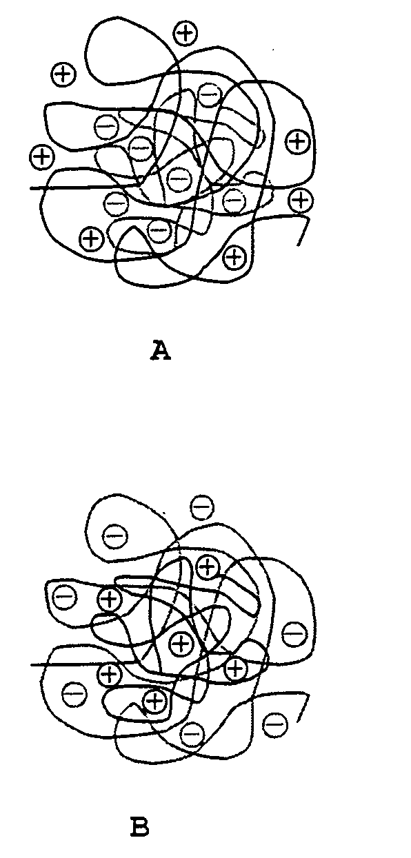
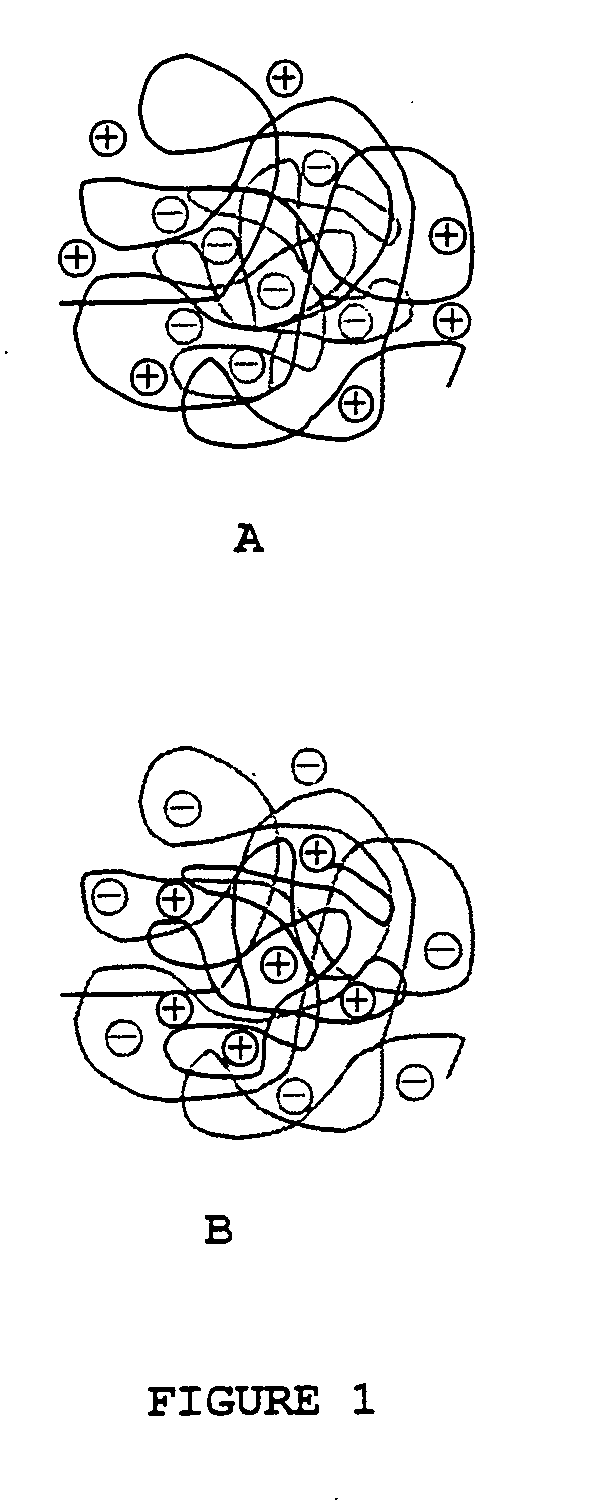
[0025]PAA with Mw=200 kDa and poly(2-methacryloxyethyltrimethylammonium bromide) were each separately dissolved in water at a concentration of 1 mg / ml. The pH value of solutions was adjusted to pH=3 by 0.10 mol / dm3 sodium hydroxide. To the solution of PAA under gentile stirring was added the solution of PAMM. After 1 hour the pH was increased to 7 resulting in a stable nanosystem with particle size of 50 to 350 nm measured by laser light scattering method. The size of nanoparticles may be varied and in a range of 10-1,000 nm by using polymers with different molecular weight. Also the particle size increases at higher pH due to the repulsion of negative charges.
example 2
Nanoparticles Formed from Chitosan (CHIT) and Poly Gamma Glutamic Acid (PGA)
[0026]Chitosan is a linear polysaccharide of randomly distributed β-(1-4)-linked D-glucosamine (deacetylated unit) and N-acetyl-D-glucosamine (acetylated unit).
[0027]In the present example, CHIT with MW=320 kDa and PGA with Mw=11.3 MDa were each separately dissolved in water. The concentration of the solutions was varied in the range 0.1 mg / ml-2 mg / ml. The pH value of solutions was adjusted to pH=3 by 0.10 mol / dm3 hydrochloric acid. The ratio of polyelectrolyte and the order of mixing was modulated. After 1 hour mixing the pH was increased by 0.1 M sodium hydroxide solution resulting stable nanosystems. The hydrodynamic diameter of nanoparticles was in the range of 40-480 nm at pH=3, and at pH=7 was 470-1300 nm measured by laser light scattering method. There was some precipitation at higher pH caused by flocculation and coagulation. The size of nanoparticles may varied by using polymers with different molec...
example 3
Nanoparticles Formed from CHIT and Hyaluronic Acid (HYAL)
[0028]CHIT with Mv=320 kDa and HYAL with Mw=2.5 MDa were dissolved in water. The concentration of CHIT was varied in the range 0.1 mg / ml-1 mg / ml, and of HYAL 0.04-0.2 mg / ml. The pH value of solutions was adjusted to pH=3 by 0.10 mol / dm3 hydrochloric acid. The ratio of polyelectrolyte and the order of mixing was modulated. After 1 hour mixing the pH was increased by 0.1 M sodium hydroxide solution resulting stable nanosystems. The hydrodynamic diameter of nanoparticles was in the range of 130-350 nm at pH=3, and was higher than 600 nm at pH=7 measured by laser light scattering method. There are some precipitation at higher pH caused by flocculation and coagulation.
[0029]The size of nanoparticles may varied by using polymers with different molecular weight.
PUM
| Property | Measurement | Unit |
|---|---|---|
| Atomic weight | aaaaa | aaaaa |
| Atomic weight | aaaaa | aaaaa |
| Atomic weight | aaaaa | aaaaa |
Abstract
Description
Claims
Application Information
 - R&D
- Intellectual Property
- Life Sciences
- Materials
- Tech Scout
- Unparalleled Data Quality
- Higher Quality Content
- 60% Fewer Hallucinations
Browse by: Latest US Patents, China's latest patents, Technical Efficacy Thesaurus, Application Domain, Technology Topic, Popular Technical Reports.
© 2025 PatSnap. All rights reserved.Legal|Privacy policy|Modern Slavery Act Transparency Statement|Sitemap|About US| Contact US: help@patsnap.com


