Microwell array chip for detecting antigen-specific lymphocytes, method of detecting and method of manufacturing antigen-specific lymphocytes, and method of cloning antigen-specific lymphocyte antigen receptor genes

Active Publication Date: 2009-07-16
BLINK BIOMEDICAL SAS
View PDF10 Cites 83 Cited by
  • Summary
  • Abstract
  • Description
  • Claims
  • Application Information

AI Technical Summary

Problems solved by technology

However, they are incapable of identifying individual antigen-specific lymphocytes present in the lymphocyte population.
(1) It is difficult to set the separating conditions of the device, requiring skills for operating device to separate out cells;
(2) The background is high, precluding the detection of antigen-specific lymphocytes at frequencies of less than or equal to 0.1 percent;
(3) cell separation is inefficient;
(4) time is required to separate out cells of low frequency; and
(5) although antigen binding can be determined, it is difficult to analyze the reaction of the lymphocyte that has bound the antigen.
However, it is not possible to analyze the reaction of the lymphocyte in binding the antigen (the metabolic or physiological reaction of the cell, such as intracellular signal transduction, RNA synthesis, or protein synthesis).
Further, the antigen-specific lymphocyte cannot be detected when the frequency of the antigen-specific lymphocyte is less than or equal to 0.1 percent.
(1) In humans, there exists the method in which peripheral B lymphocytes are transformed with EB virus, antigen-specific antibody-producing cells are screened from the colonized cells, and antibody genes are cloned from the antigen-specific lymphocytes (Roome, A. J., Reading, C. L. The use of Epstein-Barr virus transformation for the production of human monoclonal antibodies. Exp. Biol. 43:35-55, 1984 (Nonpatent Reference 5), Carson, D. A., Freimark, B. D. Human lymphocyte hybridomas and monoclonal antibodies. Adv. Immunol. 38:275-311, 1986 (Nonpatent Reference 6)). This method is bothersome in that the screening of antigen-specific lymphocyte cell colonies is inefficient and a whole month is required to culture the cells. Although hybridomas can be produced for mice, no efficient human hybridoma system has yet been produced.
(2) A further method exists in which bacteriophage is employed to clone antigen-specific antibody genes (Huston, J. S., Levinson D., Mudgett-Hunter, M., Tai, M. S., Novotny, J., Margolies, M. N., Ridge, R. J., Bruccoleri, R. E., Haber, E., Crea, R., et al. Protein engineering of antibody binding sites: recovery of specific activity in an anti-digoxin single-chain Fv analogue produced in Escherichia Coli. Proc. Natl. Acad. Sci., U.S.A. 85:5879-5883, 1988 (Nonpatent Reference 7)). In this case, mRNA is extracted from human lymphocytes, cDNA libraries consisting of the H and L chains of immunoglobulin, respectively, are prepared, the two are combined into single phage DNA, and the H and L chains are expressed by the phage. Antigen specificity is determined by the combination of H and L chains. However, in this system, the combinations are random and the phages producing antibody binding to the antigen are screened with antigen. As a result, when a phage producing antibody binding to antigen is produced, an antigen-specific antibody gene can be cloned. However, the random combination of H and L chains renders the screening of antigen-specific antibody genes highly inefficient. For example, assuming that the H chain cDNA and L chain cDNA of antibody for a given antigen are each present in the library at a frequency of one part in 104, the combination of an H chain and L chain capable of binding the antibody will be present at a frequency of one part in 108. Further, in this system, it is not known whether the combinations of H and L chains obtained are actually produced in the human body.
As set forth above, conventional methods of cloning antigen-specific antibody genes are highly inefficient.
Even so, with considerable effort, it is possible to clone antigen-specific antibody genes by these methods.
However, it is not possible to identify and select low-frequency antigen-specific lymphocytes using conventional methods.

Method used


Image

  • Microwell array chip for detecting antigen-specific lymphocytes, method of detecting and method of manufacturing antigen-specific lymphocytes, and method of cloning antigen-specific lymphocyte antigen receptor genes
  • Microwell array chip for detecting antigen-specific lymphocytes, method of detecting and method of manufacturing antigen-specific lymphocytes, and method of cloning antigen-specific lymphocyte antigen receptor genes
  • Microwell array chip for detecting antigen-specific lymphocytes, method of detecting and method of manufacturing antigen-specific lymphocytes, and method of cloning antigen-specific lymphocyte antigen receptor genes

Examples


embodiment 1

1. Separation of B Lymphocytes

[0179]Employing Ficoll-Paque (Pharmacia, Uppsala, Sweden), the lymphocyte fraction was separated from peripheral blood. The B lymphocyte fraction was then further separated and purified from the lymphocyte fraction using an AutoMACS (Miltenyi Biotec, Bergisch Gladbach, Germany)

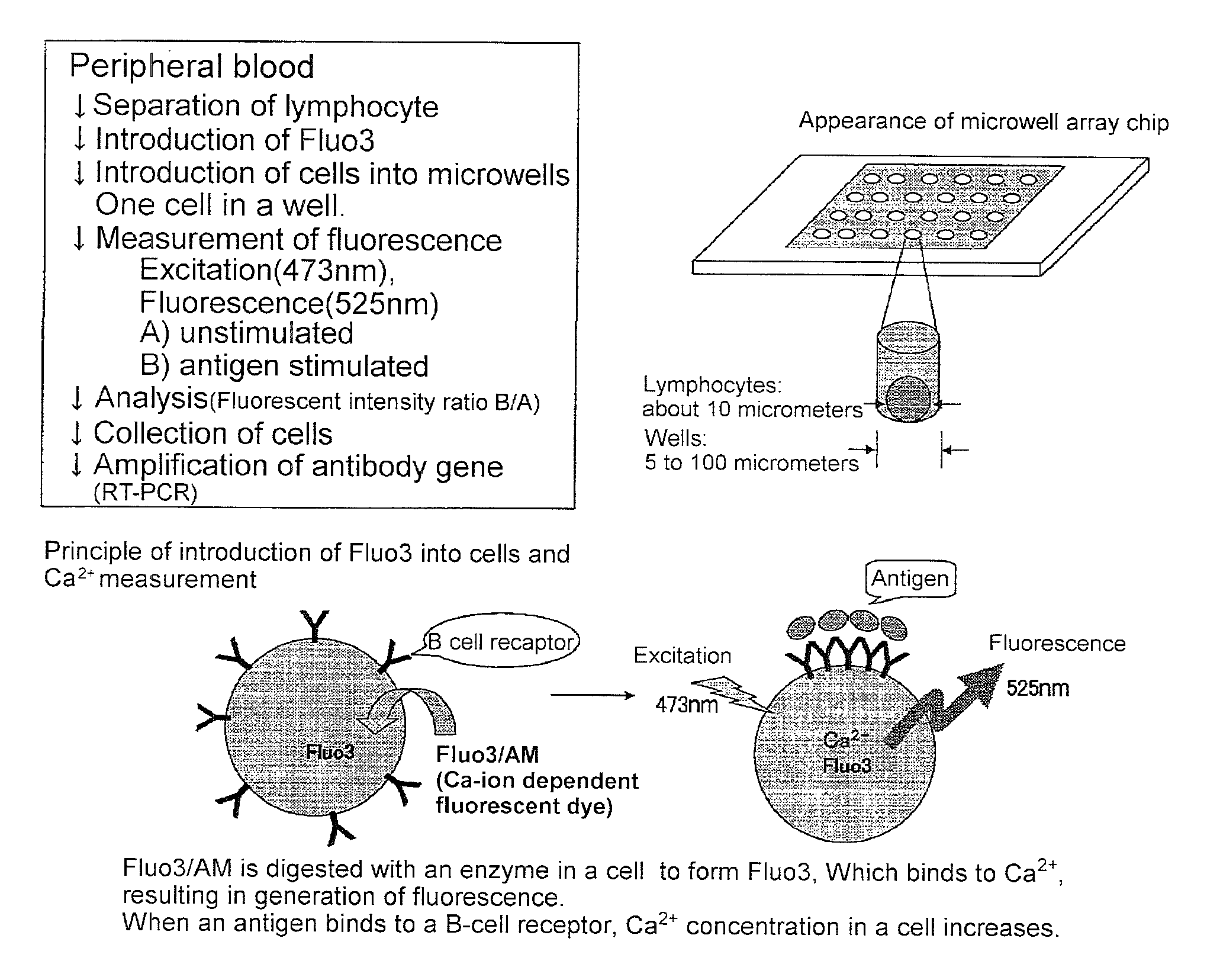
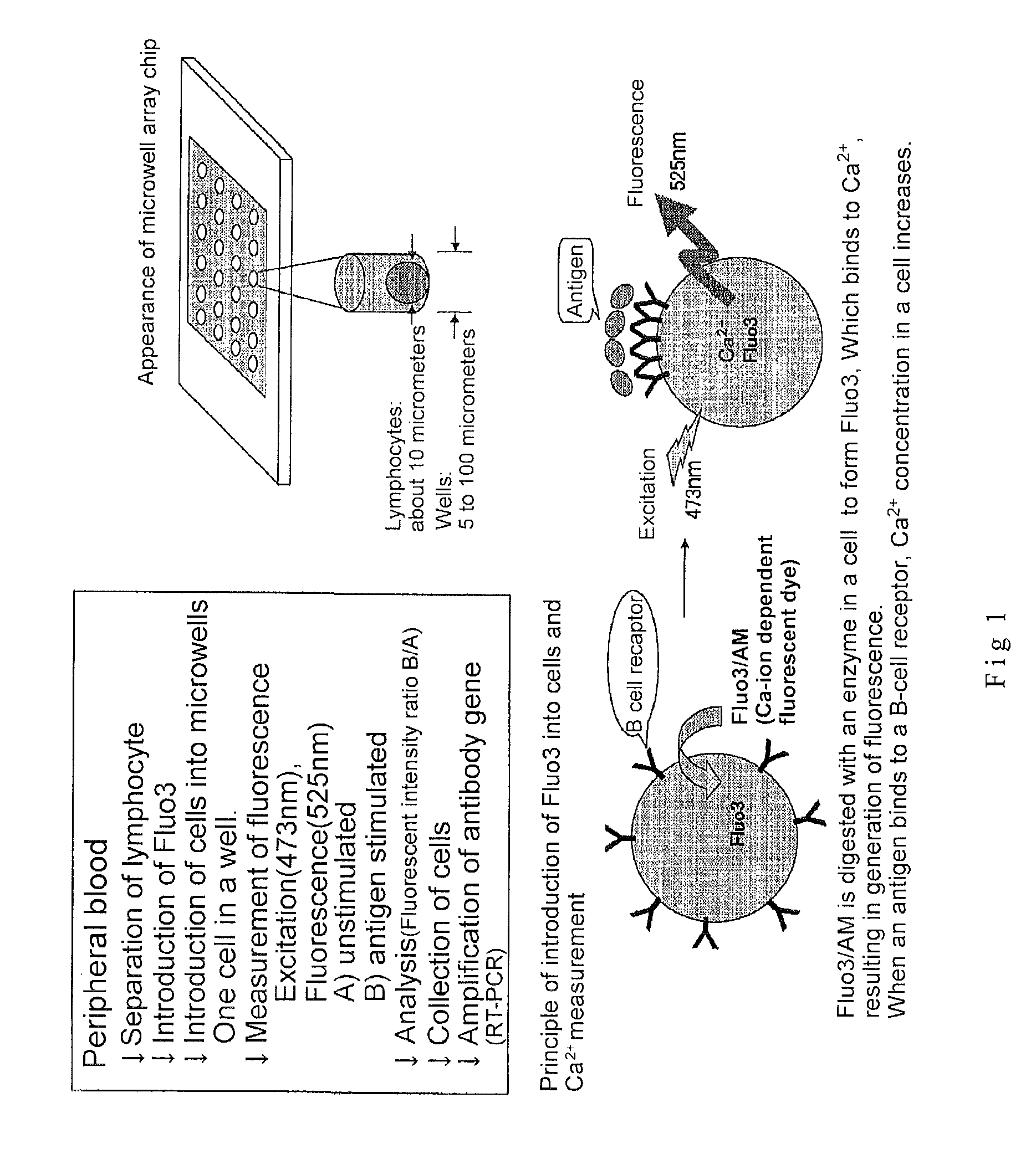
2. Introduction of Fluo3 into Cells (See FIG. 1)

[0180]2×10⁶ cells of B lymphocyte were suspended in RPMI 1640 / 10 percent FCS solution containing 2 micromoles of Fluo3 / AM (Dojin, Kumamoto) and incubated for 30 min at room temperature. The cells were washed with RPMI 1640 / 10 percent FCS to remove the Fluo3 / AM that had not been incorporated into the cells. Subsequently, the cells were suspended in RPMI 1640 / 10 percent FCS solution.

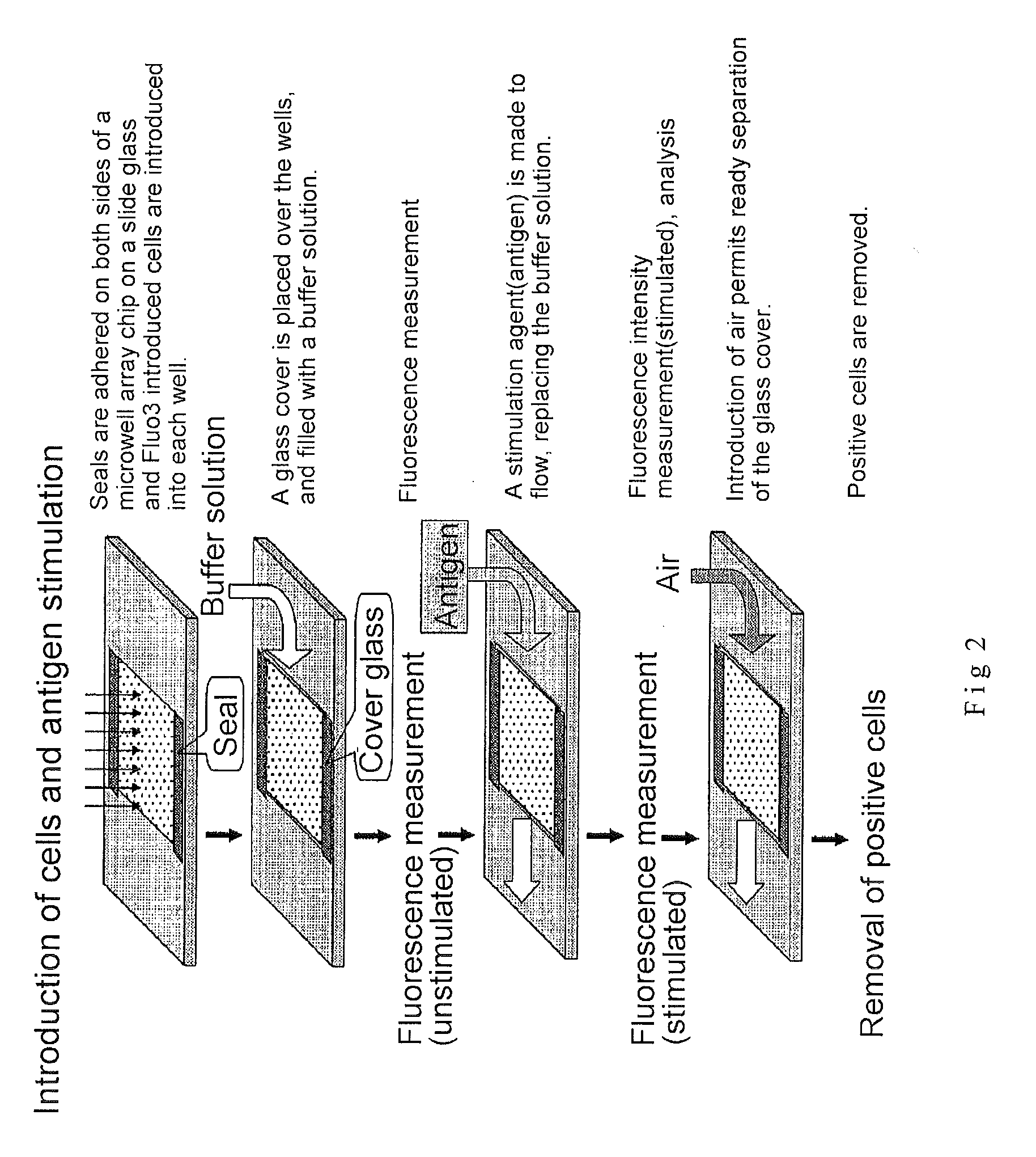
3. Microwell Array Chip (See FIG. 2)

[0181]The microwell array chip was made of poly(dimethylsiloxane) (PDMS) and had microwells of 10 micrometers in diameter and 32 micrometers in depth arranged horizontally and vertically at a spacing of 30 micrometers (th...

embodiment 2

[0188]The efficiency of introduction of cells into the microwells was examined by fluorescence microscopy or microarray scanning.

[0189]Mouse lymphocytes were fluorescently labeled with CellTracker Orange (Molecular Probe Corp.).

[0190]The mouse lymphocytes were obtained as follows.

[0191]Spleens were extracted from mice and transferred to plastic Petri dishes containing PBS. The spleens were sandwiched between two pieces of mesh and crushed to remove lymphocytes. The lymphocytes that were removed were suspended in RPMI 1640 / 10 percent FCS solution and the number of lymphocytes was counted.

[0192]The fluorescent labeling of the murine lymphocytes was conducted as follows.

[0193]2×10⁶ cells of B lymphocytes were suspended in loading buffer (20 mM HEPES, pH 7.4, 137 mM NaCl, 2.7 mM KCl, 1.8 mM CaCl2, 1 mM MgCl2, 1 mg / mL glucose, 1 mg / mL BSA) containing 1 micromole of CellTracker Orange (Molecular Probes, USA), 0.02 percent of Pluronic F-127 (Molecular Probes, USA) and incubated for 30 min ...

embodiment 3

Detection of Antigen-Specific B Lymphocytes by Microarray Scanner

[0200]A healthy volunteer was inoculated with hepatitis B virus vaccine and B lymphocytes were prepared from peripheral blood on days 4 and 6 by the usual methods. Human B lymphocytes were suspended in buffer (loading buffer (20 mM HEPES, pH 7.4, 137 mM NaCl, 2.7 mM KCl, 1.8 mM CaCl22H2O, 1 mM MgCl2, 1 mg / mL glucose, 1 mg / mL BSA)) containing 4 micromoles of the calcium fluorescence indicator Fluo-4 / AM (Molecular Probes Corp.), 1 micromole of CellTracker Orange (Molecular Probes Corp.), and 0.02 percent of Pluronic F-127 (Molecular Probes Corp.) to introduce Fluo-4 and CellTracker Orange into the cytoplasm. B lymphocytes loaded with Fluo-4 and CellTracker Orange were applied by the same method as in Embodiment 2 onto a microwell array chip identical to that in Embodiment 2.

[0201]Subsequently, the B lymphocytes loaded with Fluo-4 and CellTracker Orange that had been seeded in the microwell array chip were stimulated with...


PUM

PropertyMeasurementUnit
frequencyaaaaaaaaaa
shapeaaaaaaaaaa
dimensionsaaaaaaaaaa

Abstract

A microwell array chip that has multiple microwells and is employed to contain a single lymphocyte specimen in each microwell and detect antigen-specific lymphocytes in single units; wherein the microwell array chip is of a shape and of dimensions where only one lymphocyte is contained in each microwell. A method of detecting antigen-specific lymphocytes comprising the steps of adding antigen to each microwell in the above microwell array chip, stimulating the lymphocyte specimen, and detecting lymphocyte specimens reacting with the antigen.

Description

[0001]This application is a divisional application of U.S. patent application Ser. No. 10 / 534,800 filed May 12, 2005, which is the National Phase of PCT / JP03 / 12500, filed Sep. 30, 2003, which claims priority to Japanese patent applications JP 2002-331031, filed Nov. 14, 2002, and JP 2002-346728 filed Nov. 29, 2002. The entire contents of all of the above are herein incorporated by reference.TECHNICAL FIELD[0002]The present invention relates to a microwell array chip employed in detecting antigen-specific lymphocytes, a method of detecting antigen-specific lymphocytes, and a method of manufacturing antigen-specific lymphocytes. The present invention further relates to a method of cloning antigen-specific lymphocyte antigen receptor genes.TECHNICAL BACKGROUND[0003]In the past, antigen-specific lymphocytes have been detected by placing about 200,000 individual lymphocytes per well in a 96-well plate such as that shown in FIG. 3 and culturing them for from three days to a week (“Lymphoc...

Claims


Application Information

Patent Timeline
Patent Type & Authority Applications(United States)
IPC IPC(8): C40B30/06C07K16/08C40B60/14G01N33/53G01N33/543G01N33/569
CPCB01J2219/00317B01J2219/00743C07K16/082G01N33/56972C40B60/14G01N33/5302G01N33/54366C07K2317/21
Inventor MURAGUCHI, ATSUSHIKISHI, HIROYUKITAMIYA, EIICHISUZUKI, MASAYASU
Owner BLINK BIOMEDICAL SAS
Why Eureka
Try Eureka