
Method for analyzing and processing fluorescent images

a fluorescent image and microscope technology, applied in image data processing, diagnostics using light, sensors, etc., can solve the problems of inability to optimize resection based on an objective determination of tumor borders, inability to identify tumor borders independently of surgeons, and inability to remove only tumor cells with minimal damage to normal tissue, etc., to improve patient life expectancy, prevent additional neurological disorders, and improve the quality of life

Inactive Publication Date: 2009-08-13
KANTONSSPITAL AARAU
View PDF1 Cites 34 Cited by
  • Summary
  • Abstract
  • Description
  • Claims
  • Application Information

AI Technical Summary

Benefits of technology

[0016]The inventive method achieves this object and provides the associated support in resection of tumor tissue, so that only a minimal amount of normal tissue adjacent to the tumor is removed and thus additional neurological disorders can be better prevented.
[0017]Another object of this invention is to improve the quality of life and to increase the life expectancy of patients through virtually complete tumor removal that can be achieved.

Problems solved by technology

The tumor to be removed often has a wide cell border infiltrating the normal tissue, or tumor cell clusters are surrounded by normal tissue, so it is difficult to remove only the tumor cells with minimal damage to the normal tissue.
Optimized resection based on an objective determination of the tumor borders is impossible with the methods known so far.
Because color perception varies from one surgeon to the next and the subjective perception of a fluorescent image depends on the environment and lighting in the operating room, no objective method of determining tumor borders independently of the surgeon has thus been disclosed.

Method used


Image

  • Method for analyzing and processing fluorescent images
  • Method for analyzing and processing fluorescent images
  • Method for analyzing and processing fluorescent images

Examples


Embodiment Construction

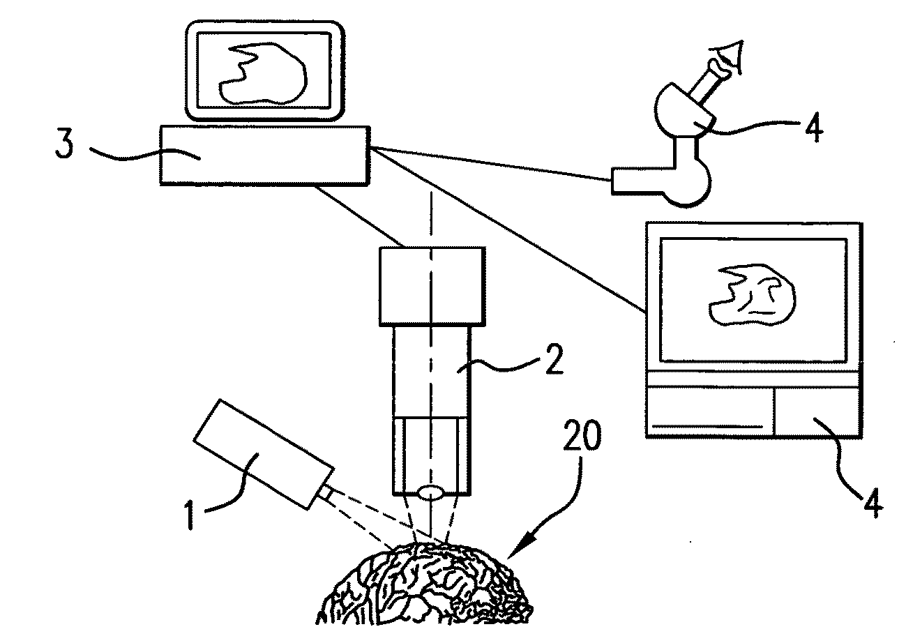
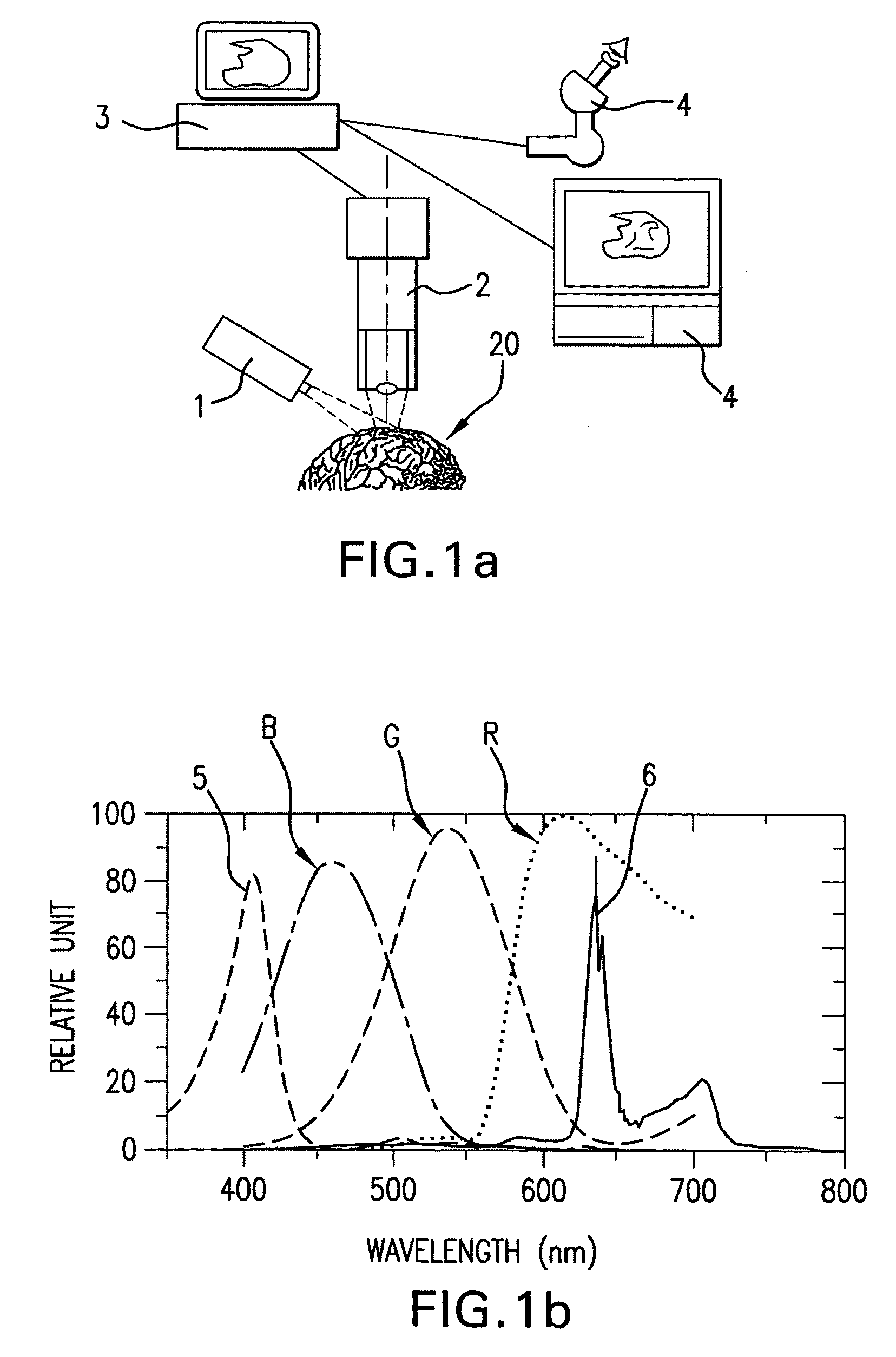
[0026]After a substance for generating a tumor detection feature, preferably 5-aminolevulinic acid has been administered orally to a glioma patient to generate a protoporphyrin fluorescence, the patient has been prepared for surgery, anesthetized and a surgical area 20, where the operation is to be performed has been made accessible, resection of tumor tissue 22 is performed using a surgical microscope.

[0027]One possible arrangement of the surgical microscope is diagrammed schematically in FIG. 1a. The operating area 20 is illuminated with UV radiation, which is in the wavelength range of approx. 400 nm and thus is in the visible blue wavelength range, from an excitation device 1. The fluorescent UV radiation may originate from a xenon light source that has filters or from a laser, for example. The 5-aminolevulinic acid synthesizes protoporphyrin IX, which has fluorescent properties and which accumulates selectively in pathologically altered cells.

[0028]In addition to an excitation ...


PUM


Abstract

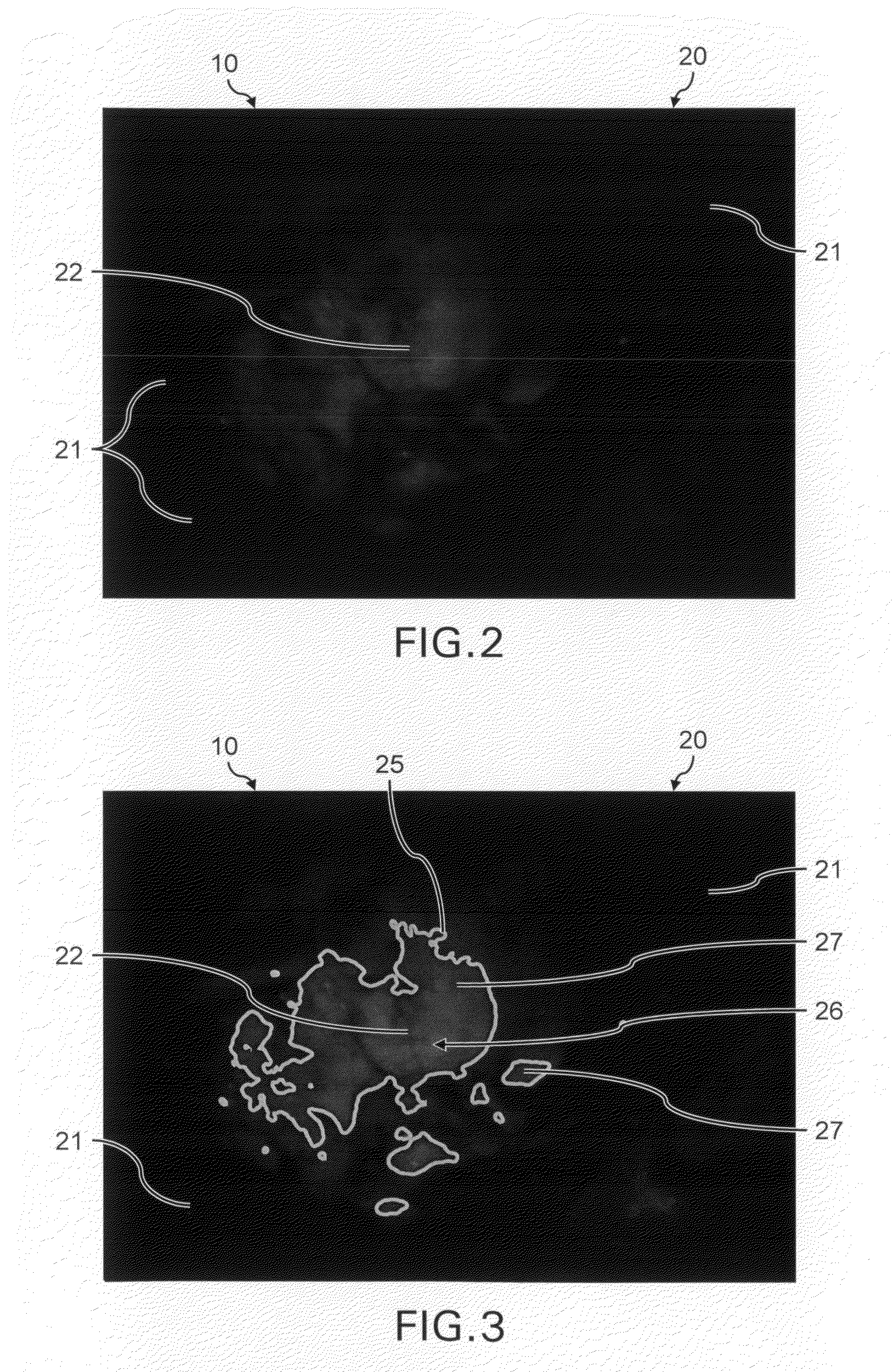
A method for analysis and processing of fluorescent images. Fluorescent light is emitted by tumor tissue in an illuminated surgical area detected by an image detection device and is forwarded to an image processing system. After determination of a maximal intensity, the image processing system determines a threshold value as a predefined fraction of the maximal intensity. With basic morphological operations of image processing, limit intensity lines which separate the tumor tissue of intensities above the threshold value and normal tissue of intensities below the threshold value are generated and imaged in a line profile image, wherein the line profile image can be superimposed on the fluorescent image.

Description

BACKGROUND OF THE INVENTION[0001]1. Field of the Invention[0002]This invention describes a method for analyzing and processing fluorescent images of a medical surgical microscope.[0003]2. Discussion of Related Art[0004]The fluorescence of tumor tissue, which is achieved by enrichment of the tumor tissue with special contrast media and by subsequent illumination with UV light, has been used in medicine for a long time, such as to support resection of portions of tissue in the operating room.[0005]In this method, known as intraoperative fluorescence detection, a natural endogenous substance (5-aminolevulinic acid, abbreviated 5-ALA), is administered to a patient with a gliobastoma before operating on the brain tumor. 5-ALA is formed in the body as part of the synthesis of hemoglobin, the pigment in blood, and is indispensable in hematopoiesis. It has the property of being converted to the pigment protoporphyrin IX in malignant brain tumor tissue. Protoporphyrin IX accumulates in neopl...

Claims


Application Information

Patent Timeline
Patent Type & Authority Applications(United States)
IPC IPC(8): G06K9/00A61B6/00
CPCA61B5/0059G02B21/0012G02B21/16G02B2207/113G06T7/0012A61B1/043G06T7/0085G06T2207/10056G06T2207/10064G06T2207/20036G06T2207/30096G06T7/0081A61B5/742G06T7/11G06T7/13
Inventor HEFTI, MARTINLOOSER, HERBERTLANDOLT, HANS
Owner KANTONSSPITAL AARAU
Learn More