

In Vivo Mitochondrial Labeling Using Positively-Charged Nitroxide Enhanced and Gadolinium Chelate Enhanced Magnetic Resonance Imaging
a magnetic resonance imaging and positively charged nitroxide technology, applied in the field of magnetic resonance imaging, can solve the problems of difficult to maximize the signal intensity received from the tumor, difficult to distinguish a tumor from surrounding tissue, etc., and achieve the effect of enhancing the contrast of processes, facilitating identification, and significantly increasing metabolic activity
- Summary
- Abstract
- Description
- Claims
- Application Information
AI Technical Summary
Benefits of technology
Problems solved by technology
Method used
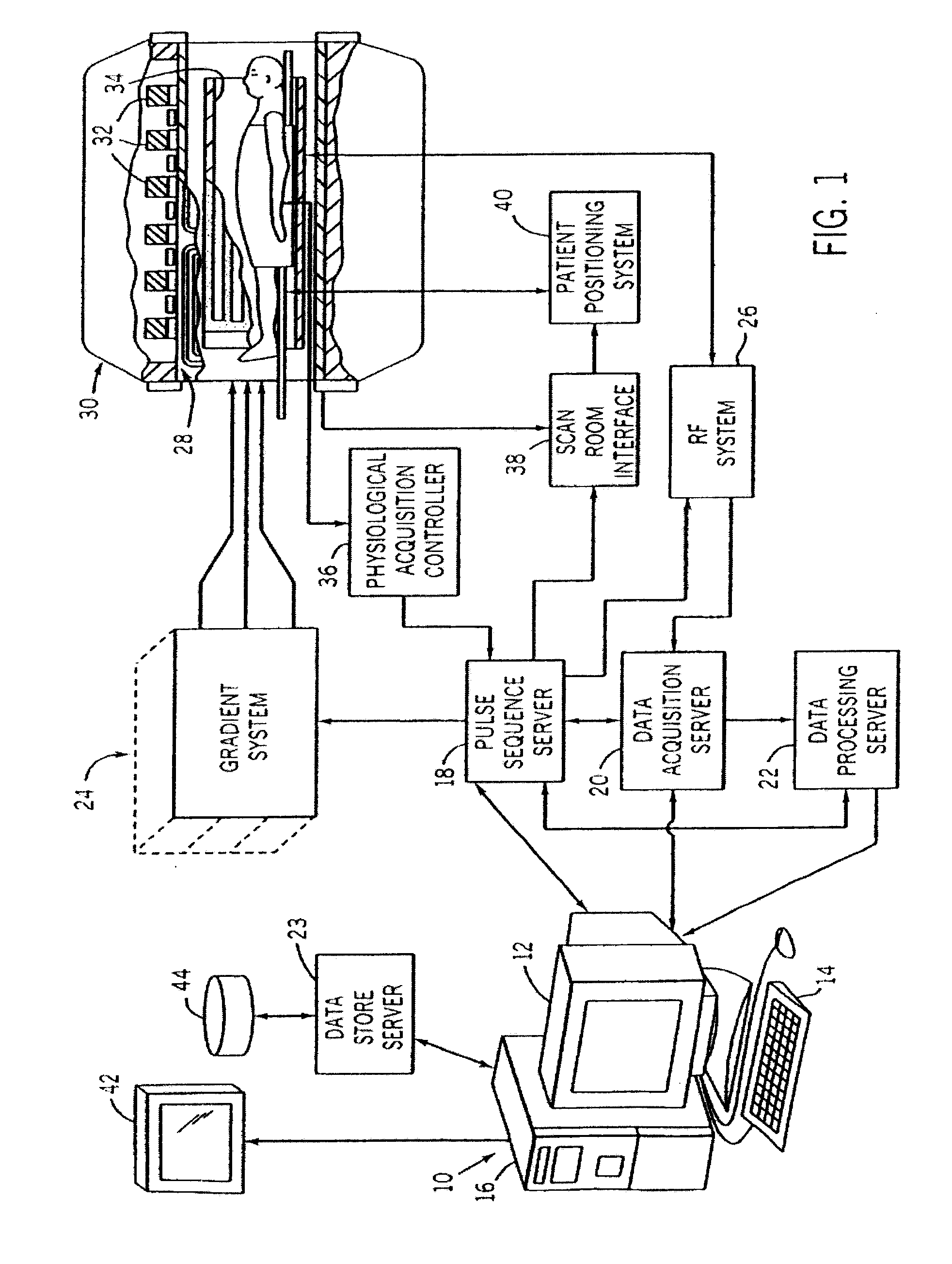
Image

Examples
example 1
Materials and Methods
[0075]All chemical and reagents were obtained from Sigma-Aldrich (Milwaukee, Wis.) unless otherwise noticed. All reagents were used as received without further purification. All cell culture materials and buffers were obtained from Invitrogen (Grand Island, N.Y.).
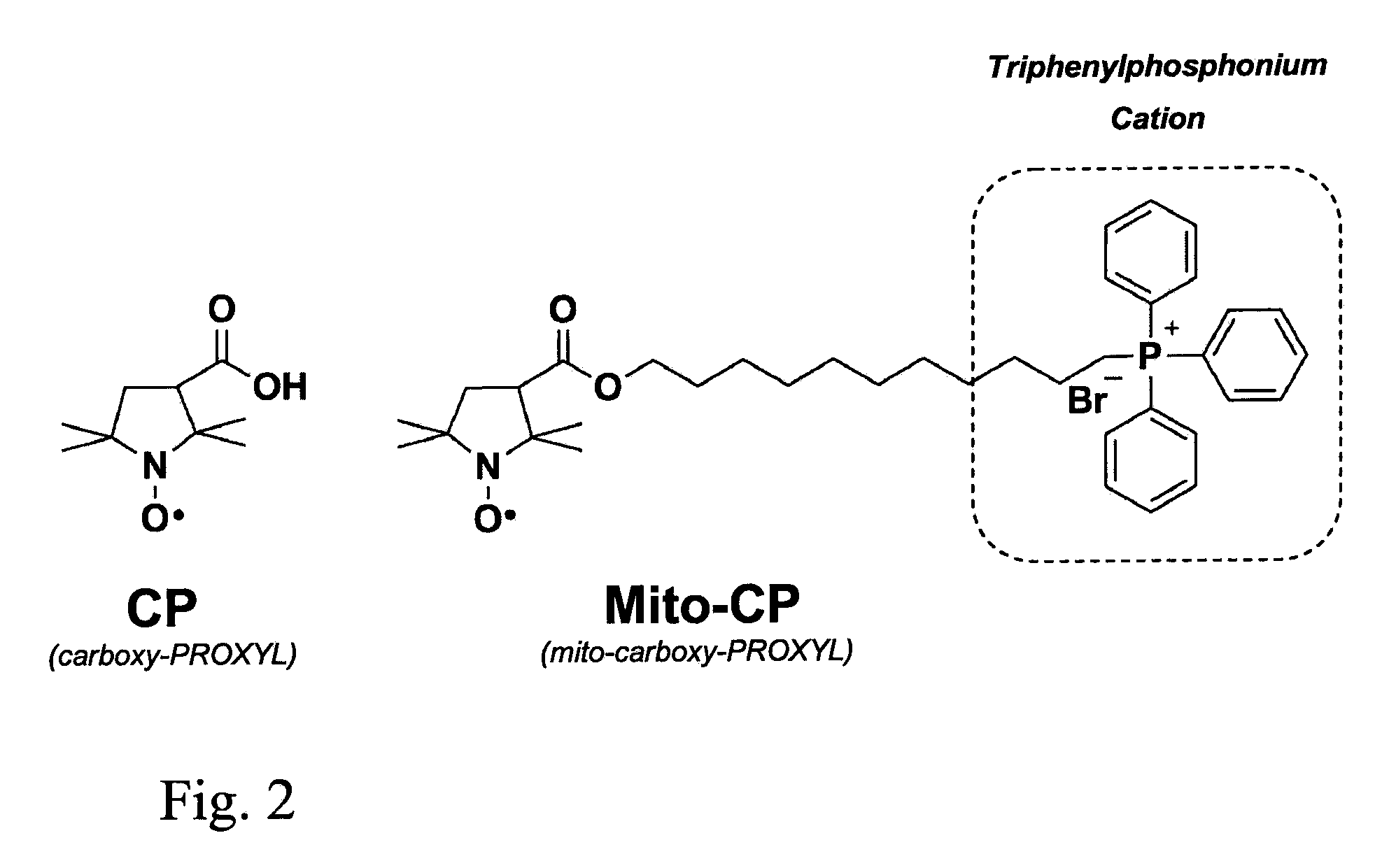
[0076]Synthesis of Mito-CP. Mito-CP, (11-(3-carboxy-2,2,5,5-tetramethyl-1-pyrrolidinyloxy)-undecyl)-triphenylphosphonium bromide was synthesized as described previously (Dhanasekaran, et al. at Free Radic Biol Med, 2005. 39(5): p. 567-8).
[0077]Cell Culture. MCF7, MDA-MB-231 and MCF10-A cells were obtained from the American Type Culture Collection (ATCC) and cultured at 37° C. in humidified 95% air / 5% CO2. MCF7 cells were maintained in MEM-alpha medium containing 10% FBS, L-glutamine (4 mM), penicillin (100 μg / ml), streptomycin (100 μg / ml), non-essential amino acids and sodium pyruvate. MDA-MB-23 were grown in Dulbecco's Modified Eagle's medium (DMEM) containing 10% FBS, L-glutamine (4 mM), penicillin (1...
example 2
[0082]The MTT assay (Sigma-Aldrich, Milwaukee, Wis.) was used to measure cell survival using quantitative colorimetry (λ=570 nm). This assay is based on the capacity of mitochondrial dehydrogenases to reduce MTT to form the insoluble formazan product. Cells were plated in 12-well plates and treated at pre-confluency with Mito-CP and CP (0-30 μM) in their respective media for 48 h. The culture media was removed after treatment, washed gently with DPBS and MTT (5 mg / ml in media) was added to each well, following incubation of the plates for 1 h at 37° C. The MTT solution was removed and the formazan product in each well was dissolved with dimethyl sulfoxide (DMSO). The optical density of each well was measured using an Agilent 8453 UV-Vis spectrometer at 570 nm.
[0083]MCF7, MDA-MB-231 and non-tumorigenic MCF10-A were grown to pre-confluency and treated with either Mito-CP or parent compound CP (1-30 uM) for 48 h and cell viability was measured using the MTT assay. F...
example 3
Relaxivities of CP and Mito-CP
[0084]The longitudinal relaxivities of other nitroxides have been approximate 0.2 mmol−1 s−1. In order to assess the longitudinal relaxivity of CP and Mito-CP, concentrations were prepared with the expectation of this relaxivity. Two separate initial solutions of CP and Mito-CP were prepared. Longitudinal relaxivities were determined for both CP and Mito-CP at three field strengths. Relaxivities were obtained from non-linear fits of standard spin echo inversion recovery images calculated using Matlab.
[0085]The longitudinal relaxivity (R1) for buffered saline solutions of Mito-CP at various field strengths matched previous published values for other nitroxides, R1≈0.2 mM−1 sec−1. FIG. 4 depicts slight field dependence such that the relaxivity increased with field strength. At 1.5T, 3.0T, and 9.4T Mito-CP exhibited an R1=0.1643±0.01314, 0.2322±0.0151, and 0.2083±0.0431 mM−1 sec−1, respectively. In addition to these measurements, CP was found to exhibit an...
PUM
| Property | Measurement | Unit |
|---|---|---|
| membrane potential | aaaaa | aaaaa |
| inversion time | aaaaa | aaaaa |
| dynamic T1-weighted time | aaaaa | aaaaa |
Abstract
Description
Claims
Application Information