Methods and assays to assess cardiac risk and ischemia

a technology of applied in the field of methods and assays to assess cardiac risk and ischemia, can solve the problem that detection assays rely on signal generation technologies that lack sufficient sensitivity to d

Inactive Publication Date: 2009-09-17
NANOSPHERE INC
View PDF0 Cites 12 Cited by
  • Summary
  • Abstract
  • Description
  • Claims
  • Application Information

AI Technical Summary

Benefits of technology

[0006]In one embodiment, the method provides for detection of cardiac troponin concentrations of 10 pg / mL or less, e.g., less than about 1 pg / mL, based on calibration to the independently established NIST troponin standard (National Institute of Standards and Technology; see www.nist.gov), e.g., concentrations of 2 fg / mL to 10 pg / mL, 2 fg / mL to 100 fg / mL or 10 fg / mL to 500 fg / mL. Such a sensitive assay allows for detection of a test subject that is not at risk of or having a cardiac event, e.g., not at risk of or having acute coronary syndrome, as well as distinguishing between acute coronary syndromes, such as UA and NSTEMI. For instance, elevated cardiac troponin levels that do not substantially increase over time (e.g., 2.0 fold) likely indicate chronic conditions, while a greater rate increase in cardiac troponin levels over time may be indicative of UA or NSTEMI. The time points for comparison may be minutes apart, e.g., 5, 10, 20, or 30 minutes apart, hours apart, e.g., 1, 2, 4, 6, or 8 hours apart, or one or more days apart, or any combination thereof. In one embodiment, the rate of increase of cardiac troponin levels in a test subject having NSTEMI is greater than the rate of increase of cardiac troponin levels in a test subject having UA. In one embodiment, the complexes are detected with one or more second moieties that specifically bind cardiac troponin linked to a detectable molecule, such as a nanoparticle, an oligonucleotide or barcode. In one embodiment, to enhance the detection of the detectable molecule, the signal generated by the detectable molecule can be amplified. For instance, a silver coating (deposition) on a gold nanoparticle bound to a complex on a substrate can amplify the signal generated by the presence of the gold nanoparticle when exposed to light and a fluorescently labeled primer that hybridizes to an oligo- or poly-nucleotide bound to a complex on a substrate can be employed to amplify nucleic acid sequences in the oligo- or poly-nucleotide by enzymatic processes.
[0011]Thus, the invention includes methods that employ an epitope-specific assay format to distinguish different forms of troponin and the changes of each form over time, so as to provide a differential diagnosis between UA, NSTEMI, ACS, AMI, and the like, at an earlier time point, which may reduce the need for additional cardiac diagnostic assays. Also included are methods for detecting troponin that are more sensitive, which employ a cutoff and / or slope measurements that may be used to differentiate one population, risk group or diagnosis from another for cardiac events.

Problems solved by technology

However, current clinical cTn protein detection assays rely on signal generation technologies that lack sufficient sensitivity to detect lower amounts of cTn that may be clinically relevant.

Method used


Image

  • Methods and assays to assess cardiac risk and ischemia
  • Methods and assays to assess cardiac risk and ischemia
  • Methods and assays to assess cardiac risk and ischemia

Examples


example 1

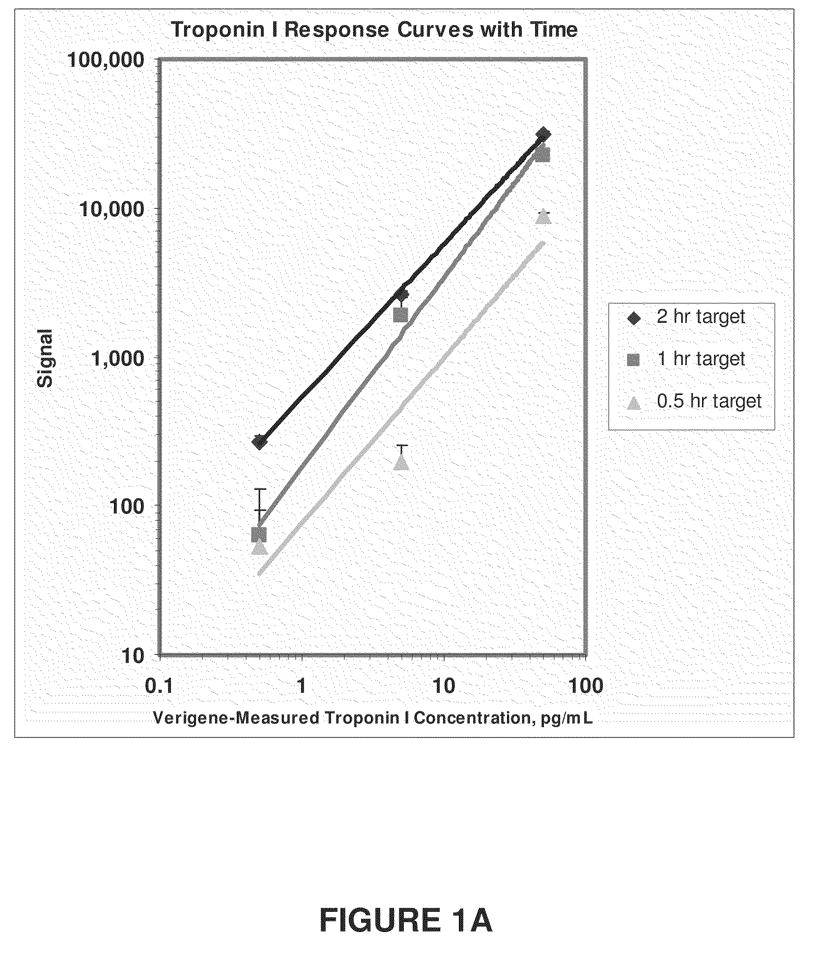
cTnI Assay Sensitivity as a Function of Time

[0108]In the example shown in FIG. 1A, known amounts of cTnI (50, 5, 0.5 and 0 pg / mL of cTnI NIST standard spiked into troponin depleted serum) were introduced to an array of anti-troponin antibodies (81-7 antibody analyzed) deposited on Codelink substrates (SurModics, Eden Prairie, Minn.) in separate assays. For each cTnI concentration, three different incubation times were tested (0.5 hours (hr), 1 hr, and 2 hrs). After capture of the cTnI molecules, a x-cTnI probe comprised of 13 nm diameter gold particle functionalized with 19C7 anti-troponin antibodies was added to each sample. Detection of the gold particle labeled troponin was achieved by catalytically reducing silver onto the surface of the gold particles followed by imaging light scattered by the silver amplified gold particles using a surface scanner such as a Tecan LS (Tecan USA) or a Verigene ID (Nanosphere, Northbrook, Ill.) detection system. These data demonstrate that differ...

example 2

Measurements on Patient Samples

Methods

[0109]The nanoparticle-based assay quantifies an optical signal resulting from selective detection of the cTn1 molecule through analyte-specific capture of gold nanoparticle probes, followed by subsequent signal enhancement. Briefly, capture antibody (mouse monoclonal, Nanogen) was contact-spotted in triplicate onto CodeLink activated microarray slides along with controls in ten sub-arrays and assembled into a hybridization chamber. Fifty microliters of sample was incubated for 90 minutes at 35° C. Wells were washed with 0.3% Tween 20 in phosphate-buffered saline (PBS) pH 7.4, then incubated with detection antibody (rabbit polyclonal, Nanogen) in 1% BSA, 0.3% Tween in PBS pH 7.4 and incubated for 20 minutes. Wells were washed again, then incubated in 150 pM gold nanoparticles for 10 minutes. Slides were washed, followed by a 150 mM sodium nitrate, pH 7.5 wash. Capture substrates were immersed in signal enhancement reaction mix for 9 minutes at 1...

example 3

Differential Measurement of cTnI Epitopes Using Selected Specific Antibodies and Diagnostic Algorithms

[0114]In this example, a cTnI sample (50 pg / mL of cTnI NIST standard spiked into troponin depleted serum) was introduced to an array of anti-troponin antibodies deposited on Codelink substrates (labeled Capture Ab), FIG. 2. The capture antibodies have known specificities for various parts of the cTnI molecule, or different forms of the cTnI molecule, FIG. 2. After capture of the various cTnI molecules, a probe or probes functionalized with antibody to complementary parts of the molecules of interest were introduced. In this example, the secondary antibodies were attached to 13 nm diameter gold particles and introduced in separate assays. Each of the anti-troponin secondary antibodies (labeled probe Ab) binds to a unique epitope of the cTnI molecule. Capture of the probe(s) and the signal generated depends on the absence, presence and / or quantity of the cTnI molecules, parts or modif...


PUM

PropertyMeasurementUnit
concentrationsaaaaaaaaaa

Abstract

The invention provides methods and apparatus to assess cardiac risk and ischemia by detecting or analyzing cardiac troponin levels. Also provided are methods to detect low levels of cardiac troponin in physiological fluid samples.

Description

CROSS-REFERENCE TO RELATED APPLICATIONS[0001]The present application claims the benefit of the filing date of U.S. application Ser. No. 60 / 998,457, filed on Oct. 10, 2007, the disclosure of which is incorporated by reference herein.BACKGROUND[0002]Cardiac troponin T (cTn) was introduced in the routine laboratory diagnostic work-up in 1989 to sensitively indicate acute myocardial ischaemia (MI). Primary causes of troponin T positivity include MI, unstable coronary heart disease, myocarditis, hypertrophic cardiomyopathy, uraemic cardiomyopathy, atrial fibrillation, congestive heart failure, hypotonia, hypovolaemia, percutaneous coronary intervention, and ASD closure. Primary causes of troponin I positivity include MI, unstable coronary heart disease, myocarditis, pericarditis, hypertrophic cardiomyopathy, tachycardia, atrial fibrillation, congestive heart failure, increased left ventricular mass, severe aortic valve disease, coronary vasospasm, cardiac contusion, cardiac tamponade, hy...

Claims


Application Information

Patent Timeline
Patent Type & Authority Applications(United States)
IPC IPC(8): G01N33/53
CPCG01N2800/324G01N33/6893
Inventor GIBBONS, WINTON G.HOLZMAN, THOMAS F.LEFEBVRE, PHILLIP A.SHIPP, GREGORY W.
Owner NANOSPHERE INC
Why Eureka
Try Eureka