Probes for In Vivo Targeting of Active Cysteine Proteases
a technology probes, which is applied in the field of in vivo targeting of active cysteine proteases, can solve the problems that none of these methods have been translated for radiological imaging, and achieve the effects of reducing the risk of cancer
- Summary
- Abstract
- Description
- Claims
- Application Information
AI Technical Summary
Benefits of technology
Problems solved by technology
Method used
Image
Examples
example 1
Preparation of Labeled Probes
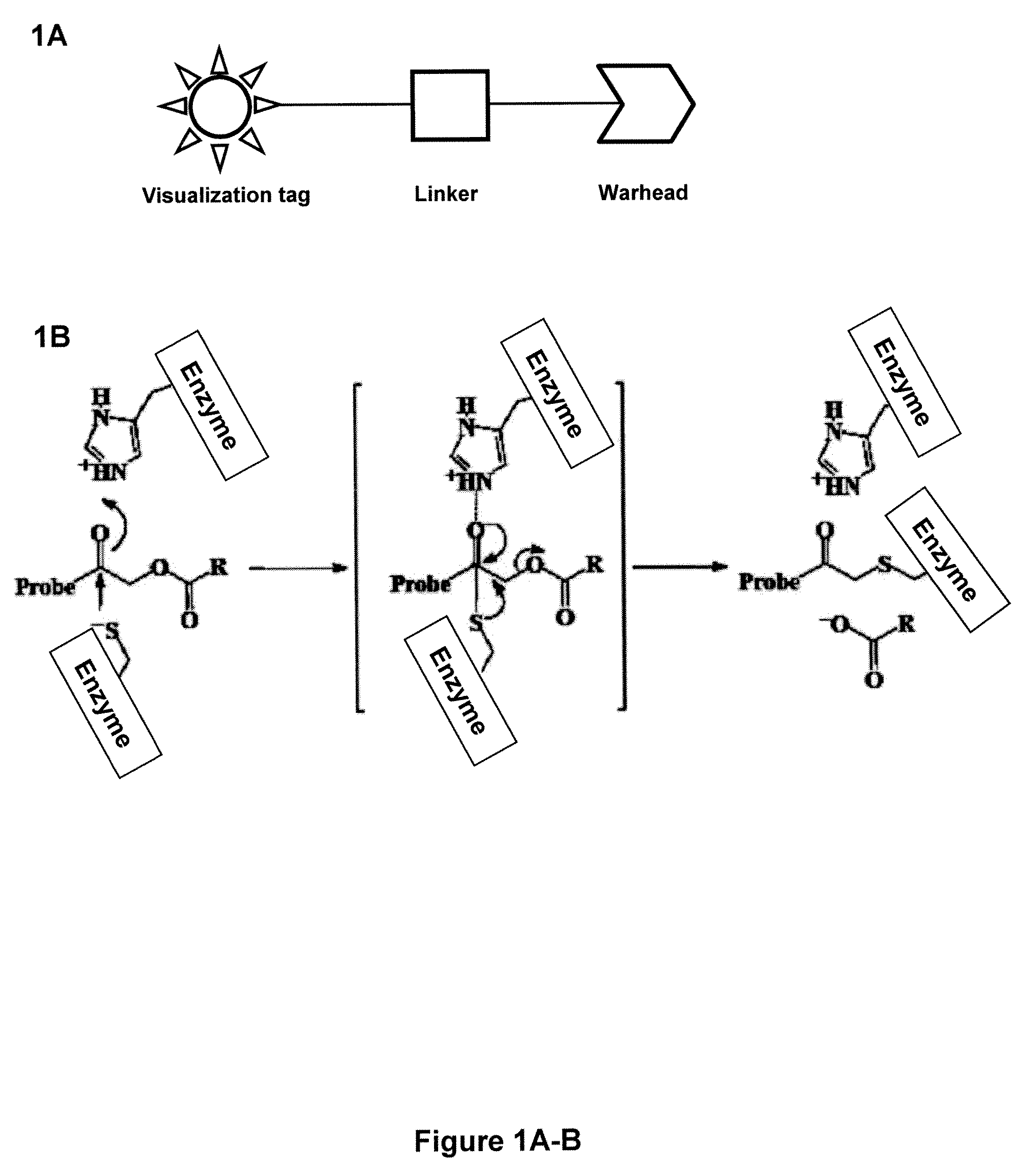
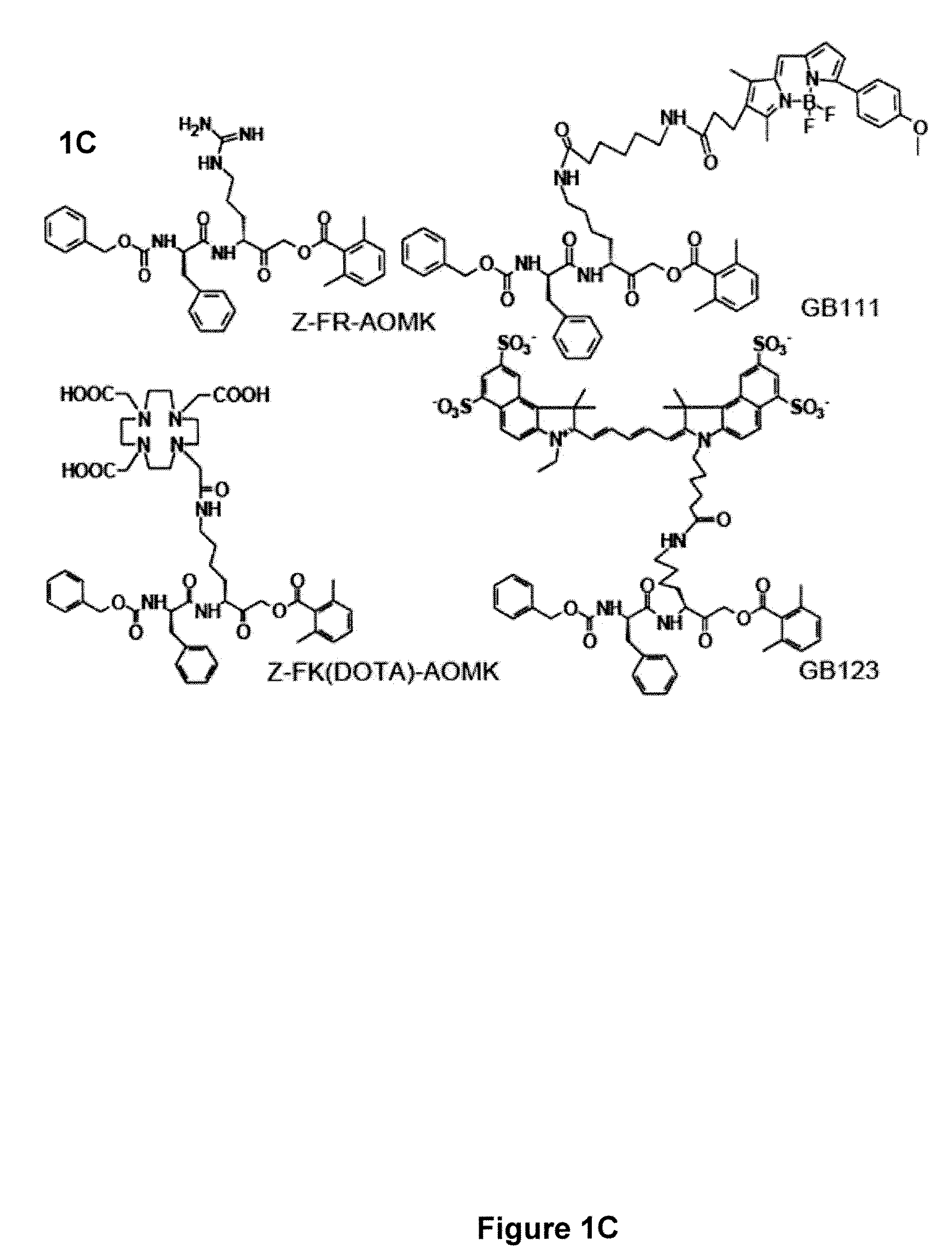
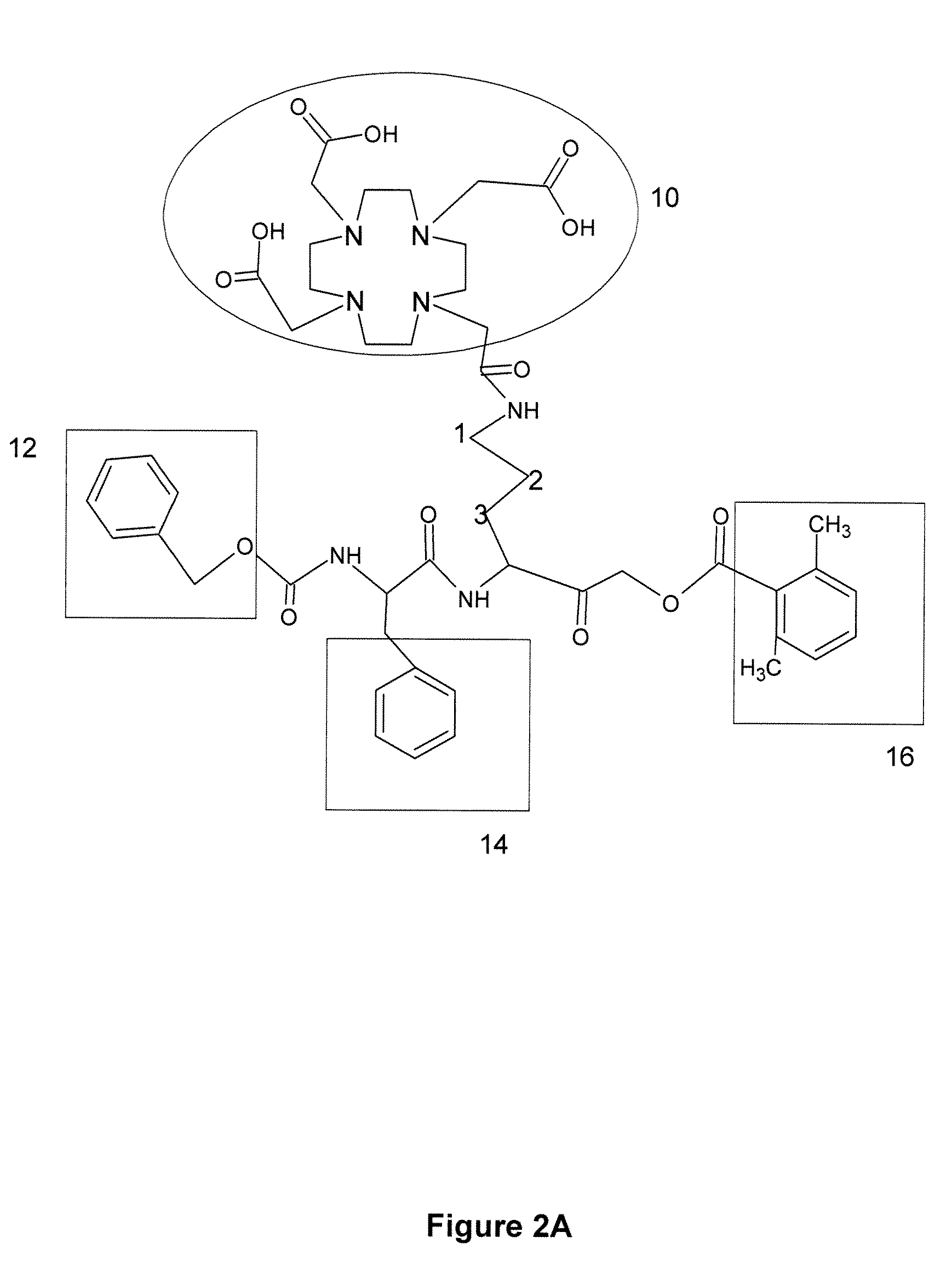
[0127]Preparation of AOMK inhibitors: 1,4,7,10-tetraaazacyclododecane-1,4,7,10 tetraacetic acid conjugated AOMK analog [Z-FR-AOMK in FIG. 1C] was prepared by methods similar to those previously described by the present inventors. See, Reference 30 US PGPUB 2007 / 0036725 by Bogyo, et al., published Feb. 15, 2007, entitled “Imaging of protease activity in live cells using activity based probes.” Methods for preparation of Z-FR-AOMK are also given in Kato et al., “Activity-based probes that target diverse cysteine protease families,” nature Chem. Biol. 1:33-38 (2005). The synthesis was carried out using a combination of solid and solution phase chemistries. This synthetic route was chosen over recently reported solid-phase methods due to the formation of an intra-molecular acyl transfer reaction on resin that was observed when an aliphatic acyloxy group was used in place of the 2,6-dimethyl benzoic acid group. The fully protected carboxyl benzoyl capped phen...
example 2
Labeling Intact Tumor Cells with 64Cu Labeled ABP
[0137]Before applying the radiolabel probe in vivo we first tested the ability of the probe to label multiple tumor cell lines grown in vitro. We initially examined the labeling of the human breast cancer cell line MBA-MB-435 and a mouse myoblastoma cell line that had been transformed by overexpression of the ras oncogene (C2C12 / Ras). Both of these cell lines were used for in vivo imaging studies with the NIRF-labeled cysteine cathepsin probes. To confirm earlier studies of these cells using the NIRF probes we labeled them with general Cy5-labeled probe GB123 (FIG. 1C). As previously reported, the cathepsin B and L activity of C2C12 / Ras was significantly (7.4 fold) higher than observed for the same targets in the MDA-MB-435 cells. Labeling of the same cells with 64Cu-Z-FK(DOTA)-AOMK confirmed that the DOTA produced a similar labeling pattern to that observed for the NIRF probe GB123. Again, the highest level of cathepsin activity was ...
example 3
Subcutaneous Tumor Model and Biodistribution Studies
[0140]All animal studies were carried out in compliance with Federal and local institutional rules for the conduct of animal experimentation. Female athymic nude mice (nu / nu) were obtained from Charles River Laboratories (Boston, Mass.) at 7-8 weeks old and kept under sterile conditions. The nude mice were inoculated subcutaneously in the right shoulder with 2×10⁶ cultured C2C12 / Ras cells or 5×10⁶ MDA-MB-435 cells. When the tumors reach 0.5-0.8 cm in diameter, the tumor bearing mice were subjected to in vivo biodistribution studies. For biodistribution studies, the mice bearing C2C12 / Ras or MDA-MB-435 tumor xenografts (n=3 for each group) were injected with 740 KBq (20 μCi) of 64Cu labeled tracer through the tail vein and sacrificed at different time points post injection (p.i.). Tumor and normal tissues of interest were removed and weighed, and their radioactivity was measured in a gamma-counter. The radioactivity uptake in the tu...
PUM
| Property | Measurement | Unit |
|---|---|---|
| Capacitance | aaaaa | aaaaa |
| Digital information | aaaaa | aaaaa |
| Digital information | aaaaa | aaaaa |
Abstract
Description
Claims
Application Information
 - R&D
- Intellectual Property
- Life Sciences
- Materials
- Tech Scout
- Unparalleled Data Quality
- Higher Quality Content
- 60% Fewer Hallucinations
Browse by: Latest US Patents, China's latest patents, Technical Efficacy Thesaurus, Application Domain, Technology Topic, Popular Technical Reports.
© 2025 PatSnap. All rights reserved.Legal|Privacy policy|Modern Slavery Act Transparency Statement|Sitemap|About US| Contact US: help@patsnap.com


