Mutant Paramyxovirus and Method for Production Thereof

a technology of mutant paramyxovirus and mutant paramyxovirus, which is applied in the direction of viruses, peptides, dna/rna fragmentation, etc., can solve the problems of low cytotoxicity and immunogenicity of viral vectors, poor safety of non-viral methods, and inability to effectively induce gene introduction to cultured cells or living tissues. , to achieve the effect of reducing the amount of receptor-binding protein, reducing the adverse effect of receptor-bind

Active Publication Date: 2009-10-29
IMMUNOMEDICINE INC
View PDF0 Cites 5 Cited by
  • Summary
  • Abstract
  • Description
  • Claims
  • Application Information

AI Technical Summary

Benefits of technology

[0009]It is a first object of the present invention to provide a modified paramyxovirus that can be utilized as a highly safe vector and a method of obtaining the modified paramyxovirus conveniently.
[0011]The present inventors conducted diligent investigations to solve the above-described problems, and found that if the RNA of the HN protein of HVJ is knocked out with an siRNA, the virus produced while the suppressive effect thereof remains effective would lack HN protein, and that the virus lacking HN protein has reduced hemagglutination activity.
[0015]Because a modified paramyxovirus of the present invention contains a reduced amount of receptor-binding protein and is capable of mitigating the adverse effects of the receptor-binding protein on target cells and the like, the paramyxovirus can be utilized as a highly safe vector. According to the method of the present invention of preparing a modified paramyxovirus, such a modified paramyxovirus can be obtained conveniently.
[0016]A paramyxovirus having a chimera protein of the present invention is highly specific for a particular tissue / cell, and is useful as a vector capable of site-specific delivery of a drug. It is also possible to obtain a still more specific viral vector by deleting a fusion-related protein of the virus, particularly a receptor-binding protein. According to the method of the present invention of preparing a targeting paramyxovirus, it is possible to obtain a paramyxovirus capable of targeting a desired tissue or cell conveniently and stably.

Problems solved by technology

However, methods using viral vectors involve possible introduction of a gene derived from the parent virus and expression thereof, immunogenicity, and possible modification of the host genome structure, posing a problem with safety.
Meanwhile, many of non-viral methods, which use liposomes and the like, tend to be inferior to viral vectors in terms of the efficiency of gene introduction to a cultured cell or living tissue, though the cytotoxicity and immunogenicity thereof are lower than those of viral methods.
It is thought that viruses gradually change the functions thereof to achieve coexistence with the host by repeating mutations, even though they are initially fatally pathogenic to the host, whereby they can survive.
Isolation of a spontaneously emerging virus mutant strain takes a long time, and a desired mutant strain can be obtained only by chance.
HVJ is highly versatile for a vector, but on the other hand it has no specificity and is not suitable for the introduction of cytotoxic molecules.
However, the method of binding a protein like this antibody to a liposome is so complex that skills are required to obtain constant results.

Method used


Image

  • Mutant Paramyxovirus and Method for Production Thereof
  • Mutant Paramyxovirus and Method for Production Thereof
  • Mutant Paramyxovirus and Method for Production Thereof

Examples


example 1

(1) Preparation of siRNA Against HN mRNA

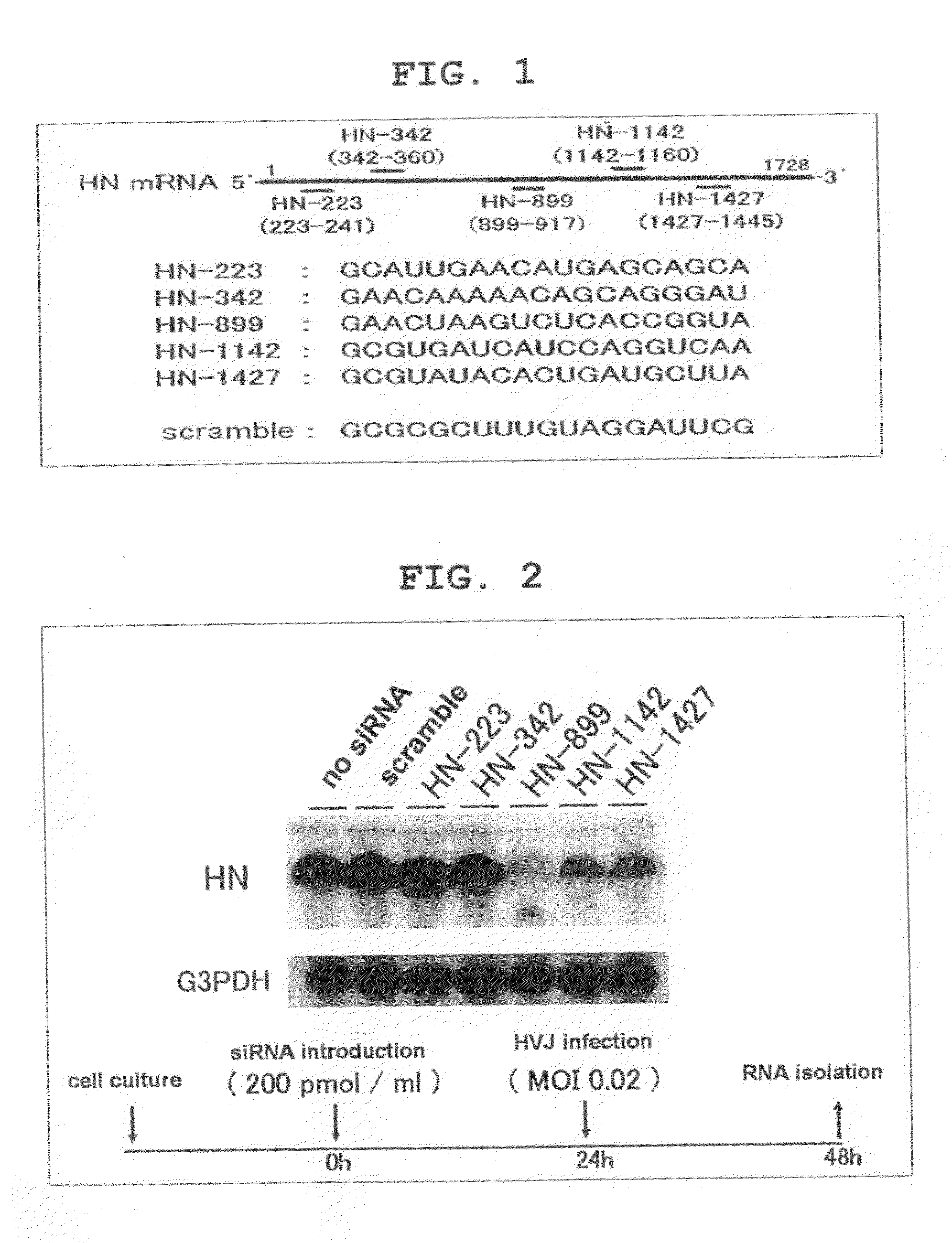
[0143]The five siRNAs for HN mRNA knock down shown in FIG. 1 were designed and synthesized using SMART siRNA Technology™ (Dharmacon Research). The five siRNAs (HN-223, HN-342, HN-899, HN-1142, HN-1427) target GCAUUGAACAUGAGCAGCA (223-241: SEQ ID NO:1), GAACAAAAACAGCAGGGAU (342-360: SEQ ID NO:2), GAACUAAGUCUCACCGGUA (899-917: SEQ ID NO:3), GCGUGAUCAUCCAGGUCAA (1142-1160: SEQ ID NO:4), and GCGUAUACACUGAUGCUUA (1427-1445: SEQ ID NO:5), respectively, on the HN mRNA. The scramble siRNA used as the control consists of a random sequence (GCGCGCUUUGUAGGAUUCG: SEQ ID NO:6).

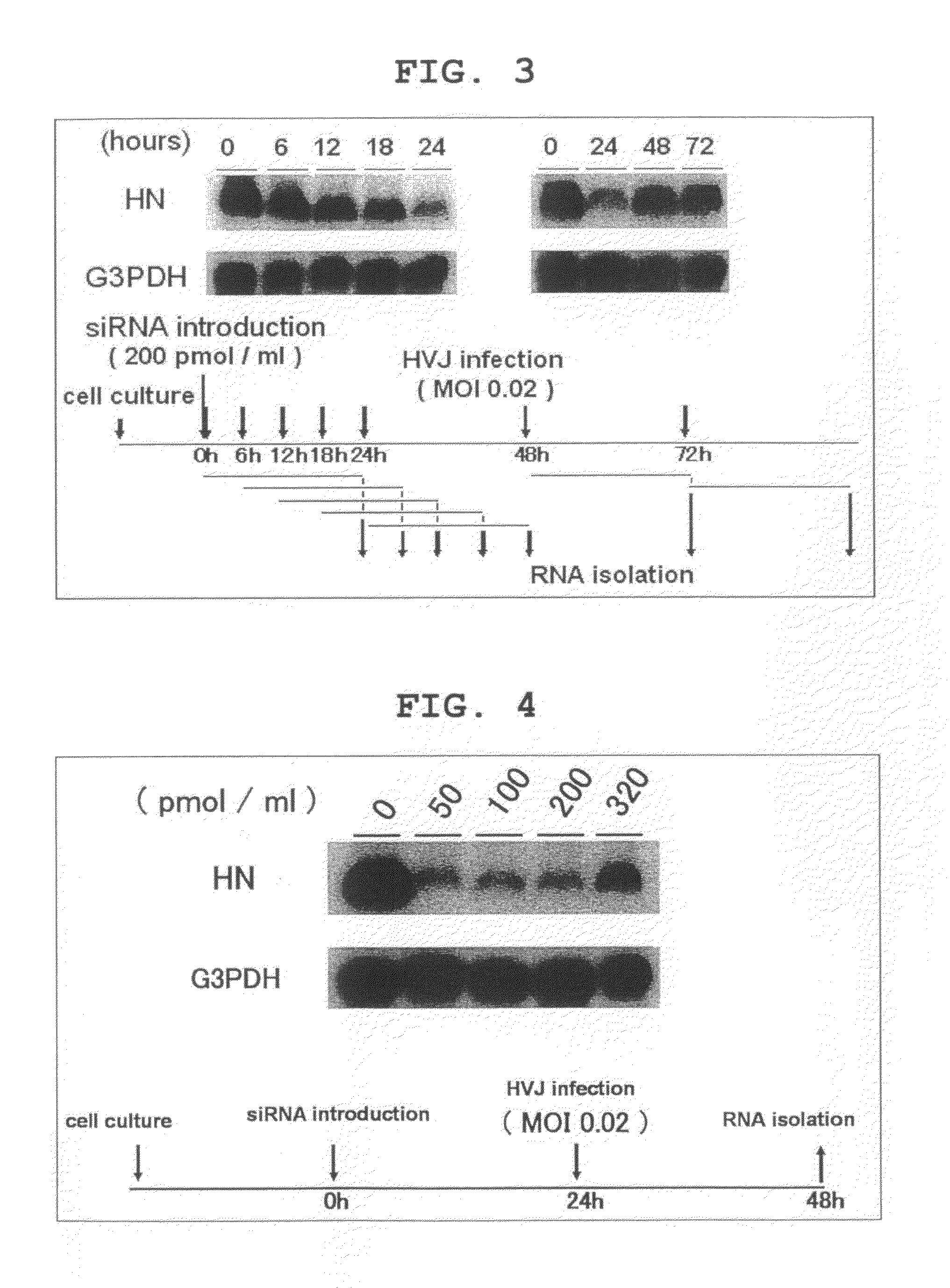
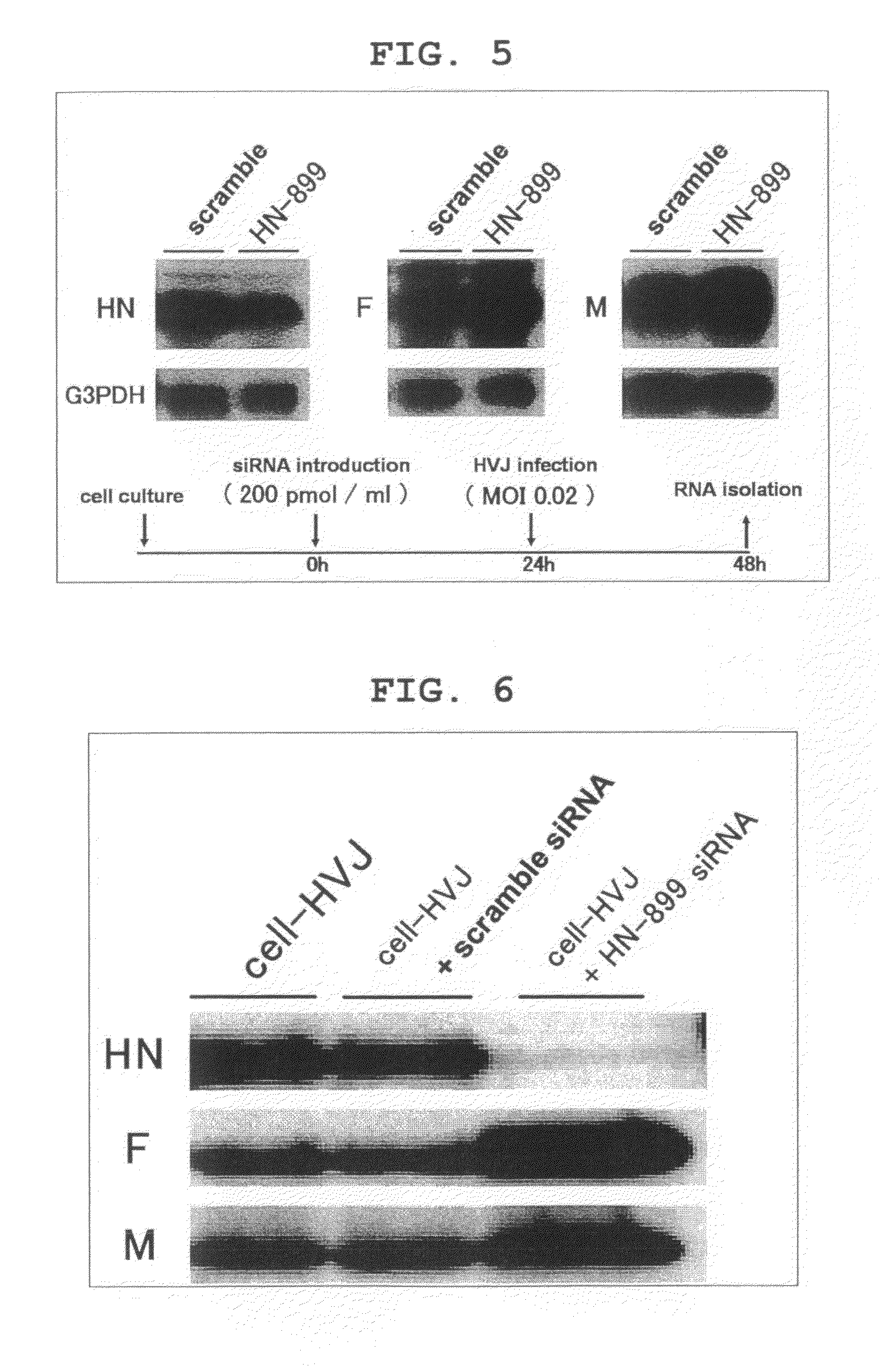
(2) Detection of Effects of siRNAs

[0144]The synthesized siRNAs were introduced to LLCMK2 cells at various concentrations by utilizing Lipofectamin Reagent (Invitrogen™, California, USA) and Plus Reagent (Invitrogen™) in accordance with the respective protocols.

[0145]24 hours after introduction, cultured cells were washed with Dulbecco's Phosphate Buffered Saline (−) (PBS) (Nacalai Te...

example 2

(1) Preparation of Expression Vector for Chimera F Protein of HVJ-F and GFP or Dsg-3-scFv

[0158]Vectors for expressing the chimera EGFP-F proteins shown in FIG. 10 (i) to (iv) were prepared.

[0159]Using Mutagenesis (Promega), the pcDNA3.1-F plasmid was mutated to delete the intrinsically possessed BglII recognition site (279-284), and an AgeI recognition site (82-87) and a BglII recognition site (346-351) were newly prepared, to yield a new plasmid vector (pcDNA3.1-Fmut). This was cleaved with HindIII / XhoI, and the F fragment obtained was inserted into similarly cleaved pCY4B to yield pCY4B-Fmut. To insert EGFP into each of the BglII cleavage site ((i) in FIG. 10), AgeI-BglII site ((ii) in FIG. 1), AgeI-Eco47III site ((iii) in FIG. 10), and AgeI-EcoNI site ((iv) in FIG. 10) in pCY4B-Fmut, amplification from pEGFP-C1 (BD Biosciences Clontech) was performed by PCR to yield EGFP cDNA fragments.

[0160]The PCR primers used here are as follows:

(SEQ ID NO: 12)GFP-BglII-f:5′-CATGGTGAGCAAGGGCGA...

example 3

(1) Preparation of HVJ Presenting Transferrin on the Virus Particle Surface Thereof, and Exhibiting a Reduced Expression of HN Protein

[0194]As shown in FIG. 21, gene that encodes the F protein of HVJ was modified, and inserted into the expression plasmid pCY4B. To express a fusion protein of the transmembrane domain (TMD) of the F protein and human transferrin wherein human transferrin has been joined to the N-terminus side of the TMD, recombinant pCY4B was constructed. cDNA of human transferrin is shown by SEQ ID NO:22. In this operation, an insertion site was designed for the myc tag to attach to the N-terminus of the fusion protein to facilitate subsequent analysis. The sequence of the fusion protein inserted here is shown by SEQ ID NO:25.

[0195]In the amino acid sequence shown by SEQ ID NO:25, amino acid numbers 1 to 29 correspond to the signal sequence of HVJ-derived F protein, amino acid numbers 30 to 39 correspond to the myc tag, amino acid numbers 40 to 718 correspond to the ...


PUM


Abstract

The present invention provides a modified paramyxovirus containing a reduced amount of receptor-binding protein compared with the wild type; a method of preparing a modified paramyxovirus, comprising the following steps: (1) a step for introducing a nucleic acid that suppresses the expression of a receptor-binding protein of a paramyxovirus into an animal cell, (2) a step for infecting the paramyxovirus to the cell, and (3) a step for isolating paramyxovirus particles replicated in the cell; and a modified paramyxovirus prepared by the method of preparation mentioned above.The present invention also provides a chimera protein wherein a fusion protein of a virus has been joined or bound to a peptide that binds specifically to a cell surface marker; a nucleic acid that encodes the chimera protein; an animal cell capable of expressing the chimera protein on the cell surface thereof; a modified paramyxovirus expressing the chimera protein on the virus particle surface thereof; and a method of preparing a tissue targeting paramyxovirus, comprising: (1) a step for supplying a nucleic acid that encodes a chimera protein wherein a fusion protein of a virus has been joined or bound to a peptide that binds specifically to a cell surface marker of the target cells, (2) a step for introducing the nucleic acid supplied in (1) into an animal cell in an expressible state, and expressing the same, (3) a step for infecting a paramyxovirus to the cell, and (4) a step for isolating paramyxovirus particles replicated in the cell.

Description

TECHNICAL FIELD[0001]The present invention relates to a modified paramyxovirus containing a reduced amount of a receptor-binding protein, and a method of preparing a modified paramyxovirus using a nucleic acid that suppresses the expression of the receptor-binding protein. Specifically, the present invention relates to a modified HVJ containing a reduced amount of HN protein, and a method of preparing a modified HVJ using an siRNA.[0002]The present invention also relates to a vector capable of site-specific delivery of a drug and a method of preparing the same.BACKGROUND ART[0003]In order to introduce a gene to a cultured cell or living tissue for gene function analysis or gene therapy, many viral methods and non-viral methods have been developed (Mulligan, Science, vol. 260, p. 926-932, (1993); Ledley, Human Gene Therapy, vol. 6, p. 1129-1144 (1995)). Generally, in introducing genes into cells, viral methods are effective. However, methods using viral vectors involve possible intro...

Claims


Application Information

Patent Timeline
IPC IPC(8): C12N15/86C12N7/01C12N5/10C07K14/00C07K16/00C07H21/02C07H21/04
CPCC07K14/005C07K2319/30C07K2319/33C12N7/00C12N15/1131C12N2810/859C12N2310/14C12N2760/18821C12N2760/18822C12N2760/18843C12N2760/18845C12N15/86
Inventor KANEDA, YASUFUMITAMAI, KATSUTOSAGA, KOTAROKAWACHI, MASAKO
Owner IMMUNOMEDICINE INC
Why Eureka
Try Eureka