
Sers-based methods for detection of bioagents

a bioagent and detection method technology, applied in the field of bioagent detection system, can solve the problems of inability to detect bioagents in complex biochemical backgrounds, high cost, and high sample preparation requirements, and achieve the effect of avoiding fluorescence signals and avoiding toxicity

Inactive Publication Date: 2009-12-03
OXONICA MATERIALS +1
View PDF0 Cites 22 Cited by
  • Summary
  • Abstract
  • Description
  • Claims
  • Application Information

AI Technical Summary

Benefits of technology

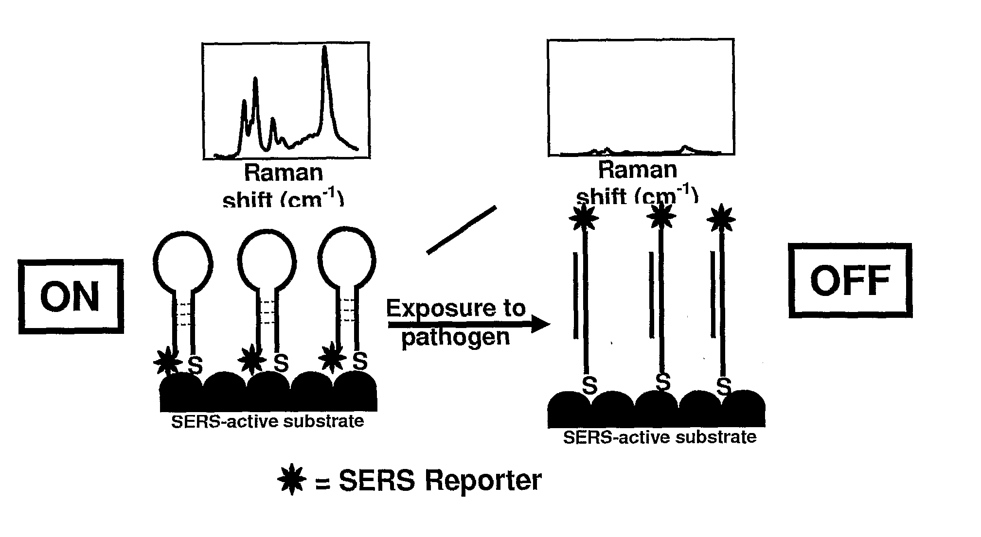
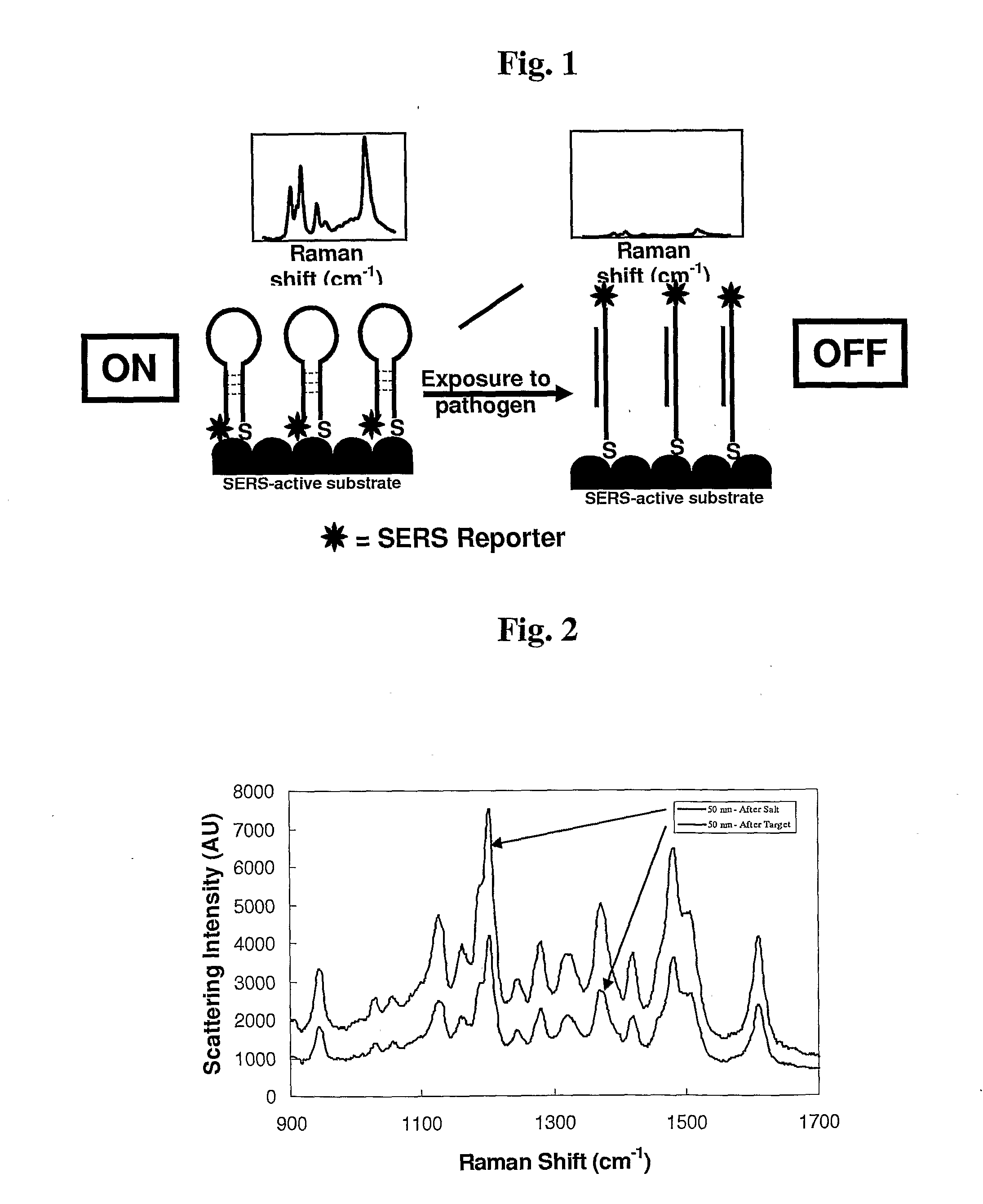
The present invention is about a way to detect certain substances in a sample using a special technique called surface enhanced Raman scattering (SERS). The invention can detect these substances using a special molecule that can bind to them and produce a signal that can be detected using a special laser. The invention can also detect multiple substances at once using a multiplexed format. This can be useful for identifying specific molecules in a sample or for detecting multiple targets at the same time.

Problems solved by technology

However, separating the fluorophore and the quencher results in a fluorescent signal.
However, when hybridized, the now double stranded oligonucleotides are rigid such that the fluorescent dye cannot interact with the surface.
While there are a few analytical techniques that can directly detect the native molecule, such as mass spectrometry and nuclear magnetic resonance spectroscopy, these often require very specific sample preparation, highly sophisticated and expensive equipment, and often do not work in complex biochemical backgrounds.
However, labeling the molecule being interrogated adds a level of complexity to an assay, thereby making it more difficult to perform properly and consistently, more difficult to turn into a “kit” or product, and more difficult to make the assay field portable and robust due to the additional steps involved.
However, normal Raman is very weak, limiting its utility for use in bioanalytical chemistry.

Method used


Image

  • Sers-based methods for detection of bioagents
  • Sers-based methods for detection of bioagents
  • Sers-based methods for detection of bioagents

Examples


example 1

SERS Beacon Probe Design

[0061]The stem-loop structures of the molecular beacons were designed using the software program MFold. The HCV probe sequence was designed from 5′ UTR region. The sequence was: 5′ thiol (CH2)6 gcgag CAT AGT GGT CTG COG AAC CGG TGA ctcgc (CH2)7 Cy5-3′ (SEQ ID NO: 1). The HCV target sequence was: TCA CCG GTT CCG CAG ACC ACT ATG (SEQ ID NO: 2). All probes and targets were purchased from BioSource. The HCV viral RNA was ordered from Ambion Diagnostics.

example 2

SERS Molecular Beacons Using Gold Colloid Substrates

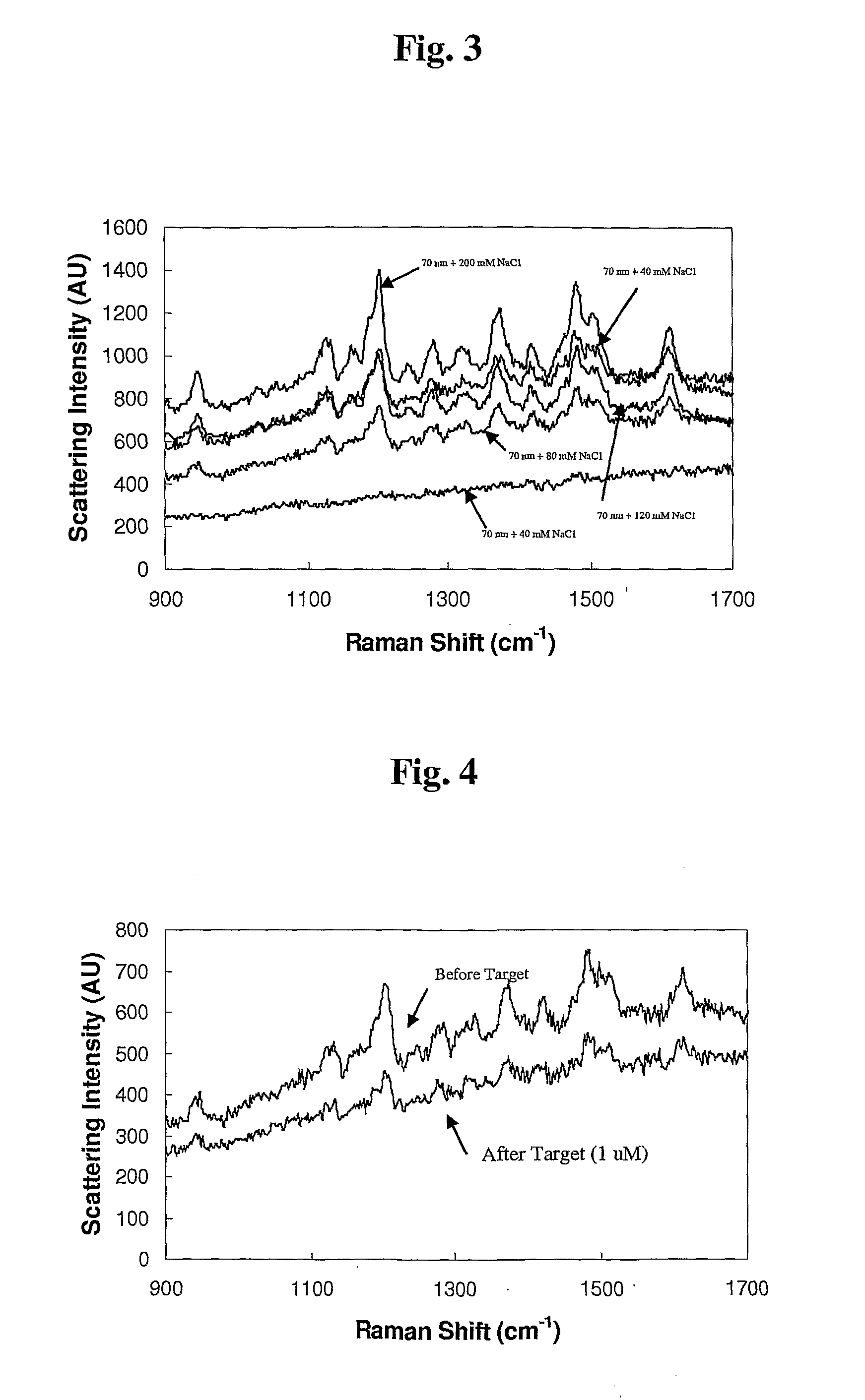
[0062]A 100 μL aliquot of the Cy5 molecular beacon (Cy5-MB) was prepared in ultrapure water. Next, 250 μL of 50 or 70 nm colloidal gold (0.01% Au by weight) was added to the beacon solution. These were incubated for approximately 6 hours before addition of 5 μL of 2.0 M NaCl. After 30 minutes, another 5 μL of NaCl was added. Another 30 minutes was allowed before excess beacon was purified by centrifugation (˜1500 RCF for 12 minutes, repeated 3 times). Particles were resuspended in TE buffer (10 mM TRIS, 0.1 mM EDTA, pH 7.5).

[0063]HCV probe assembled colloids were placed into sample wells on a quartz slide. Each well in the gasket was approximately 2 mm in diameter and depth, and held up to 10 μL of solution. Aliquots (5 μL) of each conjugate were placed into separate wells and their Raman spectra interrogated. No SERS peaks were visible using the maximum laser power setting with a 1 second integration time and a 5× objective. It wa...

example 3

SERS Beacon Assay for Detection of Oligonucleotide Targets, Using Nanowire Substrates

[0065]A. Preparation of Nanowires. Gold and silver nanowires (Nanobarcodes® Particles) have been used previously to quench fluorescence based molecular beacons (WO 2005 / 020890). The advantage of using these substrates it is possible to determine both the SERS response and the fluorescence response, thereby providing an ability to confirm the results using an orthogonal method. FIG. 5 shows a cartoon depiction of this assay format. Nanowires (Nanobarcodes® Particles) were manufactured as previous described (Nicewarner-Pena, S. R., et al., (2001) Science 294, 137-141; Reiss, et al. (2002) J. Electranal. Chem. 522, 95-103; Walton, et al. (2002) Anal. Chem. 74, 2240-2247). Briefly, alternating layers of gold and silver are electroplated into the pores of an alumina template, the template is dissolved using strong base, resulting in the formation of striped nanowires. The nanowires used in this study wer...


PUM

PropertyMeasurementUnit
sizeaaaaaaaaaa
temperatureaaaaaaaaaa
pHaaaaaaaaaa

Abstract

An assay and method of assay for optical detection of bioagents, a target nucleic acid or a target protein using a surface enhanced Raman scattering (SERS) active biomolecule molecular beacon. The present invention also provides the assay and method in a multiplexed format.

Description

FIELD OF THE INVENTION[0001]The invention relates to a bioagent detection system employing SERS (surface-enhanced Raman scattering)-based methods and systems.BACKGROUND OF THE INVENTION[0002]The use of fluorescence quenching as a detection method in biological assays is widespread and includes the use of molecular beacons, a technology first described in 1996. Tyagi, S. and Kramer, F. R., “Molecular Beacons: probes that fluoresce upon hybridization”Nature Biotechnol. 1996, 14, 303-308. Molecular beacons typically use a fluorophore reporter dye and a non-fluorescent quencher chromophore. While in close proximity, the fluorophore is quenched by the energy transfer to the non-fluorescent chromophore. However, separating the fluorophore and the quencher results in a fluorescent signal. Molecular beacons have been used in a variety of assay formats, including the monitoring of nucleus activity, the detection of pathogens and SNP detection.[0003]An assay using a fluorescent energy transfe...

Claims


Application Information

Patent Timeline
Patent Type & Authority Applications(United States)
IPC IPC(8): G01N33/566
CPCC12Q1/6818C12Q2565/632C12Q2565/549C12Q2565/1015
Inventor NATAN, MICHAEL J.SHA, MICHAELDOERING, WILLIAM E.
Owner OXONICA MATERIALS
Learn More