Muscle regeneration promoter
a muscle regeneration and promoter technology, applied in the field of muscle regeneration promoters, can solve the problems of inability to clarify the factors that promote satellite cell recruitment, growth, differentiation in vivo, and so as to promote muscle regeneration, promote proliferation, promote adhesion and differentiation of satellite cells, and no specific effect of fiber atrophy or decrease the number of satellite cells
- Summary
- Abstract
- Description
- Claims
- Application Information
AI Technical Summary
Benefits of technology
Problems solved by technology
Method used
Image
Examples
example 1
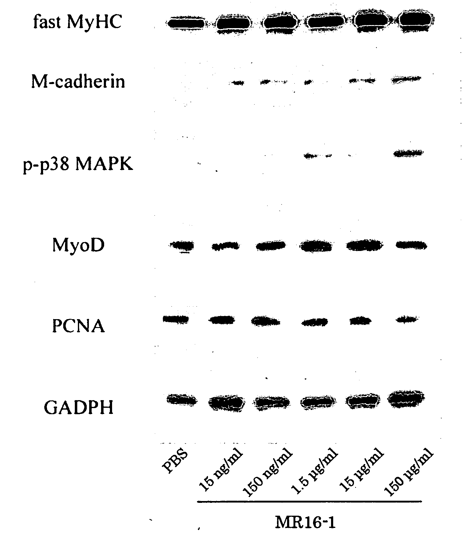
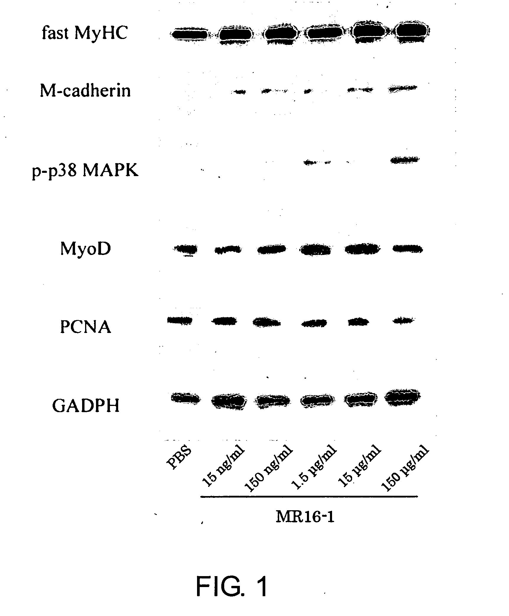
[0205]Decreased regulation of the immune mechanism during exposure to the space environment is a serious problem for astronauts. C2C12 cells were cultured in a differentiation medium containing MR16-1 (an anti-mouse IL-6 receptor monoclonal antibody) at a concentration of 15 ng / ml, 150 ng / ml, 1.5 μg / ml, 15 μg / ml, or 150 μg / ml in phosphate-buffered saline (PBS) to assess the effect of inhibiting the IL-6 signaling pathway on muscle cell growth. Control cells were cultured in a medium without MR16-1.
[0206]After 3 days of culture, half of the cells were fixed with 10% formalin and proteins involved in muscle regeneration (MyoD, myogenin, myogenic regulatory factor proteins, and myosin heavy chain) were detected immunohistochemically. The remaining cells were lysed in lysis buffer containing 1% Triton, and expressions of M-cadherin, phospho-p38, and MyoD, which are muscle differentiation markers, were confirmed by Western blot analyses.
[0207]As a result, the proliferation of C2C12 cells...
example 2
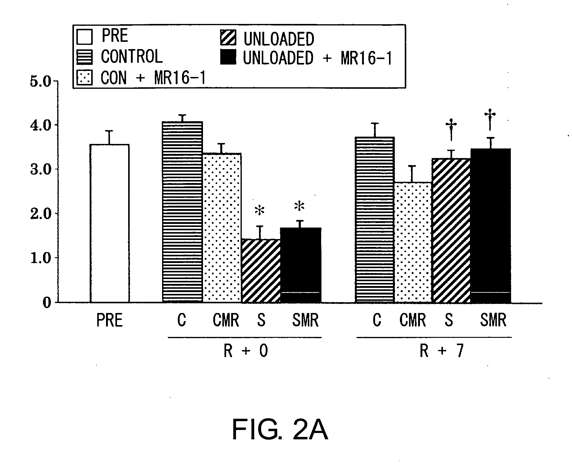
[0208]Next, changes in the properties of satellite cells in whole single fibers of soleus muscle, sampled from tendon to tendon, following MR16-1 treatment with or without gravitational loading were investigated in male mice (C57BL / 6J Jcl).
[0209]MR16-1 or PBS was intraperitoneally (i.p.) injected into mice at a concentration of 2 mg / mouse before seven days of hind-limb suspension or seven days of reloading. The collected muscles were dipped in cellbanker (Nihon Zenyaku) and frozen at −80° C., and then thawed at 35° C. Then, single muscle fibers were collected following collagenase digestion in Dulbecco's Modified Eagle's Medium supplemented with 20 μM 5′-bromo-2′-deoxyuridine (BrdU), 0.2% type I collagenase, 1% antibiotics, and 10% new-born calf serum (35° C.) for 4 hours. The muscle fibers were incubated with an M-cadherin- or BrdU-specific antibody, and stained with fluorescein or rhodamine, respectively. M-cadherin-positive (quiescent, resting stage) or BrdU-positive (mitotic act...
example 3
[0212]Satellite cells grown in MR16-1-administered culture medium are labeled with green fluorescent protein (GFP), and the cells are injected into muscle tissues or veins of animals with damaged or atrophied muscles. The effects of administering the cells to animals on the recovery or regeneration of their muscle tissues are assessed by biochemical and / or immunohistochemical analyses.
PUM
| Property | Measurement | Unit |
|---|---|---|
| molecular weight | aaaaa | aaaaa |
| molecular weight | aaaaa | aaaaa |
| pH | aaaaa | aaaaa |
Abstract
Description
Claims
Application Information
 - R&D
- Intellectual Property
- Life Sciences
- Materials
- Tech Scout
- Unparalleled Data Quality
- Higher Quality Content
- 60% Fewer Hallucinations
Browse by: Latest US Patents, China's latest patents, Technical Efficacy Thesaurus, Application Domain, Technology Topic, Popular Technical Reports.
© 2025 PatSnap. All rights reserved.Legal|Privacy policy|Modern Slavery Act Transparency Statement|Sitemap|About US| Contact US: help@patsnap.com


