Method for therapy of heart valves with a robot-based x-ray device

a robot-based x-ray and heart valve technology, applied in the field of cardiological therapy, can solve the problems of heart valve damage, long recovery (or convalescence) time for patients, and high risks associated with i

Inactive Publication Date: 2010-05-06
SIEMENS HEALTHCARE GMBH
View PDF8 Cites 46 Cited by
  • Summary
  • Abstract
  • Description
  • Claims
  • Application Information

AI Technical Summary

Benefits of technology

[0037]The combination of a minimally-invasive interventional procedures of the type described and the use of the improved imaging system support provides many advantages beyond the separate application of these features. The patient need not be moved to a separate imaging location, a high definition three dimensional image of the relevant portions of the patient is available, and the imaging apparatus is not in the way while the medical professionals carry out the procedure.

Problems solved by technology

Such procedures have high risks associated therewith as well as long recovery (or convalescence) times for the patient.
The heart valves can be damaged by other illnesses, for example by inflammation, influenza or cardiac infarction, to the extent that the valve must be replaced or surgically modeled.
Such procedures also were associated with high risks and long recovery times (up to six weeks) for the patient.
In principle, all four heart valves are accessible for a balloon dilation (referred to as valvuloplasty), but dilation of the tricuspid valve is only rarely implemented, due to the relative rarity of tricuspid stenosis.
This procedure is implemented under anesthesia, since filling of the balloon leads to a temporary interruption of circulation.
A significant disadvantage of all of the recently developed minimally-invasive procedures is that they must be implemented using x-ray fluoroscopy, which shows only a 2D (two-dimensional) image of the heart and the catheter and tools located therein or proximate thereto.
It is very difficult for a surgeon or cardiologist to mentally form spatial associations from such 2D images.
A further disadvantage associated with such known procedures is that when the catheter is clearly visible in such an x-ray image, the opening of the heart valve in question is only poorly visible, or vice versa.
The opening can be shown more clearly by the injection of a contrast agent, but a significant number of patients are at risk of having an allergic reaction to conventional contrast agents.
Due to these limitations in the content of the displayed images, a risk exists that the stenosis will not be correctly opened, or an artificial (replacement) heart valve may not be correctly placed.
It is also known to conduct some of these procedures, or portions thereof, supported by extracorporeal or intracorporeal (ICE, TEE) ultrasound imaging, but generally this does not provide a sufficient image quality.
However, the aforementioned commercially available C-arm x-ray device, as well as other commercially available C-arm x-ray devices, do not provide adequate access to the patient in order to permit percutaneous heart valve procedures to be implemented by operating personnel using known workflows or methods.

Method used


Image

  • Method for therapy of heart valves with a robot-based x-ray device
  • Method for therapy of heart valves with a robot-based x-ray device
  • Method for therapy of heart valves with a robot-based x-ray device

Examples


Embodiment Construction

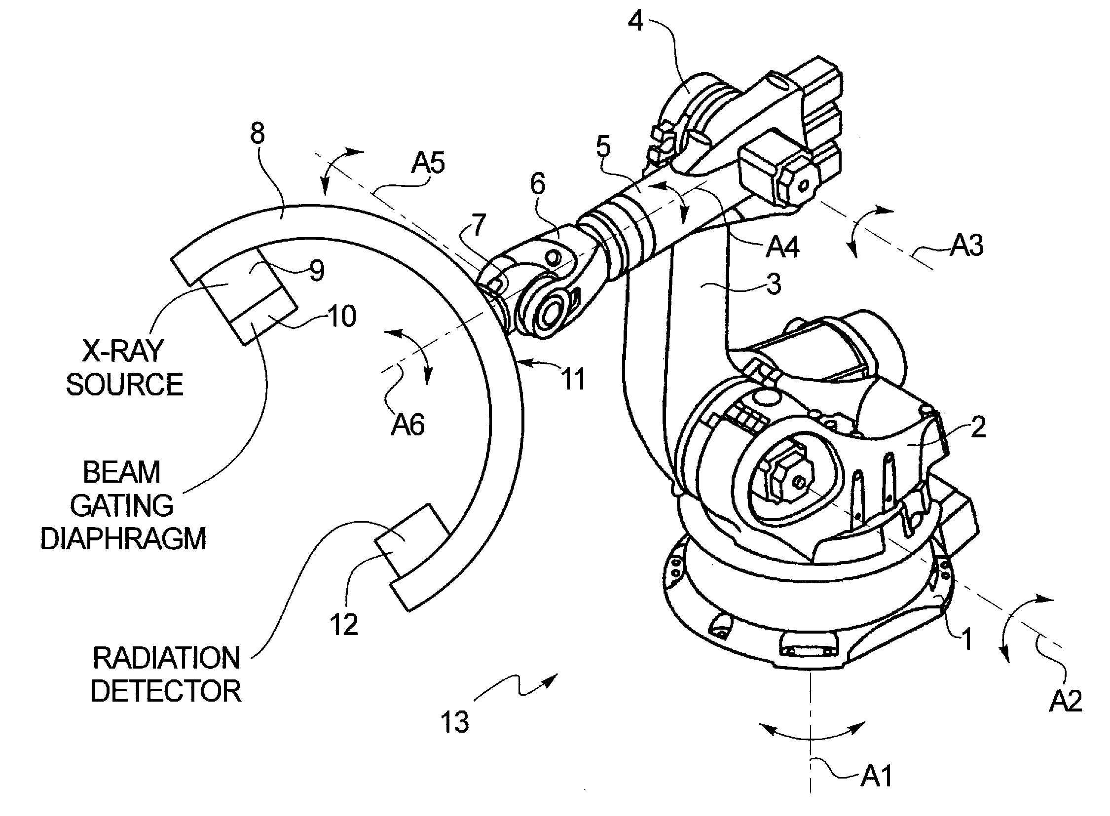
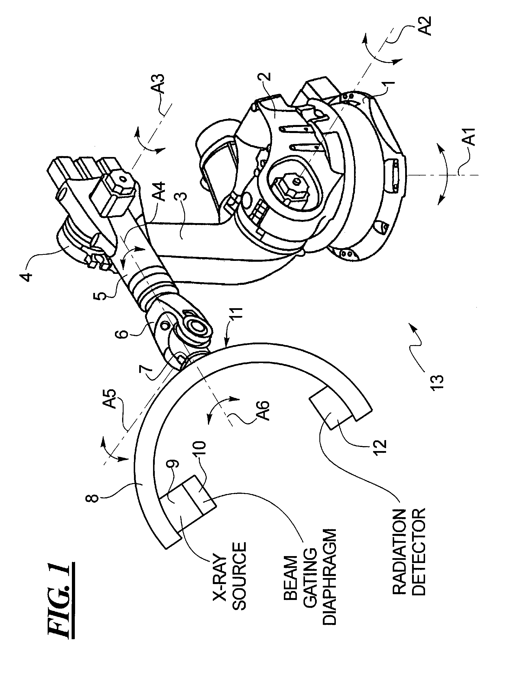
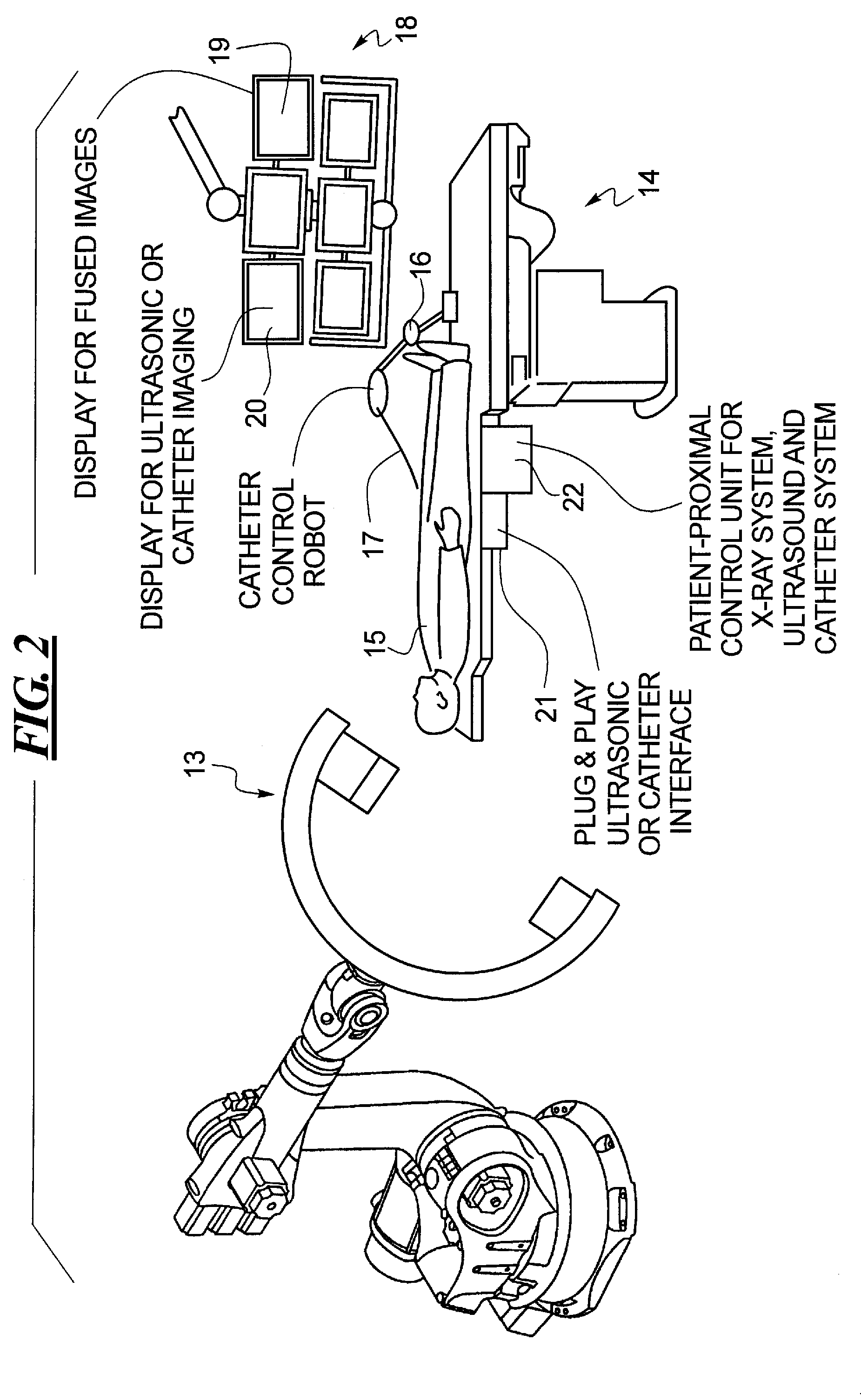
[0049]A method is provided for placing an artificial heart valve in a patient. The method of a preferred embodiment utilizes a robot arm x-ray image acquisition system 13 as shown in FIG. 1 of the type described in DE 10 2005 012 700 A1 for purposes other than implementing minimally invasive procedures involving heart valves. The robotic portion of the device 13 is mounted to a base 1 which, in this embodiment, is shown as a base affixed to the floor of a room in which the device 13 is used. The device 13, however, may also be wall-mounted or ceiling-mounted. A shoulder articulation 2 is rotatably mounted on the base 1, so as to be rotatable around a substantially vertical axis A1. The shoulder articulation is connected to a first arm portion 3 of an arm articulation, so that the first arm portion 3 is rotatable around a substantially horizontal axis A2. The first arm portion 3 is connected via an elbow articulation 4 to a second arm portion 5. The first arm portion 3 and the second...


PUM


Abstract

A method or workflow for heart valve replacement, or more precisely emplacement of a prosthetic heart valve, using minimally invasive procedures includes imaging of the patient's heart during the procedure using a multi-access articulated x-ray imaging robot that allows a radiation detector carried by the robot to be moved in arbitrary paths around a patient in order to generate multiple projection exposures of the relevant region of the patient during the procedure. The imaging system is used to generate two dimensional image data during movement of the catheter and prosthetic heart valve into place and to generate three dimensional image data of the prosthetic heart valve within the patient's heart. The two dimensional image data and the three dimensional image data are registered and superimposed for use in positioning of the prosthetic heart valve. Additional imaging may be performed once the prosthetic heart valve is in position.

Description

BACKGROUND OF THE INVENTION[0001]1. Field of the Invention[0002]The present invention relates generally to a method for cardiological therapy and in particular to a method for using imaging during an interventional heart valve therapy using a robotic imaging device.[0003]2. Description of the Related Art[0004]The reduction of the pumping power of one or both heart chambers is generally designated as a cardiac insufficiency. Cardiac insufficiency is not an actual illness, but rather is the result of various diseases and / or pathology symptoms. As a result of cardiac insufficiency, the body and its organs do not receive the necessary amount of blood per unit of time. The vital organs are supplied with only an insufficient quantity of oxygen and nutrients.[0005]Among the most important causes of cardiac insufficiency are illness of the coronary vessels (often the cause of extended infarctions), hypertension that is insufficiently medically regulated, heart muscle illness, heart muscle i...

Claims


Application Information

Patent Timeline
Patent Type & Authority Applications(United States)
IPC IPC(8): A61F2/24
CPCA61B6/12A61B6/4441A61B6/462A61B6/464A61B6/466A61B6/5247A61B8/0833A61B8/12A61B8/4416A61F2/2418A61F2/2427A61F2/2433A61B6/4458A61B6/4464
Inventor MASCHKE, MICHAEL
Owner SIEMENS HEALTHCARE GMBH
Why Eureka
Try Eureka