Method for isolation of cell, serum-free culture medium for cell, and method for culture of cell
- Summary
- Abstract
- Description
- Claims
- Application Information
AI Technical Summary
Benefits of technology
Problems solved by technology
Method used
Image

Examples
example 1
Isolation of Corneal Epithelial Stem Cells
[0104]The production of a suspension culture vessel for stem cells was carried out in accordance with the method described in Japanese Patent Application No. 2005-369270 (Japanese Laid-open Patent Application (Kokai) No. 2007-166977) by dissolving polyhydroxyethyl methacrylate described in the centrifuge tube for living cell separation in ethanol, and coating the resultant to a commercially available plastic culture vessel for suspension culture, followed by air-dry. This suspension culture vessel for stem cells may be used for the purpose of preventing adhesion of the stem cells that is not intended by the researchers.
[0105]After conjunctival tissues were removed from imported human cornea for research obtained from the Eye Bank Association of America, corneal limbal epithelial tissues where corneal epithelial stem cells were considered to exist were excised with scissors. The excised corneal limbal epithelial tissues were placed in the sus...
example 2
Test for Differentiation into the Corneal Epithelial Cells by a Growth Factor
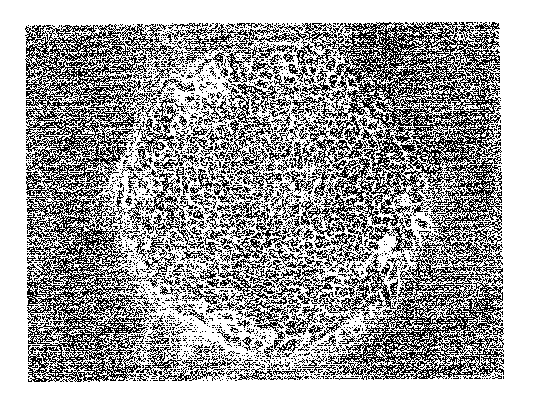
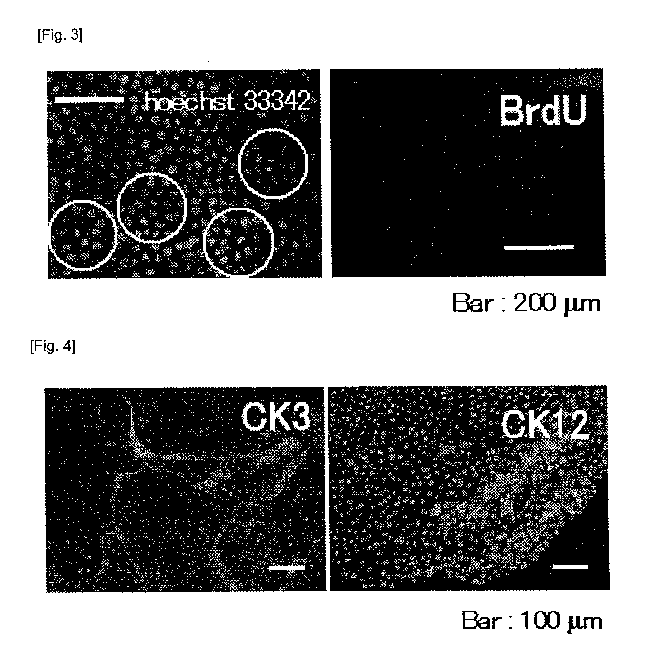
[0109]Epidermal growth factor (EGF: obtained from Wako Pure Chemical Industries, Ltd.) was added to the medium for stem cell culture (A) so as to attain a concentration of 20 ng / mL. The culturing of the isolated corneal epithelial stem cells was carried out under a condition at 37° C. and 5% CO2. The isolated corneal epithelial stem cells began vigorously to divide to form a round shaped cell population (colony) originated from a single cell in about one week as shown in FIG. 2. BrdU (Roche) uptake test (BrdU method) and nuclear staining with Hoechst33342 (Invitrogen), both by which the ability to proliferate can be checked, were carried out to this colony. The results of those are shown in FIG. 3. It can be seen that corneal epithelia cells are vigorously proliferating because the colony is positive for a FITC-Conjugated anti-BrdU antibody (Roche), which is an antibody for detecting BrdU taken up by cells ...
example 3
Test for Differentiation into the Goblet Cells by a Growth Factor
[0110]A fibroblast growth factor (FGF-2: obtained from Wako Pure Chemical Industries, Ltd.) was added to the medium for stem cell culture (A) so as to attain a concentration of 40 ng / mL. The culturing of the isolated corneal epithelial stem cells was carried out under a condition at 37° C. and 5% CO2.
[0111]The isolated corneal epithelial stem cells began vigorously to proliferate and, in about 3 to 5 days, a middle portion elevated to form a protrusion. And a colony originated from a single cell with a specific morphology appeared (FIG. 5a). As seen in a cross-sectional view of this protrusion in the middle, it contained a small amount of cell components. Thus, the colony was proven not to be a cell clump and to be rather a cluster of extracellular matrices (FIG. 5b). And then PAS staining which shows positive in the presence of the goblet cells was carried out, and the cells were found to be positive (FIG. 5c). Thus, ...
PUM
 Abstract
Description
Claims
Application Information
 - R&D Engineer
- R&D Manager
- IP Professional
- Industry Leading Data Capabilities
- Powerful AI technology
- Patent DNA Extraction
Browse by: Latest US Patents, China's latest patents, Technical Efficacy Thesaurus, Application Domain, Technology Topic.
© 2024 PatSnap. All rights reserved.Legal|Privacy policy|Modern Slavery Act Transparency Statement|Sitemap