

Ligands for imaging cardiac innervation
- Summary
- Abstract
- Description
- Claims
- Application Information
AI Technical Summary
Benefits of technology
Problems solved by technology
Method used
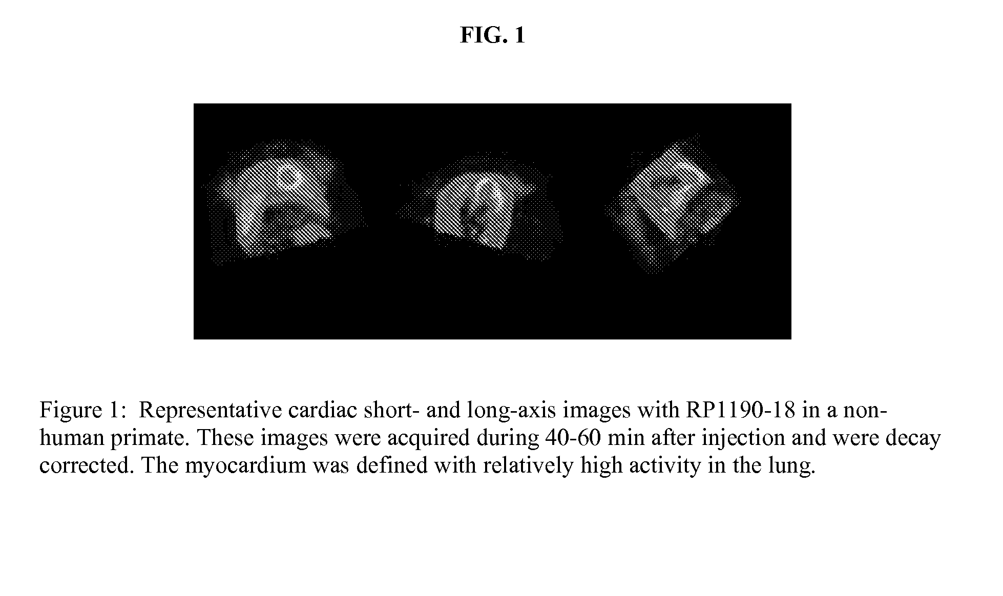
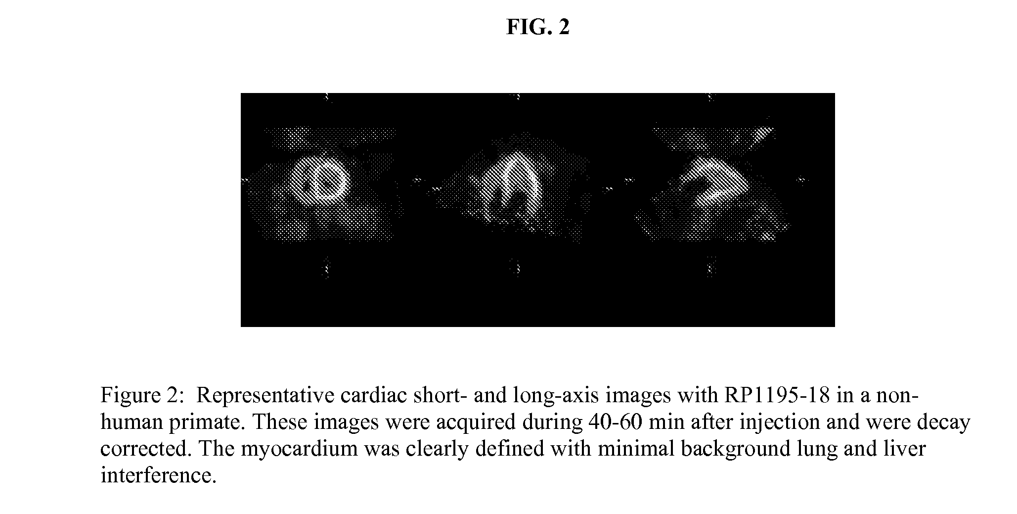
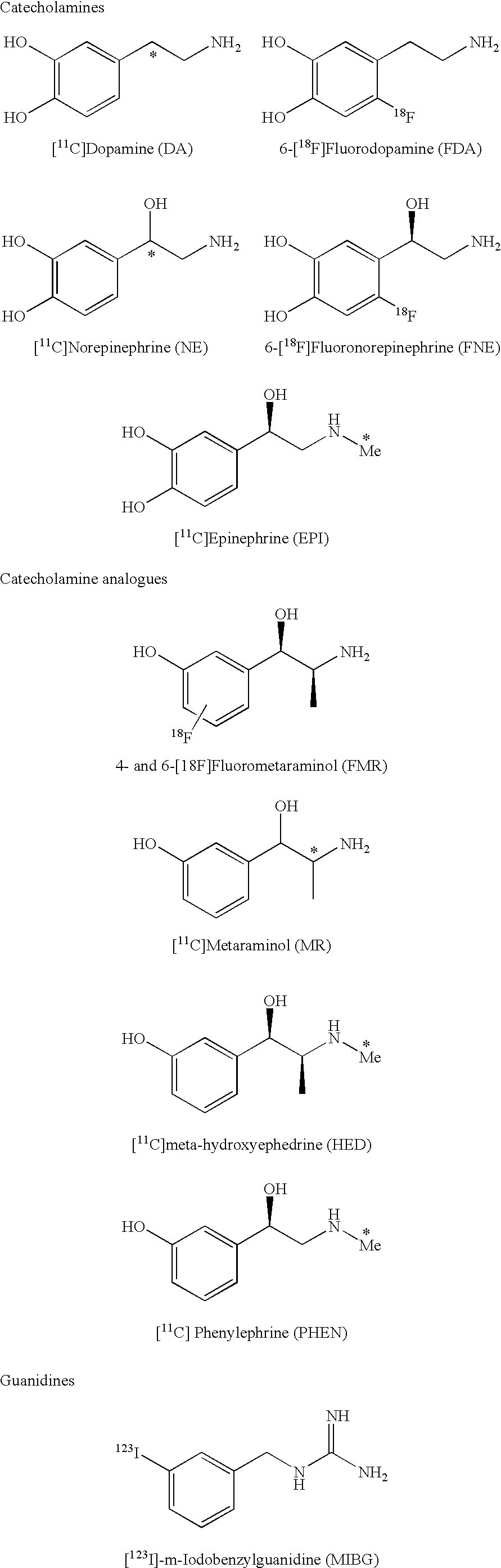
Image

Examples
example 1
Synthesis of N-(4-Fluoro-3-(trifluoromethyl)benzyl)-1H-imidazol-2-amine
[0072]
Part A
Preparation of N-(4-Fluoro-3-(trifluoromethyl)benzyl)-1-trityl-1H-imidazol-2-amine
[0073]
[0074]A solution of 4-fluoro-3-(trifluoromethyl)benzaldehyde (227 mg, 1.18 mmol) and 1-trityl-LH-imidazole-2-amine (462.3 mg, 1.42 mmol) in toluene (40 mL) was heated at reflux for 6 h while using a Dean-Stark apparatus to remove water. The mixture was cooled to room temperature, treated with sodium triacetoxyborohydride (1.00 g, 4.70 mmol), and stirred overnight. The reaction was quenched by the addition of water (150 mL), and the layers were separated. The aqueous layer was extracted with ethyl acetate (2×50 mL). The combined organic layers were dried (MgSO4) and concentrated, and the resulting residue was purified by flash chromatography (40:60 EtOAc / hexanes) to yield the title compound as a pale yellow solid (266 mg, 45%). 1H NMR (CDCl3, 300 MHz): δ 7.38-7.30 (m, 9H), 7.24-7.14 (m, 6H), 7.14-6.93 (m, 3H), 6.71 ...
example 2
Synthesis of 1-(2-(4-(2-fluoroethoxy)phenyl)-2-hydroxyethyl)guanidinium Chloride
[0076]
Part A
Preparation of 1-(2-Hydroxy-2-(4-hydroxyphenyl)ethyl)-2,3-bis(tert-butoxycarbonyl)guanidine
[0077]
[0078]A solution of (+ / −)-octopamine hydrochloride (500 mg, 2.89 mmol) and N,N′-bis(Boc)-1H-pyrazole-1-carboxamidine (1.13 g, 3.60 mmol) in DMF (10 mL) was stirred for 1 h at ambient temperatures. The reaction mixture was concentrated, and the residue was dissolved in EtOAc (60 mL). The solution was washed with 1 N KHSO4 (2×30 mL) and 5% Na2CO3 (30 mL). The organic layer was dried (Na2SO4), concentrated, and purified by flash chromatography (EtOAc / hexane 30 / 70→50 / 50) to yield the title compound as a colorless solid (836 mg, 73%). 1H NMR (CDCl3, 600 MHz): δ 11.45 (bs, 1H), 8.76 (s, 1H), 7.15 (d, J=8.4 Hz, 2H), 6.77 (d, J=8.4 Hz, 2H), 6.52 (bs, 1H), 4.81-4.78 (m, 1H), 3.66-3.50 (m, 2H), 1.51 (s, 9H), 1.49 (s, 9H); 13C NMR (CDCl3, 150 MHz): δ 162.86, 157.52, 156.04, 153.19, 133.52, 127.34, 115.65, 83...
example 3
Synthesis of 1-(4-(2-Fluoroethoxy)phenethyl) guanidinium Chloride
[0082]
[0083]The product of Example 2, Part B (88.4 mg, 0.20 mmol) was dissolved in a solution of TFA (1.9 mL), triisopropylsilane (0.05 mL), and water (0.05 mL). The reaction solution was heated at 55° C. for 10 min and concentrated. The crude mixture was purified by HPLC using the procedure of Example 2, Part B. The 20, product fraction was lyophilized yielding a hygroscopic solid.
[0084]Relyophilization from 0.5 N HCl gave the title compound as a dry colorless solid (12.4 mg, 24%). 1H NMR (1:1 CD3CN / D2O, 600 MHz): δ 7.18-7.14 (m, 2H), 6.90-6.87 (m, 2H), 4.75-4.65 (m, 2H), 4.22-4.15 (m, 2H), 3.31 (t, J=72 Hz, 2H), 2.76 (t, J=7.2 Hz, 2H); 13C NMR (1:1 CD3CN / D2O, 150 MHz): δ 158,08, 157.75, 132.00, 131.09, 115.82, 83.65 (d, J=164.6 Hz), 68.44 (d, J=18.8 Hz), 43.57, 34.36. HRMS calculated for C11H17FN3O (M+H): 226.1350; Found: 226.1352.
PUM
| Property | Measurement | Unit |
|---|---|---|
| Capacitance | aaaaa | aaaaa |
| Fraction | aaaaa | aaaaa |
| Fraction | aaaaa | aaaaa |
Abstract
Description
Claims
Application Information