
Biosensor Using Whispering Gallery Modes in Microspheres

a microsphere and biosensor technology, applied in the field of analyte detection, can solve the problems of limited ability for multiplexing measurements, specific and expensive instruments for spr, etc., and achieve the effect of sensitive detection

Inactive Publication Date: 2010-09-09
GENERA BIOSYSTEMS LTD +1
View PDF15 Cites 7 Cited by
  • Summary
  • Abstract
  • Description
  • Claims
  • Application Information

AI Technical Summary

Benefits of technology

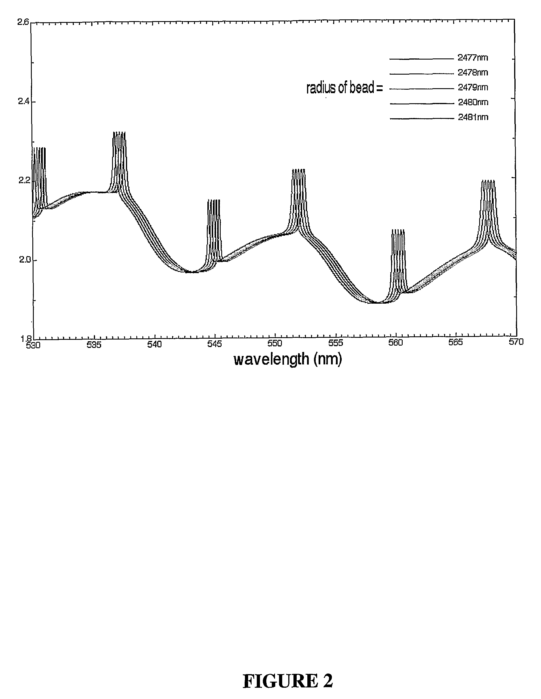
[0010]The present invention provides methods and reagents for, inter alia, detecting molecules in a sample. These molecules are referred to herein as analytes. The presence or structure of the analytes need not be known and hence the subject method is ideal for detecting hitherto unknown binding partners of orphan receptors and potential modulators of nucleic acid expression or protein including enzyme activity, folding, antigenicity or function. The methods of the present invention are predicated, in part, on the phenomenon that optically detectable labels embedded within or onto a microspheroidal particle will display a distinctive Whispering Gallery Mode (WGM) profile. Reference to “optically detectable” includes reference to detection by spectrometric means. When a target analyte interacts with a binding partner immobilized to the microspheroidal particle, the WGM profile changes enabling very sensitive detection of even rare binding events.
[0013]The methods of the present invention may be applied to detect modulation in the WGM profile of a microspheroidal particle wherein said modulation results from detection of binding or other association of molecules in a sample to potential binding particles immobilized to the surface of the microspheroidal particle. Detection of binding reactions between an analyte and its binding partner based on sensitive changes in WGM profiles enables the identification and isolation of the analytes.

Problems solved by technology

However, SPR requires specific and expensive instrumentation and the ability for multiplexed measurements is limited.

Method used


Image

  • Biosensor Using Whispering Gallery Modes in Microspheres
  • Biosensor Using Whispering Gallery Modes in Microspheres
  • Biosensor Using Whispering Gallery Modes in Microspheres

Examples


example 1

Production and Labeling of Quantum Dot Labeled Microspheroidal Particles

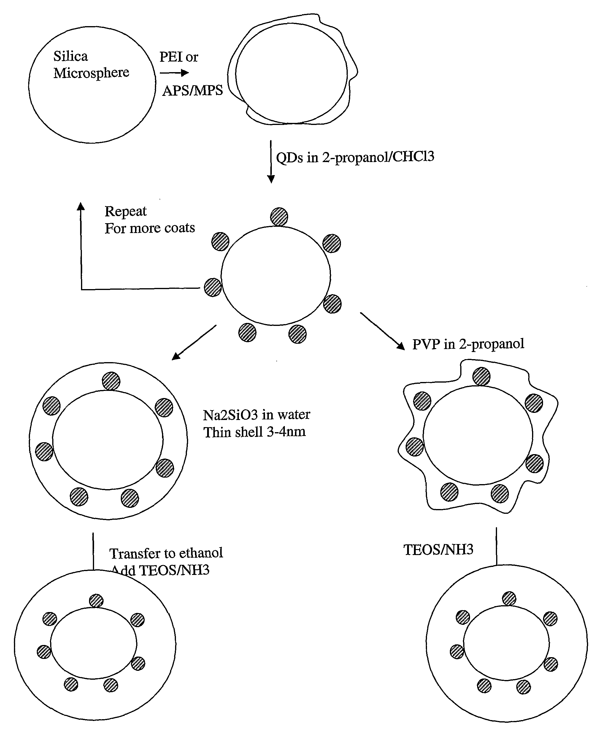
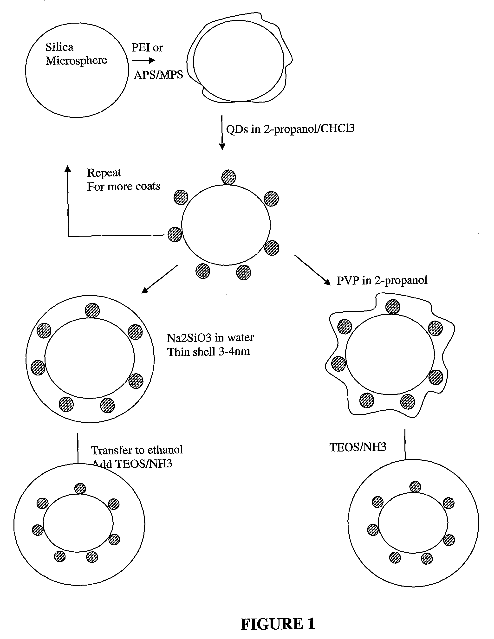
[0153]Typically, microspheroidal particles may be prepared by overlaying commercial beads of any desired composition or size with quantum dots. For more robust materials, they should be overcoated with another silica layer to minimize interactions with the medium or adsorbates.

[0154]Without limiting the many possibilities, described herein is a typical process used to prepare microspheroidal particles using glass / silica beads.

[0155]0.1 g of five micron diameter commercial silica beads were placed in 20 ml 2-propanol and 20 microlitres of either mercaptopropylsilane (MPS) or aminopropylsilane (APS) was added. The solution was refluxed at 80° C. to allow chemisorption of the MPS or APS, which led to the creation of mercaptan or amino groups on the bead surface. Excess MPS or APS was removed by centrifugation.

[0156]Instead of a silane functionalized bead, the beads can also be activated by adsorbing a cationic poly...

example 2

DNA Detection

[0164]Q-Sand beads were conjugated to DNA, forming a complex with a Q-Sand bead and immobilized 48-mer DNA. The fifth base position of the 48-mer was a thymidine base with an incorporated amine. This amine was used to label the DNA with a BODIPY·630 / 650 NHS ester using a standard condensation reaction. The WGM profile for the Q-Sand bead conjugated to the 48-mer DNA is shown in FIG. 5, panel a.

[0165]Panel b of FIG. 5 shows the WGM profile of the Q-Sand bead of panel a conjugated to a α-Transprobe DNA which hybridizes to bases 6-24 of the DNA conjugated to the Q-Sand bead. The resulting WGM profile, as seen in panel b of FIG. 5, was a shift of approximately 2.4 nm for all peaks. In addition, the Q-factor (the measure of the quality of the microresonator) reduction was approximately 5×.

[0166]Finally, the Q-Sand beads shown in FIG. 5, panel a, were hybridized to a PCR product containing a complementary region to DNA bases 25-48 of the DNA 48-mer. The PCR product was genera...

example 3

Surface Functionalization of Silica Microspheroidal Particles

[0167]Surface functionalization of silica microspheroidal particles can be done in the same way used for the initial silica microspheroidal particles by refluxing the QD-silica microspheroidal particles in 2-propanol containing MPS or APS or other silane to activate the surface and add functional groups which react with the target bioadsorbate.

Preparation of Conjugates

[0168]5 micron microspheroidal particles labeled with orange QDs emitting at 560 nm and overcoated with 10 nm silica were treated with MPS, centrifuged and washed. The microspheres were allowed to react in water with the conjugate, such as a nucleic acid, polypeptide, antibody, carbohydrate or the like for about 1 hour. They were then centrifuged and washed to yield Bioactive QD Microspheres (BQDM).

Binding Assays:

[0169]Several BQDM are placed on a microscope slide under a confocal microscope. A drop of reference solution is placed on the microspheres and the ...


PUM

PropertyMeasurementUnit
diameteraaaaaaaaaa
wavelength changeaaaaaaaaaa

Abstract

A biosensor for detecting the presence of a target analyte is disclosed. The biosensor is formed from microspheroidal particles which have had a binding partner for the target analyte immobilized on their surfaces. The binding partners may be nucleotides; peptides, proteins, enzymes, antibodies and so on. When the analyte binds to its partner, the whispering gallery mode (WGM) profiles of the microspheroidal particles change such that the profile peaks undergo a red-or blue-shift. The immobilised binding partners may include fluorophores and the like so that they emit fluorescence, phosphorescence, incandescence and the like. These fluorophores may take the form of a nanocrystal or quantum dot.

Description

BACKGROUND OF THE INVENTION[0001]1. Field of the Invention[0002]The present invention relates generally to the field of analyte detection. More particularly, the present invention relates to the use of changes in the Whispering Gallery Mode (WGM) profiles of microspheroidal particles induced by the analyte binding to an immobilized binding partner on the particle to thereby detect the presence of the analyte. The present invention further relates to multiplexing protocols and to analytes detected by the WGM profile changes.[0003]2. Description of the Prior Art[0004]Bibliographic details of references provided in the subject specification are listed at the end of the specification.[0005]Reference to any prior art in this specification is not, and should not be taken as, an acknowledgment or any form of suggestion that this prior art forms part of the common general knowledge in any country.[0006]Rapid advances in genomics and proteomics has highlighted the inadequancies of traditiona...

Claims


Application Information

Patent Timeline
Patent Type & Authority Applications(United States)
IPC IPC(8): C12Q1/68G01N33/53G01N21/25G01N21/63G01N21/64G01N33/543
CPCB82Y10/00B82Y20/00G01N21/6486G01N33/54346G01N21/7746
Inventor POETTER, KARLTOOHEY, BRENDANMULVANEY, PAUL
Owner GENERA BIOSYSTEMS LTD
Learn More