Method and Apparatus for Rapid Detection and Identification of Live Microorganisms Immobilized On Permeable Membrane by Antibodies

a technology of live microorganisms and antibodies, applied in the field of microorganisms, can solve the problems of fc, inability to distinguish between living and dead cells, and inability to detect living cells in the presence of antibodies, etc., and achieve the effect of rapid detection, identification and/or enumeration

Inactive Publication Date: 2010-09-09
NANOLOGIX INC
View PDF18 Cites 5 Cited by
  • Summary
  • Abstract
  • Description
  • Claims
  • Application Information

AI Technical Summary

Benefits of technology

[0011]The present invention provides an apparatus and methods for the rapid detection, identification and / or enumeration of live microorganisms in a sample after short growth on nutrient media.
[0012]In an embodiment, there is provided a method for the rapid detection, identification and / or enumeration of one or more live target microorganisms, such as bacteria, yeast, fungi or eukaryotic cells in a sample. The method comprises placing at least one target microorganism on a nutrient medium to grow at least one microcolony of the at least one target microorganism. After a variable growth period, a permeable membrane is placed on the microcolony(ies). The permeable membrane has immobilized thereon at least one type of antibody specific to an antigen on the target microorganism(s) in the microcolony(ies) in order to bind the target microorganism(s) to the antibody(ies) to form a replica of the microcolony(ies) on the permeable membrane. Any non-bound microorganisms are then washed away. The microcolony(ies) on the permeable membrane is stained to form a stained replica of the microcolony(ies) by placing the permeable membrane on a plate containing, for example, agar, and at least one chromogenic substrate, at least one fluorogenic substrate or a combination of the two dissolved therein to obtain colored and / or fluorescent spots of the target microorganism(s).
[0016]In another embodiment, there is provided a method for rapid detection, identification and / or enumeration of one or more live target microorganisms without the use of immobilized antibodies. The method comprises placing at least one target microorganism on a nutrient medium to grow at least one microcolony of the target microorganism(s). A permeable membrane is placed on the microcolony(ies) to form a replica of the microcolony(ies) on the permeable membrane. The microcolony(ies) then are stained to form a stained replica of the microcolony(ies) by using at least one chromogenic substrate, at least one fluorogenic substrate or a combination of the two to obtain colored and / or fluorescent spots of the at least one target microorganism.

Problems solved by technology

Nevertheless, some of these methods (PCR and immunology) do not distinguish between living and dead cells, which may be important depending upon the test desired.
Furthermore some tests like ELISA can be intensive in process steps and chemical reagent materials.
While FC in combination with artificial (mainly fluorogenic) substrates is able to detect live cells, FC has drawbacks such as high equipment cost, extensive training requirements, and the need for concentrated of microbiological samples.
However, analysis by Petri plate can be time and labor intensive.
Thus a relatively long time is needed to form colonies easily visible to the naked eye.
If the sample arises from a time-sensitive biohazard incident, or a hospital patient in critical care, or industrial (food, pharmaceutical) products with short “shelf life” then time is of the essence and time-consuming incubation and serial testing can be a substantial burden with potentially life-threatening or profit lose consequences.
The removal of a suspicious colony and subsequent analysis of that colony by long and cumbersome traditional methods or complicated and expensive high-tech methods and instruments led to the development of CHROMagar™ nutrient medias.
Unfortunately, the substances initiating coloration tend to collect in the cellular bodies themselves, thereby causing growth problems of the cellular bodies.
Therefore, colonies are atypically small and very often need prolonged incubation.
Only regular-sized colonies but not microcolonies can be detected because the color is weak, and small light absorption is ineffective for microscopy.
Therefore CHROMagar™ has only limited usefulness for comprehensive total viable organisms (TVO) microbial detection and enumeration.

Method used


Image

  • Method and Apparatus for Rapid Detection and Identification of Live Microorganisms Immobilized On Permeable Membrane by Antibodies
  • Method and Apparatus for Rapid Detection and Identification of Live Microorganisms Immobilized On Permeable Membrane by Antibodies
  • Method and Apparatus for Rapid Detection and Identification of Live Microorganisms Immobilized On Permeable Membrane by Antibodies

Examples


example 1

Rapid Detection and Identification of One Target

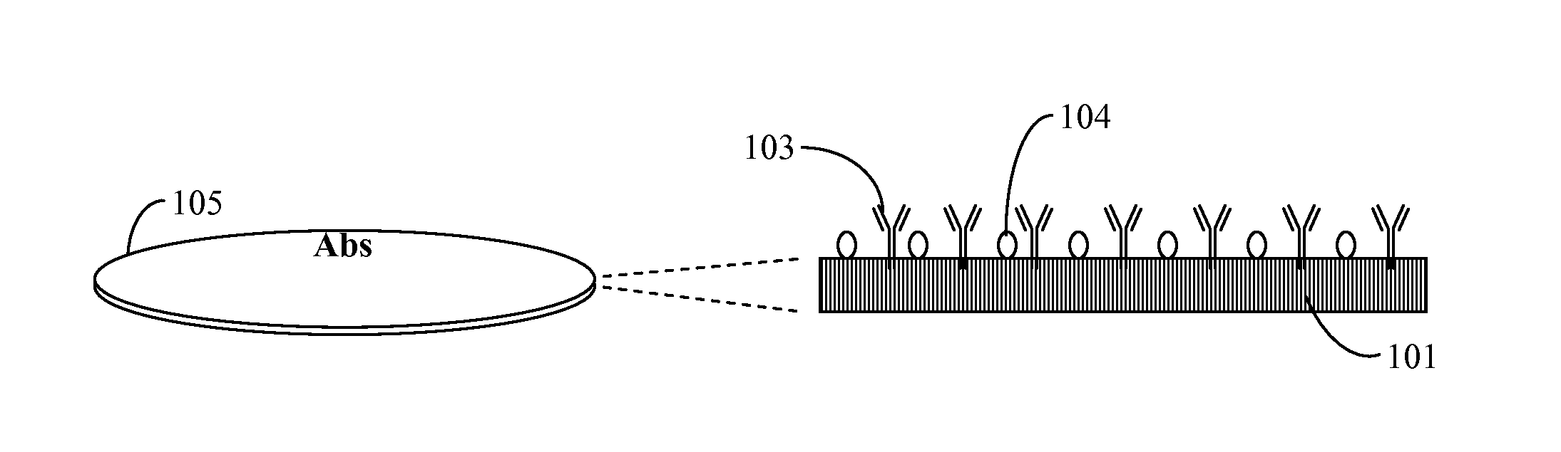
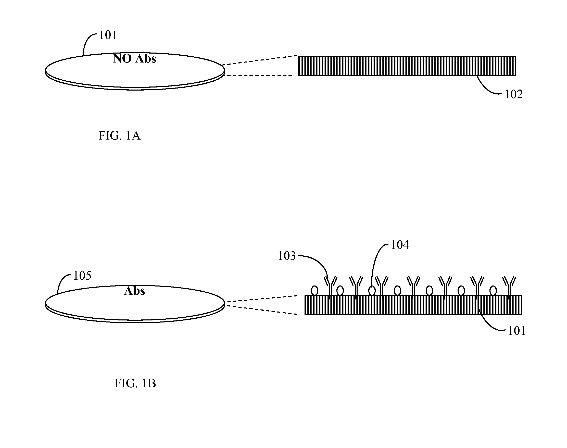
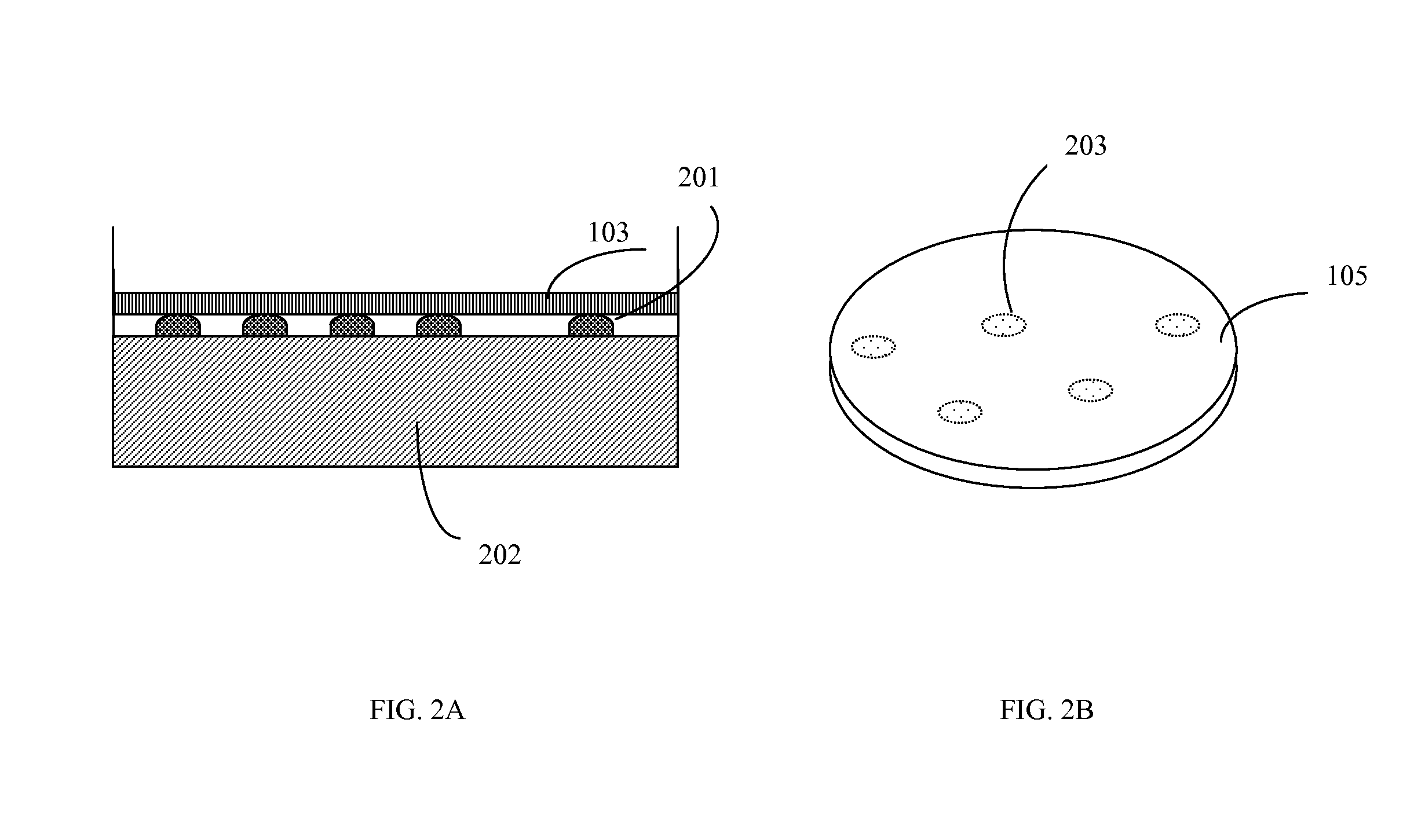
[0063]A sample containing an unknown mixture of species including a possible target microorganism such as E. coli O157 H:7 was poured on a Petri plate filled with nutrient agar. A plate with microorganisms was incubated at a temperature optimal for the target microorganism for 5 to 6 hours or more. A transfer permeable membrane containing immobilized antibodies against E. coli O157 H:7 and blocked in 1% of bovine serum albumin overnight was mounted on the surface of nutrient media and incubated for approximately 0.5 to 1 minute. This time was sufficient for an antigen-antibody reaction to occur and reliable attaching of one layer of cells to immobilized antibodies. Next, the transfer membrane was washed in washing solution (PBS pH 7.4 with 0.1% Tween 20) in a washing device at medium speed of rotation for 2 minutes. The transfer membrane was placed on a staining plate containing agar with dissolved 3-(4,5-Dimethylthiazol-2-yl)-2,5-diph...

example 2

Rapid Detection and Identification of Two Targets: E. Coli O157 and Salmonella

[0064]This procedure differed from the previous procedure by using a permeable nitrocellulose membrane with two immobilized antibodies for two different targets. Thus, two species left patterns on the transfer membrane. Differentiation of the two species (E. coli O157 and Salmonella) were performed by the use of a mixture of two antibodies immobilized on the surface of a permeable membrane and blocked after immobilization in order to prevent non-specific binding. Thus, a sample containing a mixture of species presumably including E. coli O157 and / or Salmonella was placed on a solid nutrient TSA medium in a Petri plate and grown for 7 to 8 hours at 37° C. Then, the membrane with the two antibodies was placed on the surface for 0.5 to 1 minute, washed in a washing solution for 2 minutes and placed on 1% agar filled with MTT (1 mg / ml). Patterns of E. coli O157 replicas obtained a well visible violet color wi...

example 3

Rapid Detection of All Microcolonies

[0065]In this procedure, a transfer permeable membrane contained no antibodies. All microcolonies adhered to the surface of the transfer membrane by non-covalent hydrophobic interaction but not by antibody-antigen binding. The washing out step was omitted in order to retain all replicas on the surface of the transfer permeable membrane. Next, the transfer membrane was moved to a staining plate containing MTT or another chromogen without washing. All replicas obtained a very intense dark violet color without washing and could be enumerated at a very early stage of growth: after only 4 to 6 hours. This was very important for early and rapid analysis of all live cells in a sample. The chromogenic substrate could be immobilized on the permeable membrane instead of being dissolved in the agar. This could simplify analysis because it would cut out two steps: washing in washing solution and transfer onto a staining plate (agar with a chromogenic substrat...


PUM

PropertyMeasurementUnit
timeaaaaaaaaaa
timeaaaaaaaaaa
timeaaaaaaaaaa

Abstract

An apparatus and method is provided for the rapid detection, identification and / or enumeration of one or more live target microorganisms growing on solid nutrient media or filtration membrane. Microcolonies or colonies attach to a porous transfer permeable membrane loaded with immobilized antibodies specific for target microorganisms. Non-specific cells are washed out of the permeable membrane and the membrane then is placed on a container containing a chromogenic and / or a fluorogenic substrate and incubated to allow coloration and subsequent identification and enumeration of color spots of targeted microorganisms and patterns of microcolonies or colonies.

Description

CROSS REFERENCE TO RELATED APPLICATIONS[0001]This application is a continuation-in-part of U.S. application Ser. No.12 / 532,501, filed Mar. 16, 2010, which is a Section 371 of PCT / US08 / 003826, filed Mar. 24, 2008, which claims priority to U.S. Provisional Application No. 60 / 896,321, filed Mar. 22, 2007, all of which are incorporated herein by reference.BACKGROUND OF THE INVENTION[0002]1. Field of the Invention[0003]The present invention relates generally to the field of microbiology and, in particular, to microbiological diagnostics for the rapid detection, identification and / or enumeration of colonies or microcolonies of live microorganisms.[0004]2. Description of the Prior Art[0005]Modern microbiological analysis is based on two main trends: 1) analysis without preliminary growth and 2) analysis after a preliminary growth. The first trend of analyzing without preliminary growth includes methods such as: i) immunological analyses [e.g., immunofluorescence, radioimmunoassay, enzyme i...

Claims


Application Information

Patent Timeline
Patent Type & Authority Applications(United States)
IPC IPC(8): G01N33/569
CPCC12Q1/04G01N33/56916G01N33/569C12Q1/06
Inventor GAZENKO, SERGEY
Owner NANOLOGIX INC
Why Eureka
Try Eureka