[0003]The object of the present invention is therefore represented by an
in vitro or
ex vivo method for the determination of 5-ASA efficacy in preventing and / or treating CRC in a mammalian, preferably a human, possibly affected by CRC, which method comprises measuring the inhibition of the β-catenin pathway and the activation of independent onco-suppressor genes in presence of 5-ASA.More in details, it is represented by a method for the determination of 5-ASA efficacy in preventing and / or treating CRC in a mammalian by measuring the expression of at least one
gene involved in the regulation of the β-catenin signalling pathway and the expression of other onco-suppressor genes.
[0005]The preferred genes which are suitable for the method according to the present invention are preferably selected from μ-protocadherin, E-
cadherin, β-catenin, Axin1, ICAT, p21waf-1, KLF4 and CEBPα.
[0008]Preliminary experiments performed in our laboratory confirmed that 5-ASA treatment inhibits the
proliferation activity of CaCo2 cells. To investigate the molecular mechanisms underlying this effect we performed a
microarray analysis, using the Affimetrix methodology, in order to assess the
transcriptome changes determined on CaCo2 cells by treatment with 20 mM 5-ASA for 96 h. The results obtained revealed an up-regulated expression of a number of onco-suppressor genes, potentially explaining the anti-proliferative effect elicited by 5-ASA, among which the most important was represented by μ-protocadherin. This gene codes for a member of the
cadherin superfamily and, based on previous reports by other authors, appeared as a putative inhibitor of the β-catenin signalling pathway, i.e. a
cell proliferation pathway mediated by the activity of the β-catenin
transcription factor16,17. The relevance of this finding resides in the observation that the β-catenin signalling pathway is constitutively activated in CRC. Other onco-suppressor genes up-regulated by 5-ASA treatment of the analyzed cells were represented by the KLF4 and CEBPα transcription factors, previously implicated in the proliferation inhibition of epithelial
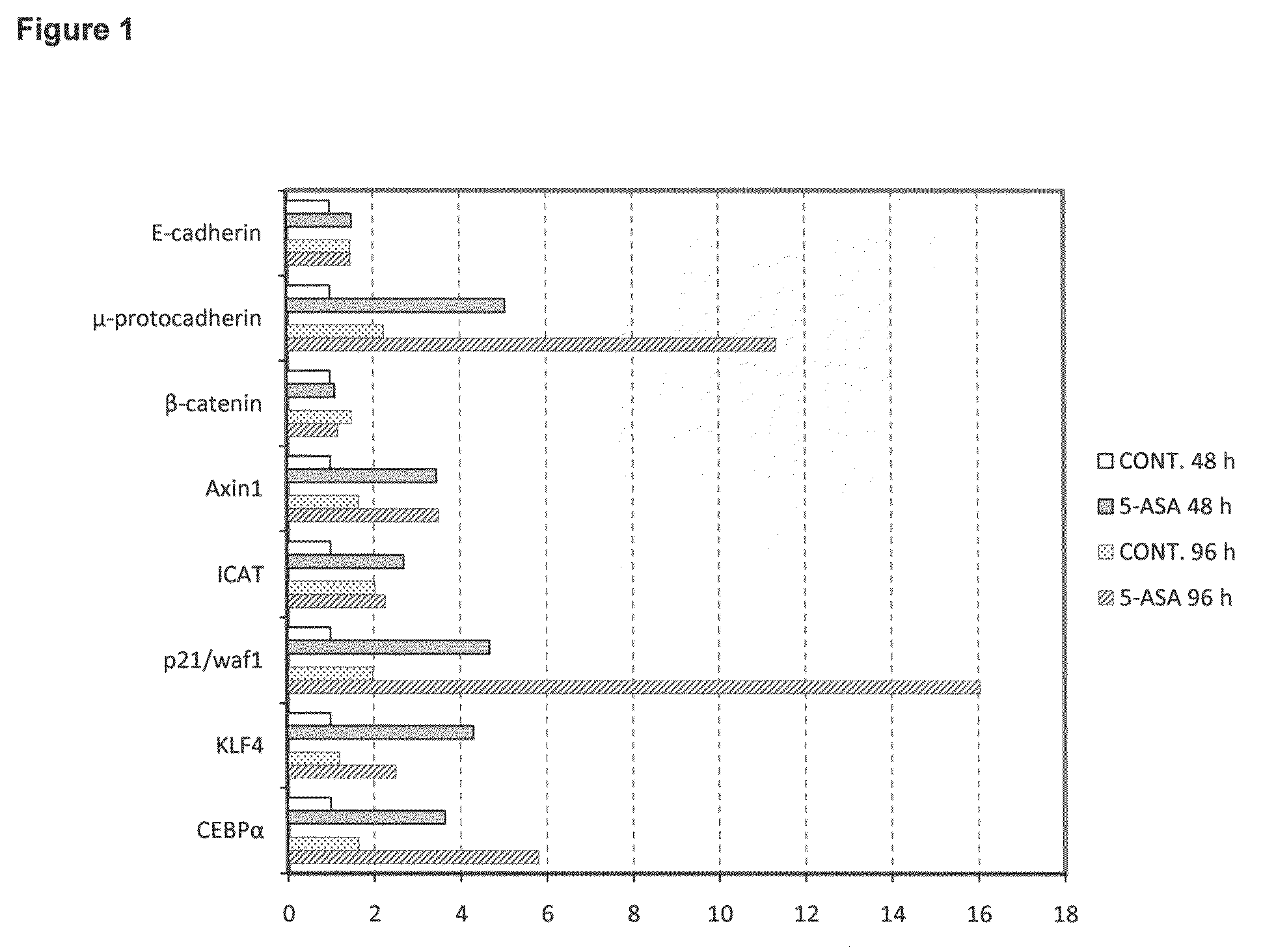
tumor cells in general and of CRC cells in particular18-20. To validate these data, the mRNA levels of the mentioned genes were analyzed by quantitative real time RT-PCR (QRT-PCR), in CaCo2 cells treated with 20 mM 5-ASA for 48 and 96 h. In addition, to better characterize the functional effect exerted by the investigated treatment on the β-catenin signalling pathway, we also included in this analysis other genes coding for the following proteins: β-catenin itself, i.e. the main component of the pathway; E-cadherin, for its ability to sequester β-catenin on the plasmatic membrane of cells; Axin1, due to its capacity to promote the activity of the β-catenin degradation complex; ICAT, responsible for the inhibition of β-catenin transcription in the nuclear compartment; p21waf-1, previously implicated in
growth arrest processes and also demonstrated to be a negative target of β-catenin. Up-regulation of
mRNA expression resulted to be 12-fold for μ-protocadherin, 16-fold for p21waf-1, 4 to 6-fold for KLF4 and CEBPα and 2 to 3-fold Axin 1 and ICAT. The most remarkable variations of expression were consequently observed for the μ-protocadherin and for the p21waf-1 genes, suggesting that the β-catenin pathway was really inhibited under the adopted experimental conditions. The up-regulation of these genes appeared also more pronounced at the end of stimulation (96 h).
mRNA expression of E-cadherin and β-catenin appeared, conversely, unaffected (FIG. 1).
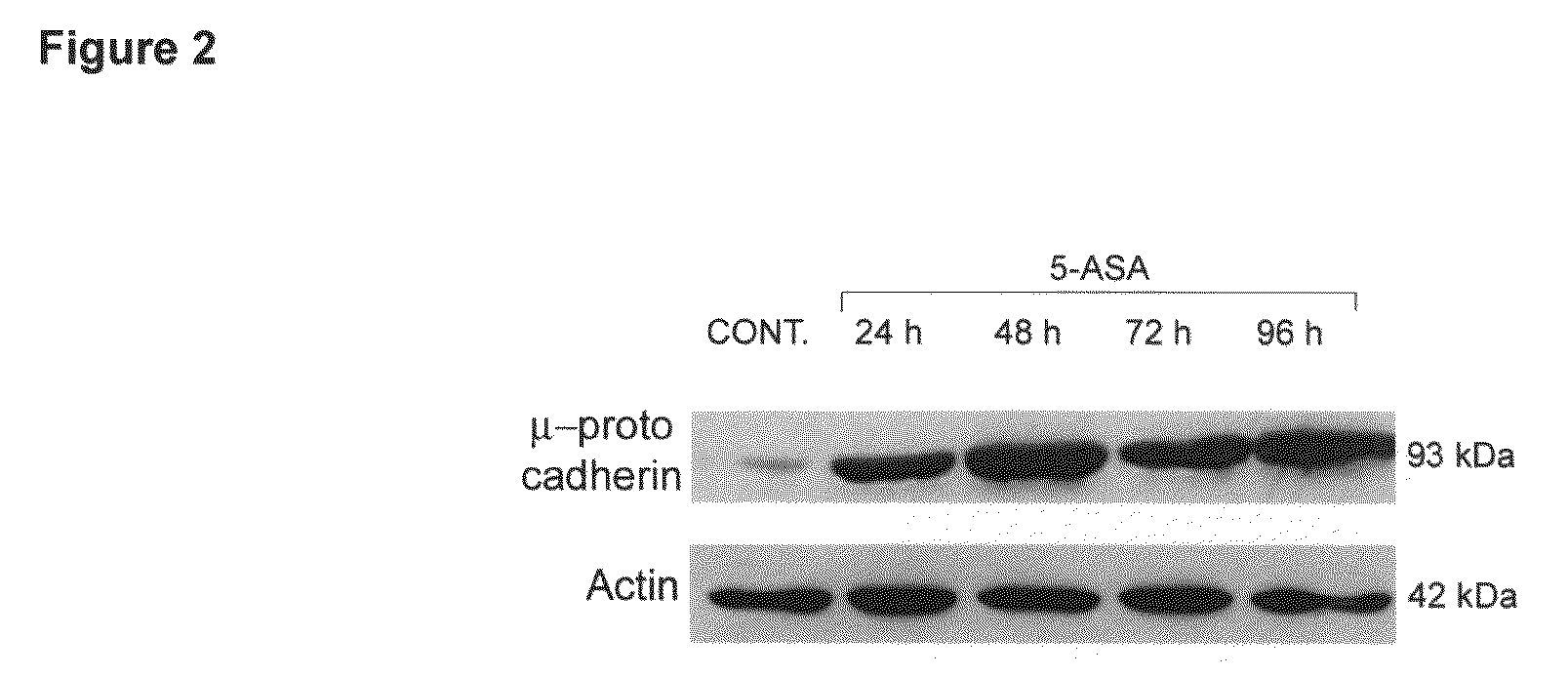
Western Blot Analysis of μ-Protocadherin
Protein Expression in CaCo2 Cells Treated with 5-ASA.


