Substrate and method for culturing breast cells

Inactive Publication Date: 2010-11-25
CORNING INC
View PDF4 Cites 6 Cited by
  • Summary
  • Abstract
  • Description
  • Claims
  • Application Information

AI Technical Summary

Problems solved by technology

However, many cells, such as non-malignant and malignant mammary cells and other differentiated cell types, rapidly lose their specific morphology and cellular functions when cultured on 2D surfaces.
While Matrigel™—based 3D cell culture has successfully demonstrated the significance of 3D culture in cancer research; it has some distinct disadvantages that signi

Method used


Image

  • Substrate and method for culturing breast cells
  • Substrate and method for culturing breast cells
  • Substrate and method for culturing breast cells

Examples


example 1

Fabrication of Porous Substrate

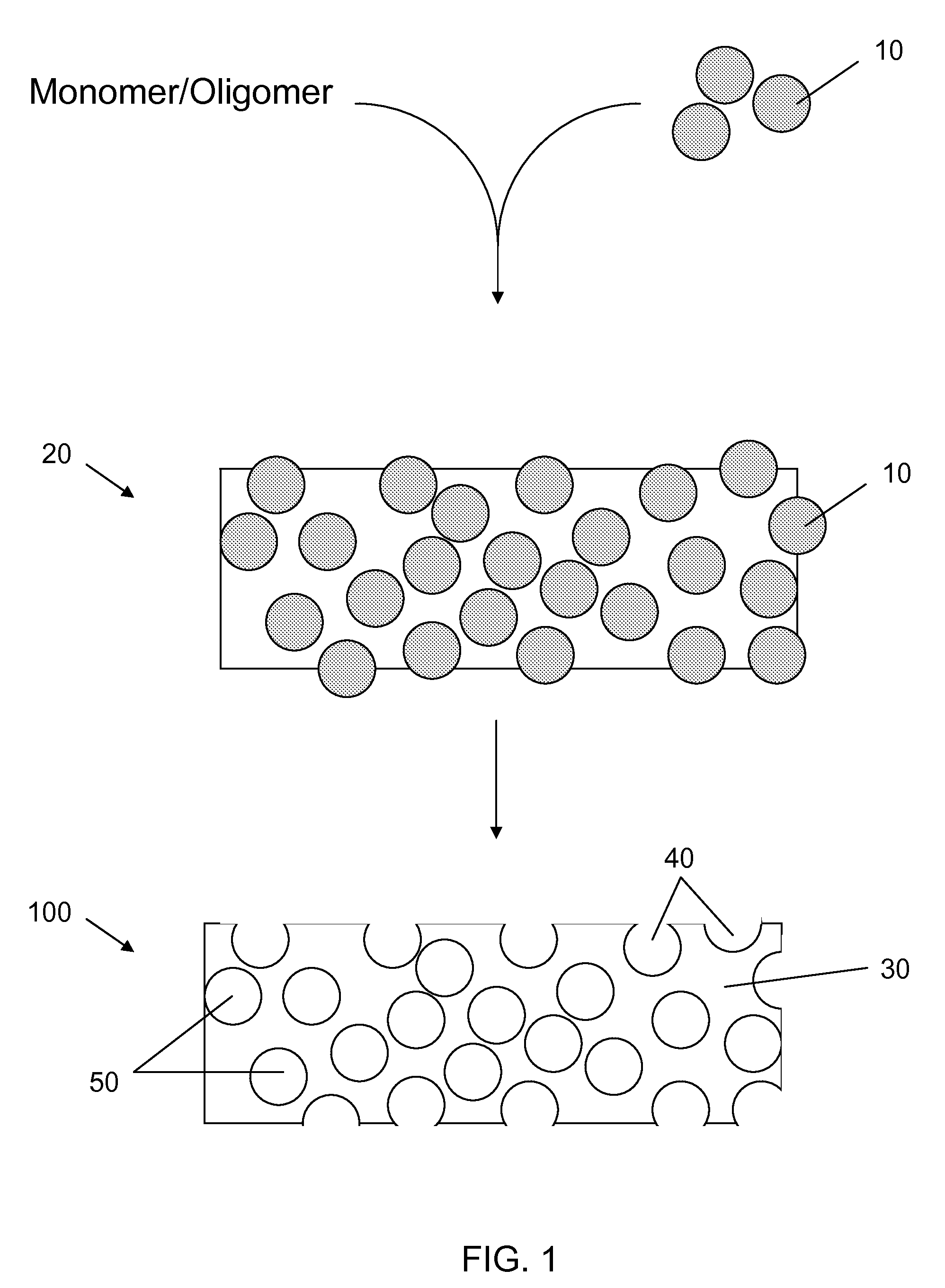
[0081]Porous polydimethylsiloxane (PDMS) substrates were generated by mixing a PDMS pre-polymer and a curing agent using the Sylgard 182 kit available from Dow Corning in a ratio of 10 to 1. The mixture was closely packed with sugar crystals of size ranging from 212-250 micrometers, which was then filled in a mold and cured at 100 degrees for one hour. The sugar in the cured polymer was washed out in an ultrasonic bath, forming porous PDMS substrates. The resulting porous PDMS was released from the mold and assembled into multi-well plate for cell culture.

[0082]Depending on the density of wells in the plates, the plates were prepared as follows: (1) for 96-well plate, porous PDMS was molded in a 96-well format on a piece of glass insert, then the insert was released from the mold and glued to the bottom of 96-well holy plate (plate without the bottom) by double-sided pressure sensitive adhesive (PSA) plate; or (2) for plates of density lower than 96 we...

example 2

Culture of Non-malignant Breast Cells on Porous Substrate

[0083]Non-malignant breast cells MCF-10A were seeded in wells having the porous PDMS substrates described in Example 1. 10,000 cells were seeded and cultured in MEBM / F12 cell culture medium with 5% horse serum, 5% pen / strep, 20 ng / ml of hEGF, 0.5 μml of hydrocortisone, 100 ng / ml of Cholera toxin and 10 μg / ml of insulin. The medium was changed on alternate days. Following two week of culture, the cells were stained with Rhodamine phalloidin (F-actin staining) and imaged with confocal fluorescence microscope. The confocal image of MCF-10A, as shown in FIGS. 8A-B (with FIG. 8B being of higher magnification), reveals that MCF-10A formed a structure of organized nuclei and robust cell-cell adhesion. The observed cell morphology closely resembled the in-vivo acini structure of non-malignant mammary epithelial cells. This is in contrast to MCF-10A cells cultured on traditional 2D surfaces, on which the cells form monolayer.

example 3

Culture of Malignant Breast Cells on Porous Substrate

[0084]Malignant breast cells MCF-7 were seeded in wells having the porous PDMS substrates described in Example 1. 10,000 cells were seeded and cultured in EMEM cell culture medium with 10% Fetal Bovine Serum, 5% pen / strep, and 10 μg / ml of insulin. The medium was changed on alternate days. Following two weeks of culture, the cells were stained with Rhodamine phalloidin (F-actin staining) and DAPI nuclei counter stain and imaged with confocal fluorescence microscope as shown in FIG. 9. The image shows that MCF-7 cells formed a mass structure of disorganized nuclei but maintain robust cell-cell adhesion.


PUM


Abstract

A cell culture article includes a porous substrate having a plurality of pores and a plurality of interstices in communication with the pores. At least some of the plurality of pores and interstices are sufficiently large for two or more mammary epithelial cells to cluster within the pores or interstices. Non-malignant mammary epithelial cells or breast cancer cells may not attach strongly to the substrate surface, which may encourage cell-cell interaction. In many cases, the article is desirably free of components of unknown origin. The articles may be capable of maintaining culture of malignant and non-malignant mammary epithelial cells and allowing for development of in vivo-like morphologies or characteristics of such cells.

Description

RELATED APPLICATION[0001]This application claims the benefit of priority to U.S. Provisional Patent Application Ser. No. 61 / 117,366, filed on Nov. 24, 2008, which application is hereby incorporated by reference in its entirety to the extent that it does not conflict with the present disclosure.FIELD[0002]The present disclosure relates to cell culture, and more particularly to culture of breast cells and to substrates that promote in-vivo like characteristics of cultured breast cells.BACKGROUND[0003]In vitro studies of human cancer including breast cancer have been primarily carried out with established cell lines on two-dimensional (2D) cell culture surfaces. However, many cells, such as non-malignant and malignant mammary cells and other differentiated cell types, rapidly lose their specific morphology and cellular functions when cultured on 2D surfaces. To overcome this deficiency of 2D surfaces, extracellular matrices (ECM) have been employed to establish and maintain functional ...

Claims


Application Information

Patent Timeline
IPC IPC(8): G01N33/574C12N5/07
CPCC12N5/0062C12N5/0075C12N5/0068C12N5/0631C12N5/0693G01N33/5011C12N2503/02C12N2506/30C12N2513/00C12N2533/30G01N33/57415C12N2535/00
Inventor DENG, HUAYUNLAHIRI, JOYDEEPSU, HUI
Owner CORNING INC
Why Eureka
Try Eureka