
Method for detecting tumor cell invasion using short diffusion times

a tumor cell and diffusion time technology, applied in the field of tumor cell invasion detection using short diffusion time, can solve the problems of water also affecting the diffusion properties, and standard magnetic resonance imaging (mri) techniques do not adequately reflect the cellular and microscopic structure of the tumor

Inactive Publication Date: 2010-11-25
IMAGING BIOMETRICS
View PDF11 Cites 12 Cited by
  • Summary
  • Abstract
  • Description
  • Claims
  • Application Information

AI Technical Summary

Benefits of technology

[0005]The present invention discloses an improved method for detecting the presence of tumor cell invasion which reduces diffusion times to as little as 2 msec. by incorporating isotropic diffusion weighing into a standard spin echo (SE) pulse sequence using a pair of balanced bipolar gradients position

Problems solved by technology

Standard magnetic resonance imaging (MRI) techniques do not adequately reflect the cellular and microscopic structure of the tumor, particularly areas of tumor cell invasion.
However, in biological tissues, diffusion is not truly random because tissue has structure which limits or restricts the amount of diffusion possible.
Moreover, chemical interactions of water and the macromolecules which may be contained in the water also may affect diffusion properties.

Method used


Image

  • Method for detecting tumor cell invasion using short diffusion times
  • Method for detecting tumor cell invasion using short diffusion times
  • Method for detecting tumor cell invasion using short diffusion times

Examples


Embodiment Construction

[0015]Current DWI techniques implemented with relatively long diffusion times have a reduced sensitivity to intracellular compartments, such as the nuclear compartment, which is known by histomorphometric techniques to be altered. The detection method of the present invention is designed to expose the intracompartmental signals that are overlooked by DW sequences using longer diffusion times and to provide new information useful for glioma localization.

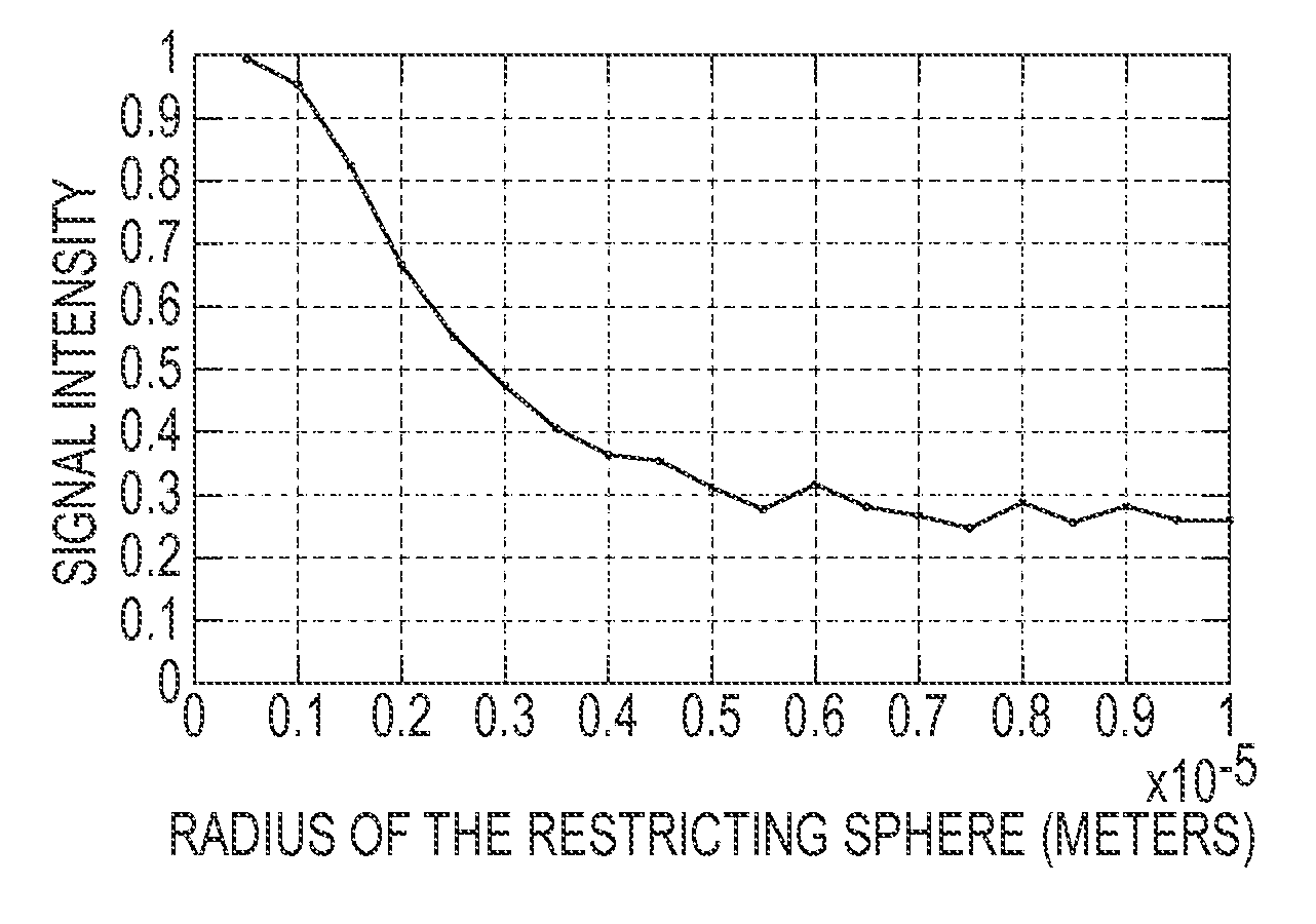
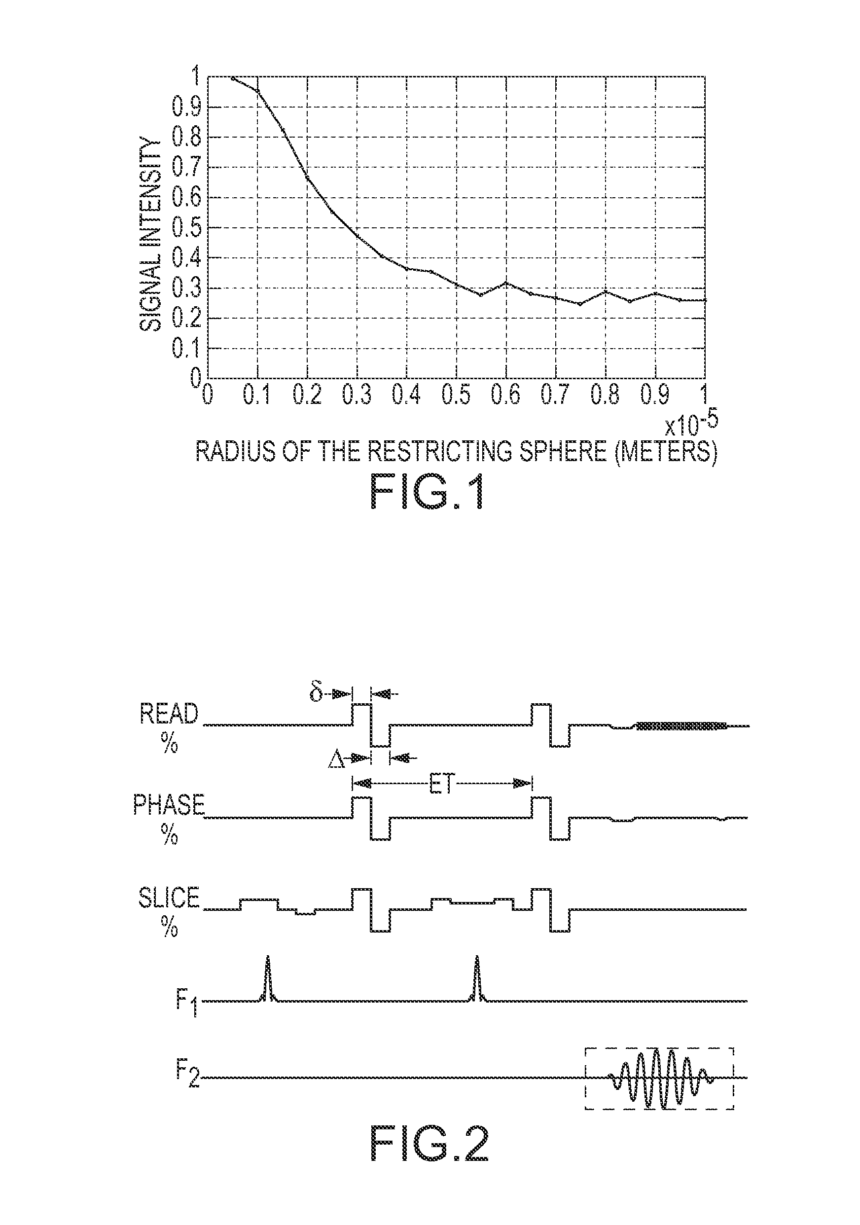
[0016]A Simulation of the Effects of Compartment Size on the Diffusion Weighted Signal.

[0017]Diffusion weighted MR obtains estimates of the diffusion coefficient by allowing ensembles of spins to course through the medium over a known time. For a given signal attenuation the diffusion coefficient can be calculated from the length of the diffusion experiment and the gradient shape. In vivo this estimate is influenced by restrictive boundaries and other objects that hinder the ensemble's translational motion. If the time given for the e...


PUM


Abstract

An improved method for detecting tumor cell invasion using diffusion times as short as two (2) msec incorporates diffusion weighing imaging techniques into a standard spin echo (SE) pulse sequence to minimize the effects of compartment boundary restrictions on diffusion values and corresponding MRI imaging data related to glioma invasion.

Description

CROSS-REFERENCE TO RELATED APPLICATIONS[0001]This application is the National Stage of International Application No. PCT / US2008 / 064599 filed May 22, 2008, which claims the benefit of U.S. Provisional Patent Application Ser. No. 60 / 939,546 filed on May 22, 2007, the entire contents of each of which are incorporated herein by reference.BACKGROUND OF THE INVENTION[0002]Remedial surgical and radiological techniques and procedures for brain tumors require precise knowledge of the boundary of the tumor cell mass. Standard magnetic resonance imaging (MRI) techniques do not adequately reflect the cellular and microscopic structure of the tumor, particularly areas of tumor cell invasion. Magnetic resonance imaging relies on the relaxation properties of excited hydrogen nuclei in water and in lipids. Water molecules are in constant motion, and the rate of movement or diffusion is temperature dependant and also depends upon the kinetic energy of the molecules. However, in biological tissues, d...

Claims


Application Information

Patent Timeline
IPC IPC(8): A61B5/055
CPCA61B5/055G01R33/56341G01R33/5617G01R33/5616
Inventor SCHMAINDA, KATHLEENPAULSON, ERIC S.PRAH, DOUGLAS E.
Owner IMAGING BIOMETRICS
Learn More