Cell assay kit and method

a cell assay and kit technology, applied in the field of cell assay kits and methods, can solve the problems of not being able to lend themselves readily to field settings, storefront clinics, field clinics, trained laboratory personnel, etc., and achieve the effect of increasing the density or magnetic susceptibility of cells

Active Publication Date: 2010-12-09
STEMCELL TECH CANADA INC
View PDF7 Cites 14 Cited by
  • Summary
  • Abstract
  • Description
  • Claims
  • Application Information

AI Technical Summary

Benefits of technology

[0010](a) reacting a cell sample containing cells of the given cell type with particles capable of specific attachment to the cells, and being effective, when attached to the cells, to increase the density or magnetic susceptibility of the cells, allowing particle-bound cells and free particles to be separated from non-particle bound cells in the sample based on their greater rate of migration through a selected-density and / or selected viscosity medium under the influence of a gravitational or applied centrifugal or magnetic-field force,
[0015]The particles, when attached to the cells in step (a), may be effective to label the cells with a detectable reporter, and said inspecting includes visually inspecting the collection chamber for the presence of cells labeled with the detectable reporter.
[0021]In one embodiment, step (a) may further include reacting cells in the sample with first particles capable of binding specifically to CD14 antigen on monocyte cells, and with second particles capable of binding specificallyCD4 antigen on T lymphocyte cells and monocyte cells, thus to confer on the T lymphocyte cells, enhanced density, and on the monocyte cells, both enhanced density and magnetic susceptibility, and step (b) may further include first removing particle-bound monocytes from the cell sample by application of a magnetic force effective to selectively remove the particle-bound monocytes from particle-bound T lymphocyte cells. Both particles may also be applied simultaneously in defined ratios, ensuring that statistically the monocyte cells are bound to at least one particle having magnetic susceptibility. Other possible approaches to remove non-desired cells having the same surface marker as the target cells are: i) Masking of the target surface markers with sterically large beads having an affinity for other, non-target surface markers on the interfering cell types (e.g. using non-dense and non-magnetically susceptible anti-CD14 beads to first coat the monocytes before exposing the sample to the anti-CD4 beads), thus preventing the anti CD4 beads to bind to the CD4 markers on the monocytes due to the masking effect of the antiCD14 beads covering the monocytes. ii) Masking the target surface markers on non desired cells by other means like using antibody mediated tetrameric antibody complexes which have specificity to both, e.g. CD14 antigen and red blood cell surface markers, thus masking the monocytes by covering them with a dense layer of red blood cells. iii) The interfering non-target cells can be removed by specifically capturing these cells by a solid matrix containing e.g. antibodies against surface markers only present on the interfering cells. For example, antiCD14 antibody can be immobilized on a filter matrix. After exposing the blood sample to that filter matrix, the monocytes are depleted from the blood sample by binding to that matrix, after which the blood sample is exposed to the antiCD4 particles.

Problems solved by technology

However, they do not lend themselves readily to field settings, such as storefront clinics, or field clinics in third-world countries, where neither trained laboratory personnel or sophisticated cell sorting or microscope equipment are present.
In poorer areas of Africa, for example, these methods may be ill suited for testing large numbers of people for CD4+ T cell counts, as an indication of the onset of AIDS following HIV infection, or for monitoring a person's response to e.g. antiretroviral drugs.

Method used


Image

  • Cell assay kit and method
  • Cell assay kit and method
  • Cell assay kit and method

Examples


Embodiment Construction

I. Definitions

[0051]Unless otherwise, the terms below have the following meaning herein:

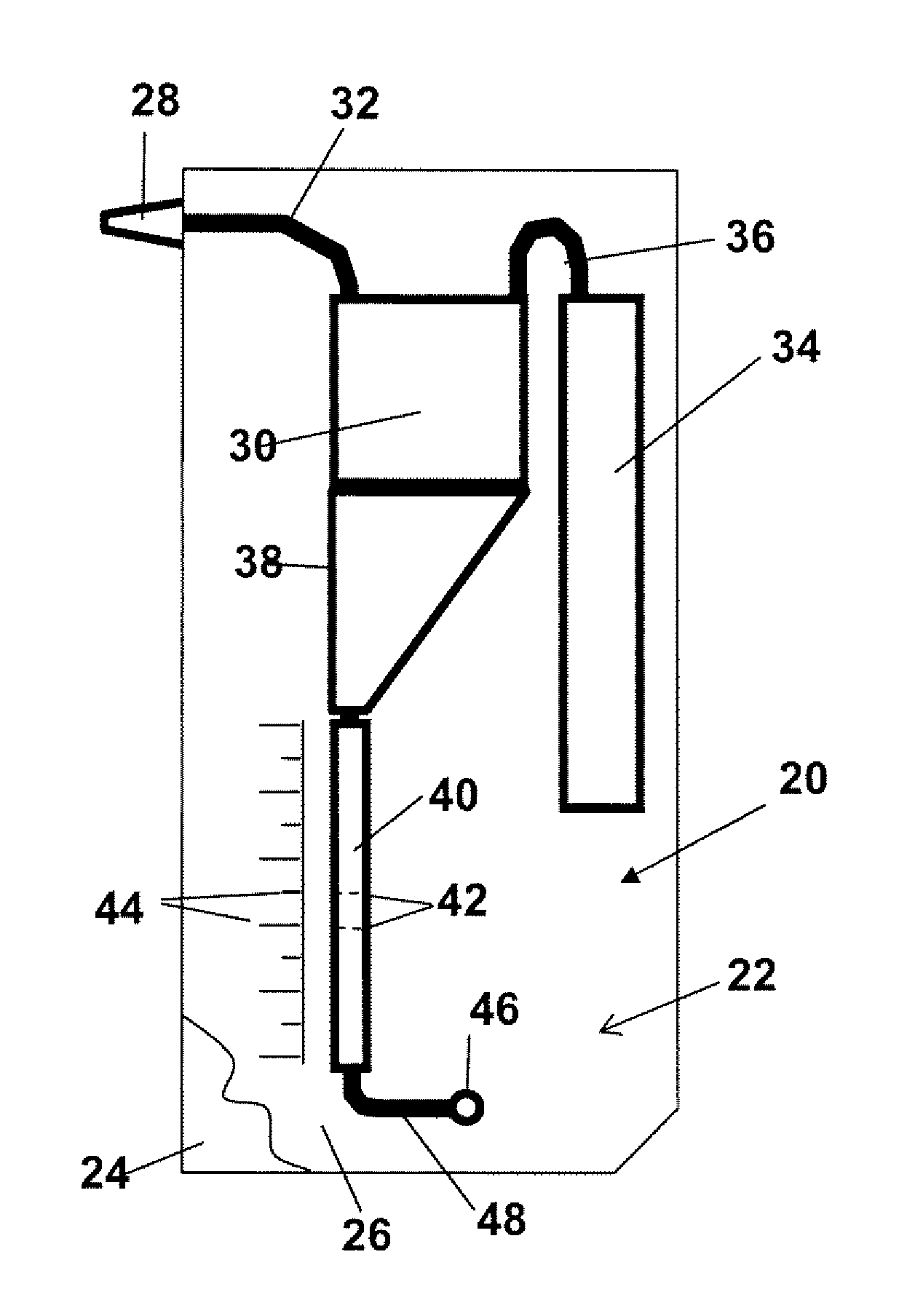
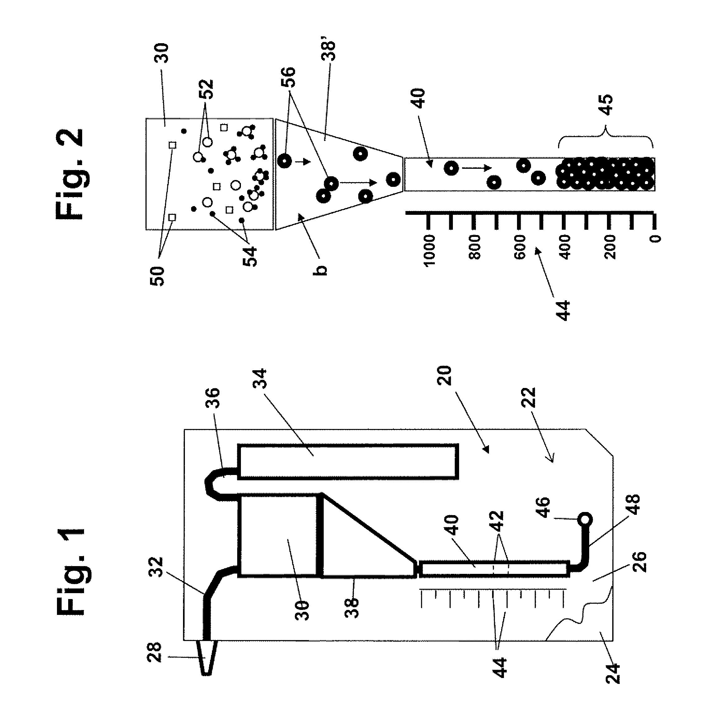
[0052]A “cell sample” refers to any liquid sample containing or suspected of containing one or more types of cells in suspension. A cell sample includes a “body-fluid sample,” referring to e.g. a blood, urine, or saliva sample obtained from a human or other animal body. A blood sample may be whole blood or processed blood or whole blood in which all or the bulk of red blood cells have been removed. Other possible cell samples include e.g. cell cultures, cell extracts obtained from tissue samples, waste-water. Cell samples that are suspected of containing cells include e.g. milk and other food that is contaminated with an unwanted e.g. cell or bacteria type.

[0053]“Concentration” of cells in a cell sample refers to the number of cells in a given cell-sample volume. The term is typically expressed as number of cells / per sample volume.

[0054]A “threshold level or concentration of cells of a given type...


PUM

PropertyMeasurementUnit
sizeaaaaaaaaaa
sizeaaaaaaaaaa
diametersaaaaaaaaaa

Abstract

A method and kit for assaying a cell sample for the presence of at least a threshold number of cells of a given type are disclosed. The kit includes an assay device having a sample chamber for receiving the cell sample and an elongate collection chamber containing a selected-density and / or viscosity medium and having along its length, a plurality of cell-collection regions, and particles which are capable of specific attachment to cells of the selected cell type, and which are effective, when attached to the cells, to increase the density or magnetic susceptibility of the cells. In operation, particle-bound cells and particles in the cell sample are drawn through the elongate collection chamber under the influence of a gravitational or selected centrifugal or magnetic-field force until the particle-bound cells and particles completely fill successive cell-collection regions in the collection chamber. Indicia associated with at least one collection regions indicates a concentration of cells of the selected type effective to at least partially fill that collection region.

Description

[0001]This application is a continuation of U.S. patent application Ser. No. 12 / 329,380 filed on Dec. 5, 2008, which claims priority to U.S. Provisional Patent Application No. 60 / 992,624 filed on Dec. 5, 2007, both of which are incorporated in their entirety herein by reference.FIELD OF THE INVENTION[0002]The present invention is directed to a cell assay kit and method for determining the presence and / or concentration of cells of a given type in a cell sample, and in particular, the presence of at least a selected threshold level of the cells in the sample.BACKGROUND OF THE INVENTION[0003]A variety of disease conditions, including the response to treatment of a disease state, can be monitored by hematology markers, using the approximate concentration of certain white blood cells in a blood sample as an indicator of the body's response to the disease. For example, the concentration of CD4+ T-lymphocyte cells in a blood sample may provide a marker for the outbreak of AIDS following HI...

Claims


Application Information

Patent Timeline
Patent Type & Authority Applications(United States)
IPC IPC(8): G01N33/50C12M1/34
CPCY10T436/25125G01N33/56972Y10T436/13G01N33/54313G01N33/491G01N33/54326Y10T436/25375G01N33/538
Inventor ZAUGG, FRANKTOBIAS, RENEEMCMANUS-MUNOZ, SILVIAKERNEN, PETERRUIZ-TAYLOR, LAURENCEWAGNER, PETER
Owner STEMCELL TECH CANADA INC
Why Eureka
Try Eureka