Method for monitoring, diagnosing, and screening cancer through measuring the concentration of des-r prothrombin activation peptide fragment f2 (des-r f2) in a serum
a prothrombin activation peptide and serum concentration technology, applied in the field of protein markers for monitoring, diagnosis and screening of cancer, can solve the problems of large amount of time and effort, still number of complex processes of diversity, and critical limitations in genome analysis methods, so as to improve the survival rate of liver cancer, reduce the number of national losses, and improve the effect of survival ra
- Summary
- Abstract
- Description
- Claims
- Application Information
AI Technical Summary
Benefits of technology
Problems solved by technology
Method used
Image
Examples
example 1
Obtainment and Preservation of Serum
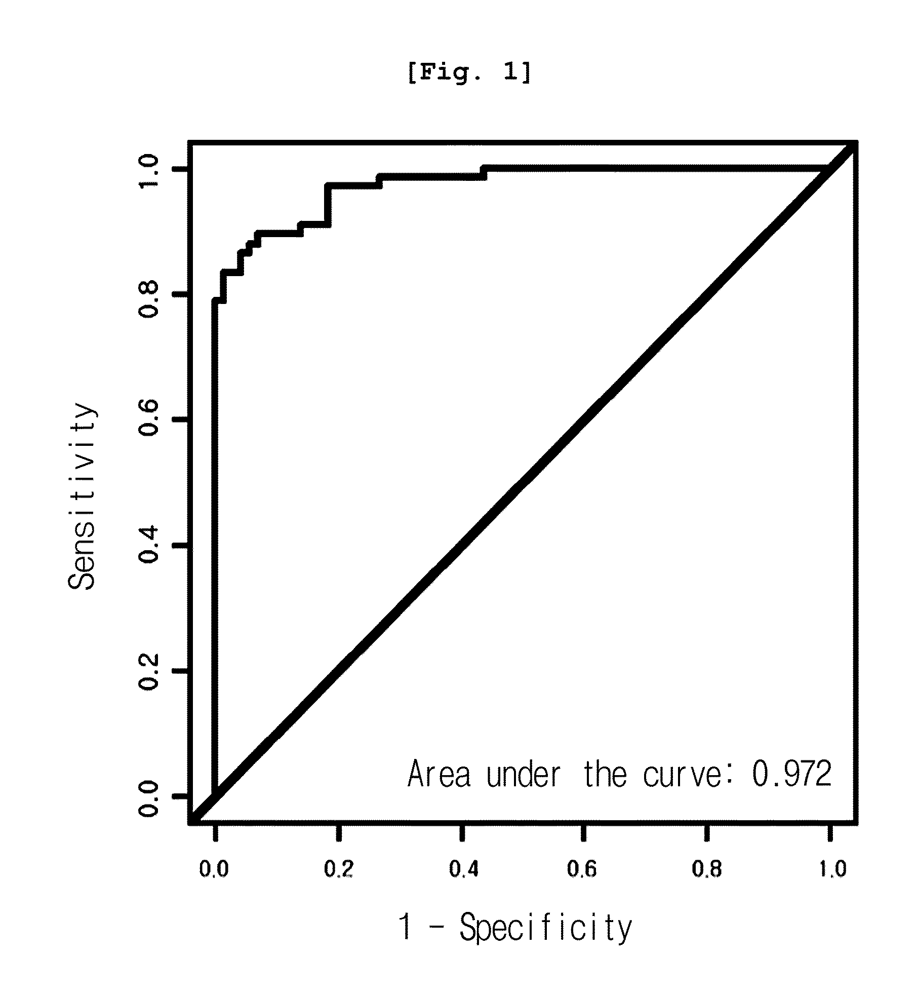
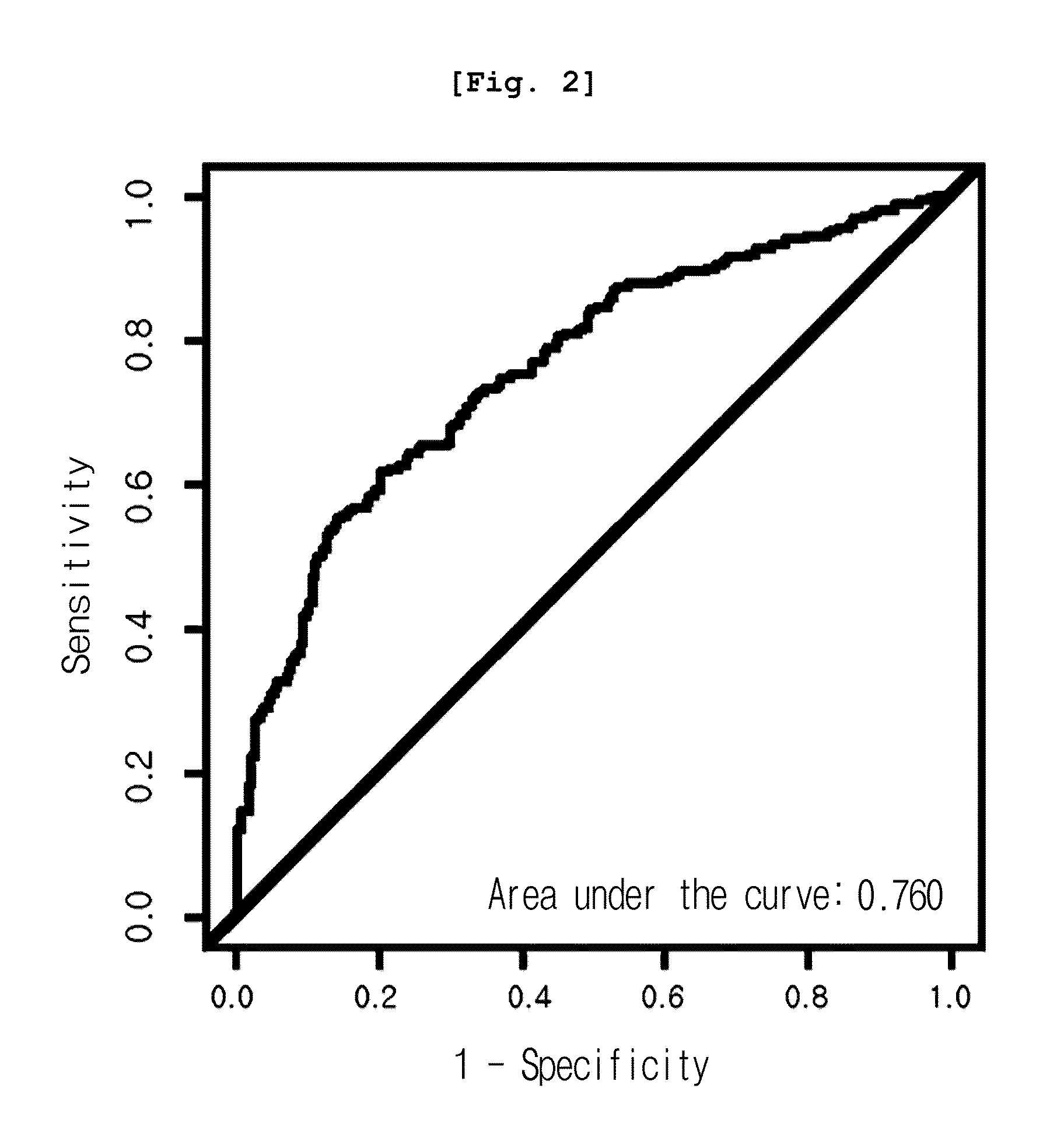
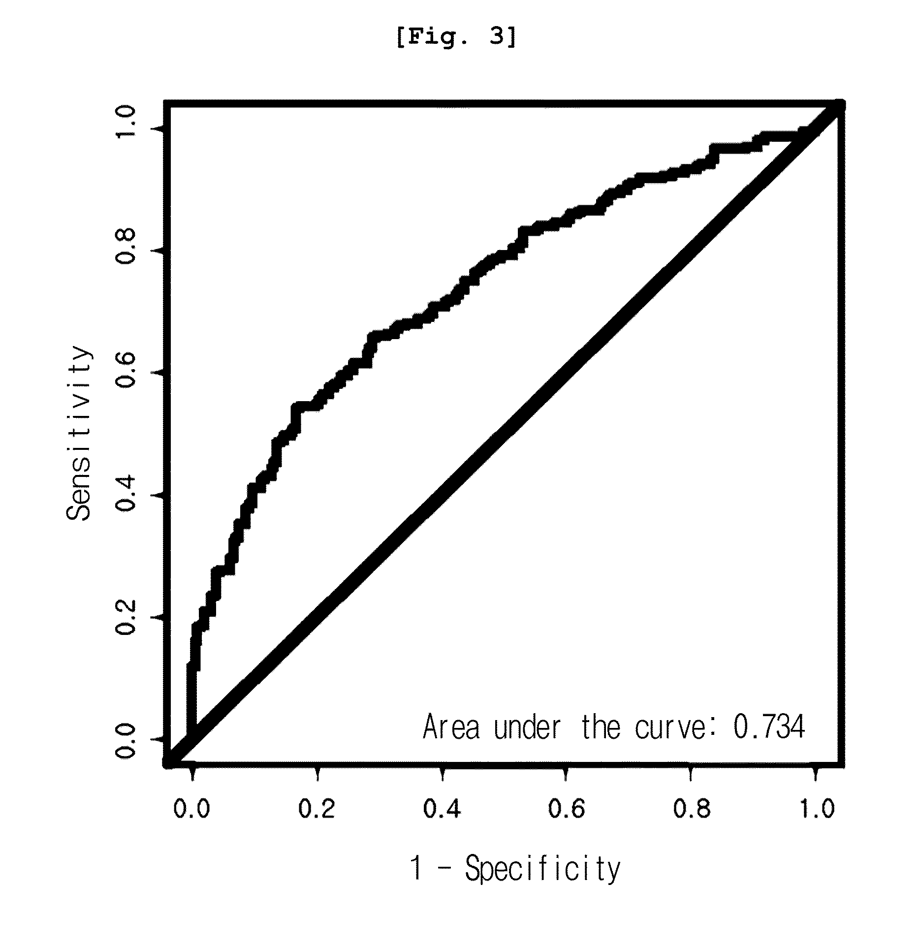
[0074]In this invention, three test groups were prepared; group 1 with 71 normal subjects (29 women, 42 men) and 66 liver cancer patients (14 women, 52 men); group 2 with 250 normal subjects (119 women, 131 men) and 245 stomach cancer patients (84 women, 161 men); and group 3 with 196 normal subjects (196 women) and 252 breast cancer patients (252 women).
[0075]The age of the normal subjects in group 1 for the liver cancer related experiment ranged from 29 to 64 (mean: 46.9, median: 47), and the age of the liver cancer patients ranged from 35 to 75 (mean: 57.1, median: 58). The age of the normal subjects in group 2 for stomach cancer related experiment ranged from 23 to 67 (mean: 45.7, median: 45) and the age of the stomach cancer patients ranged from 28 to 86 (mean: 59.1, median: 61). Cancer stages of those who had stomach cancer were as follows: stage 1, 129 patients; stage 2, 48 patients; stage 3, 36 patients; and stage 4, 32 patients. In the ca...
example 2
Serum Protein Analysis
[0077]Serum was analyzed by anion exchange (Q10) proteinchip array (Ciphergen). Particularly, 10 μl of serum was diluted with 15 μl of solubilization buffer (9 M Urea, 2% CHAPS), which stood at room temperature for 30 minutes. 45 μl of Q10 binding buffer (0.05% Triton X-100 in 50 mM HEPES, pH 7.0) was added to 5 μl of the diluted serum sample, followed by mixing with vortex. To equilibrate Q10 chip, 10 μl of binding buffer was loaded on every spot, which stood at room temperature for 5 minutes. After eliminating binding buffer, 10 μl of fresh binding buffer was loaded on each spot again, which stood at room temperature for 5 minutes. 5 μl of serum mixture (serum sample+solubilization buffer+binding buffer) was loaded on each spot, followed by reaction in a humidify chamber at room temperature for 1 hour. After eliminating serum mixture, 10 μl of binding buffer was loaded and then binding buffer was rightly eliminated (repeated 4 times). After eliminating bindin...
example 3
[0078]Mass calibration was performed according to external calibration using a calibrant such as bovine superoxide dismutase, equine myoglobin, bovine beta-lactoglobulin A and horseradish peroxidase (Ciphergen Biosystems Inc., Fremont, Calif., USA), followed by normalization using total ion current. Automatic peak detection was performed using Ciphergen Express Client ver. 3.0 software (signal-to-noise: 5 or up, valley depth: 5 or up, minimum peak threshold: 0%, cluster mass window: 0.3% MW). As a result, 203 peak clusters in total were confirmed. Mann-Whitney U-test p-value and AUC (area under the receiver operating characteristic curve) of the peaks were compared between normal and cancer groups.
[0079]As a result, among all peaks, the expression pattern of 12.5 kDa peak (12,451 m / z) was significantly different in three cancer groups, from that of the normal group. As shown in Table 1, the peak was reduced in cancer groups, even though the reduction rate varied, co...
PUM
| Property | Measurement | Unit |
|---|---|---|
| Fluorescence | aaaaa | aaaaa |
Abstract
Description
Claims
Application Information
 - R&D
- Intellectual Property
- Life Sciences
- Materials
- Tech Scout
- Unparalleled Data Quality
- Higher Quality Content
- 60% Fewer Hallucinations
Browse by: Latest US Patents, China's latest patents, Technical Efficacy Thesaurus, Application Domain, Technology Topic, Popular Technical Reports.
© 2025 PatSnap. All rights reserved.Legal|Privacy policy|Modern Slavery Act Transparency Statement|Sitemap|About US| Contact US: help@patsnap.com


