Anti-Adhesion Barrier Wound Dressing Comprising Processed Amniotic Tissue and Method of Use

a barrier wound and amniotic tissue technology, applied in the field of anti-adhesion wound dressings, can solve the problems of difficult handling and manipulation of amnion, and achieve the effect of maintaining the orientation of the amnion patch and facilitating the handling of amnion

Inactive Publication Date: 2011-06-02
ALLOSOURCE +1
View PDF25 Cites 45 Cited by
  • Summary
  • Abstract
  • Description
  • Claims
  • Application Information

AI Technical Summary

Benefits of technology

[0013]The AAB material is produced by processing human amniotic tissue. In one embodiment of the invention, the human amniotic tissue is processed by debriding the tissue to remove the amnion from the chorion and other amniotic tissue and fluids. The amnion is rinsed in a sterile saline solution, such as 0.9% NaCl solution. Preferably, multiple saline rinse steps are performed. After rinsing with sterile saline, the amnion is soaked in a glutaraldehyde solution. Preferably, the amnion is soaked in a 1% glutaraldehyde solution for a period of up to 15 minutes. Following the glutaraldehyde soak, the amnion is again rinsed with sterile saline solution. The tr...

Problems solved by technology
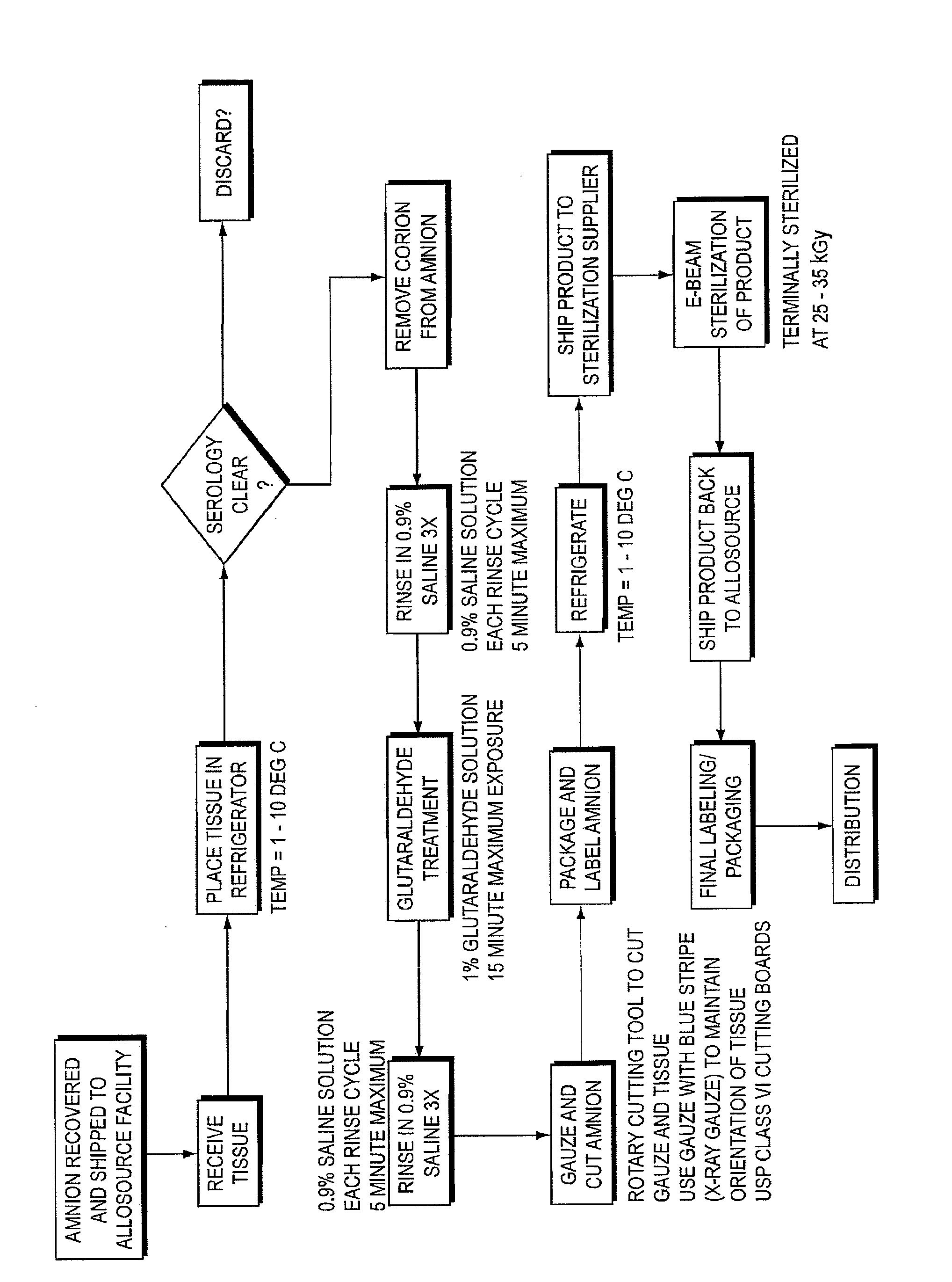
As described in detail below, amnion can be difficult to h...

Method used


Image

  • Anti-Adhesion Barrier Wound Dressing Comprising Processed Amniotic Tissue and Method of Use

Examples


example 1

Amnion Patch Treated with Glutaraldehyde Implanted with the Maternal Side Towards the Dura

[0035]Gluteraldehyde treated amnion patches were prepared using the method described above, with the patches treated in 1% gluteraldehyde solution for 15 minutes. Three saline rinses as described above were performed before and after the gluteraldehyde treatment. In this example, the amnion treated patches were implanted with the maternal side of the amnion towards the dura.

[0036]The pathologist's review revealed the following. The laminectomy site was filled completely with reactive bone, reactive fibrocartilaginous tissue and reactive fibrosis. The reactive bone was organized and extended from the ends of the adjacent vertebral bodies. Superficially, there were ligaments and skeletal muscle bundles with regional muscle fiber atrophy and endomysial fibrosis. Separated by a distinct linear clear zone was an underlying membrane with a simple layer of flattened cuboidal epithelioid cells upon a h...

example 2

Amnion Patch with Non-Treated Amnion Patches with the Fetal Side Towards the Dura

[0038]A group of sheep were implanted with amnion patches that were not treated with gluteraldehyde with the fetal side of the amnion toward the dura. The pathologist's review revealed the following. Embedded within the abundant reactive fibrosis filling the bone defect was a partial plicated membrane that was eosinophilic and acellular. It had “fragmentation” with fibrosis filling the defects. There was intimately associated mild lymphocytic inflammation forming a single lymphofollicular structure. There was extensive fibrosis of the dura that was continuous with the filling fibrosis. A summary of the findings is presented in Table 4.

TABLE 4Non-treated amnion patch, Fetal side towards the Duraadhesion-adhesion-Sectionmembraneinflammation-inflammation-fill fibrosis-Dural-sheeplam siteLevelmembraneintactlymphgranulomatousfibrosismembranemembrane2Acran1122321middle1022322caudal10223224Bcran1132322middle11...

example 3

Glutaraldehyde Treated Amnion Patch with the Fetal Side Towards the Dura

[0039]Gluteraldehyde treated amnion patches were prepared using the method described above, with the patches treated in 1% gluteraldehyde solution for 15 minutes. Three saline rinses as described above were performed before and after the gluteraldehyde treatment. In this example, the amnion treated patches were implanted with the fetal side of the amnion towards the dura.

[0040]The pathologist's review of the histology slides revealed the following. The laminectomy site was filled completely with reactive bone, reactive fibrocartilaginous tissue and reactive fibrosis. The reactive bone was organized and extended from the ends of the adjacent vertebral bodies. Superficially there were ligaments and skeletal muscle bundles with regional muscle fiber atrophy and endomysial fibrosis. There was a distinct linear eosinophilic membrane lined by a simple layer of flattened cuboidal epithelioid that was on the dural side ...


PUM


Abstract

An anti-adhesion wound barrier fabricated from amnion obtained from human birth tissue and treated with a glutaraldehyde solution is provided. The amnion is treated in 1% glutaraldehyde solution for up to 15 minutes to fix the amnion. Methods of processing the birth tissue to prepare the amnion for use as a wound barrier are also provided. Use of the amnion anti-adhesion wound barrier for dressing wounds is also described.

Description

[0001]This application claims priority under 35 U.S.C. §120 to U.S. Provisional Application No. 61 / 047,842 filed on Apr. 25, 2008, the entire contents of which are hereby incorporated by reference.FIELD OF THE INVENTION[0002]The present invention is directed, in one aspect, to anti-adhesion wound dressings fabricated from amnion obtained from human birth tissue and treated with a glutaraldehyde solution. In another aspect, the present invention is directed to methods of processing the birth tissue to prepare the amnion for use as a wound dressing. In yet another aspect, the present invention is directed to methods of using the processed amnion for dressing a wound.BACKGROUND OF THE INVENTION[0003]Postoperative fibrosis is a natural consequence of all surgical wound healing. The source of fibrotic tissue after spinal surgery was originally thought to be the disrupted intervertebral disc but a later study revealed that fibroblasts arose from the disrupted epaxial muscles in the surgic...

Claims


Application Information

Patent Timeline
IPC IPC(8): A61L15/16A61K35/48
CPCA61K35/48
Inventor BOGDANSKY, SIMONRONHOLDT, CHADSAMANIEGO, ADRIANBLOOD, KENNETHSUDDARTH, TERRELL
Owner ALLOSOURCE
Why Eureka
Try Eureka