Dual-chamber perfusion bioreactor for orthopedic tissue interfaces and methods of use

a bioreactor and orthopedic tissue technology, applied in tissue culture, tissue after-treatment, prosthesis, etc., can solve the problem of not being able to control the communication between different regions of the construct, and achieve the effect of modulating the interaction and communication

Inactive Publication Date: 2011-09-22
FLORIDA STATE UNIV RES FOUND INC
View PDF9 Cites 25 Cited by
  • Summary
  • Abstract
  • Description
  • Claims
  • Application Information

AI Technical Summary

Benefits of technology

[0009]A major advantage of the present invention is the ability to control the biomechanical and physiochemical conditions in the bioreactor growth chambers individually and the ability to modulate the interactions and communication between two compartments by directing flow. Large tissue constructs require a controlled heterogeneous environment to grow. The current bioreactor technology typically creates a homogenous growth environment by introducing media flow in one direction and is not able to control the communication between different regions of the construct. Recent studies have shown the inte

Problems solved by technology

The current bioreactor technology typically creates a homogenous growth environment by introducing media fl

Method used


Image

  • Dual-chamber perfusion bioreactor for orthopedic tissue interfaces and methods of use
  • Dual-chamber perfusion bioreactor for orthopedic tissue interfaces and methods of use
  • Dual-chamber perfusion bioreactor for orthopedic tissue interfaces and methods of use

Examples


example 1

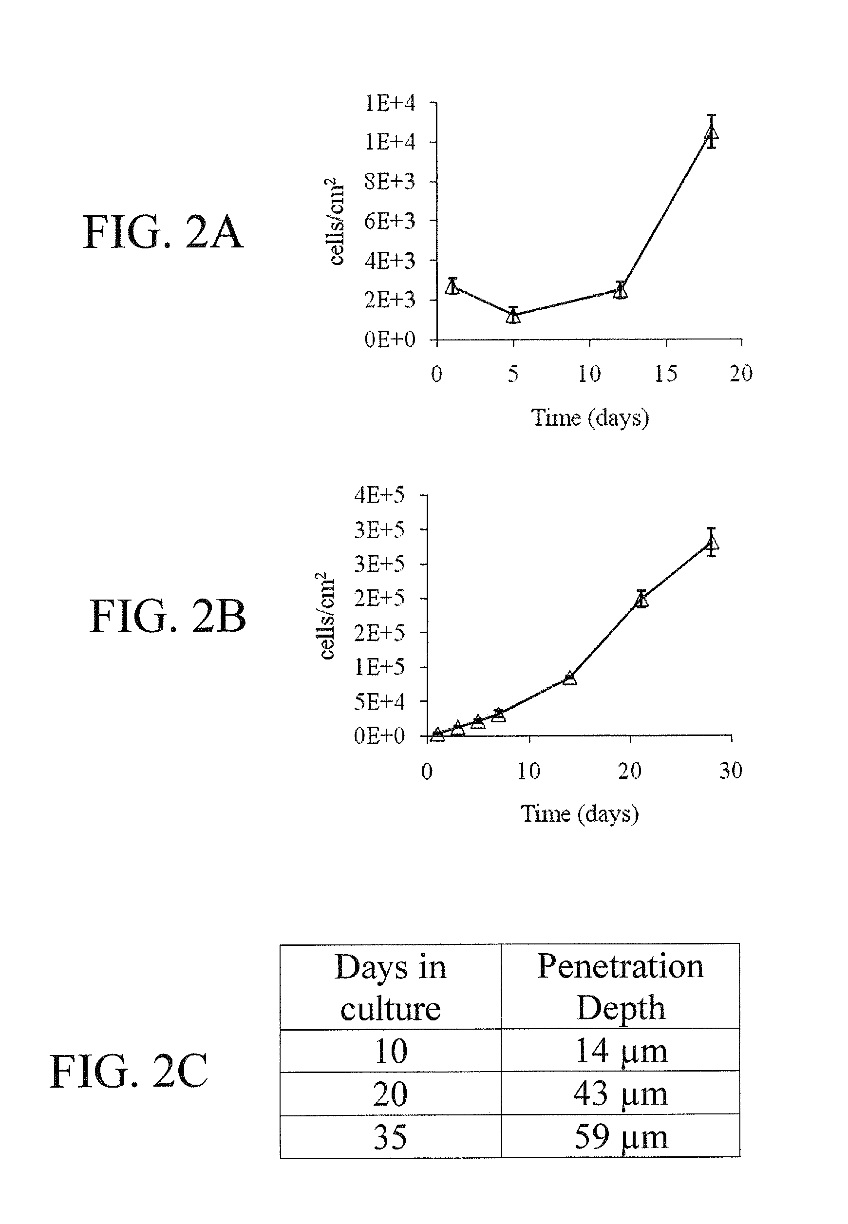
[0048]With reference to FIGS. 1-8, hMSC exhibit rapid proliferation in preincubated HCG due to cell migration into the matrices, indicating excellent biomimetic properties HCG's inherent osteoinductive properties without chemical induction is confirmed by hMSC's alkaline phosphatase (ALP) activity and osteogenic gene expression quantified by qPCR. Sustained substrate mediated transfection by incorporating chtiosan-BMP-2 nanoparticles at 28 days of culture is confirmed by q-PCR, demonstrating the potential for achieving sustained gene delivery in HCG matrices. hMSCs maintained their osteogenic differentiation ability after extensive perfusion growth in the HCG scaffolds, indicating the potential of bioreactor for streamlined construct fabrication.

Materials and Methods for Examples 2-4

[0049]Materials. Medium molecular weight chitosan and gelatin from bovine skin were purchased from Sigma Chemical Co. (St. Louis, Mo., USA). Hydroxyapatite (HA) powder with an average particle diameter o...

example 2-3d

Porous HCG Scaffold Fabrication

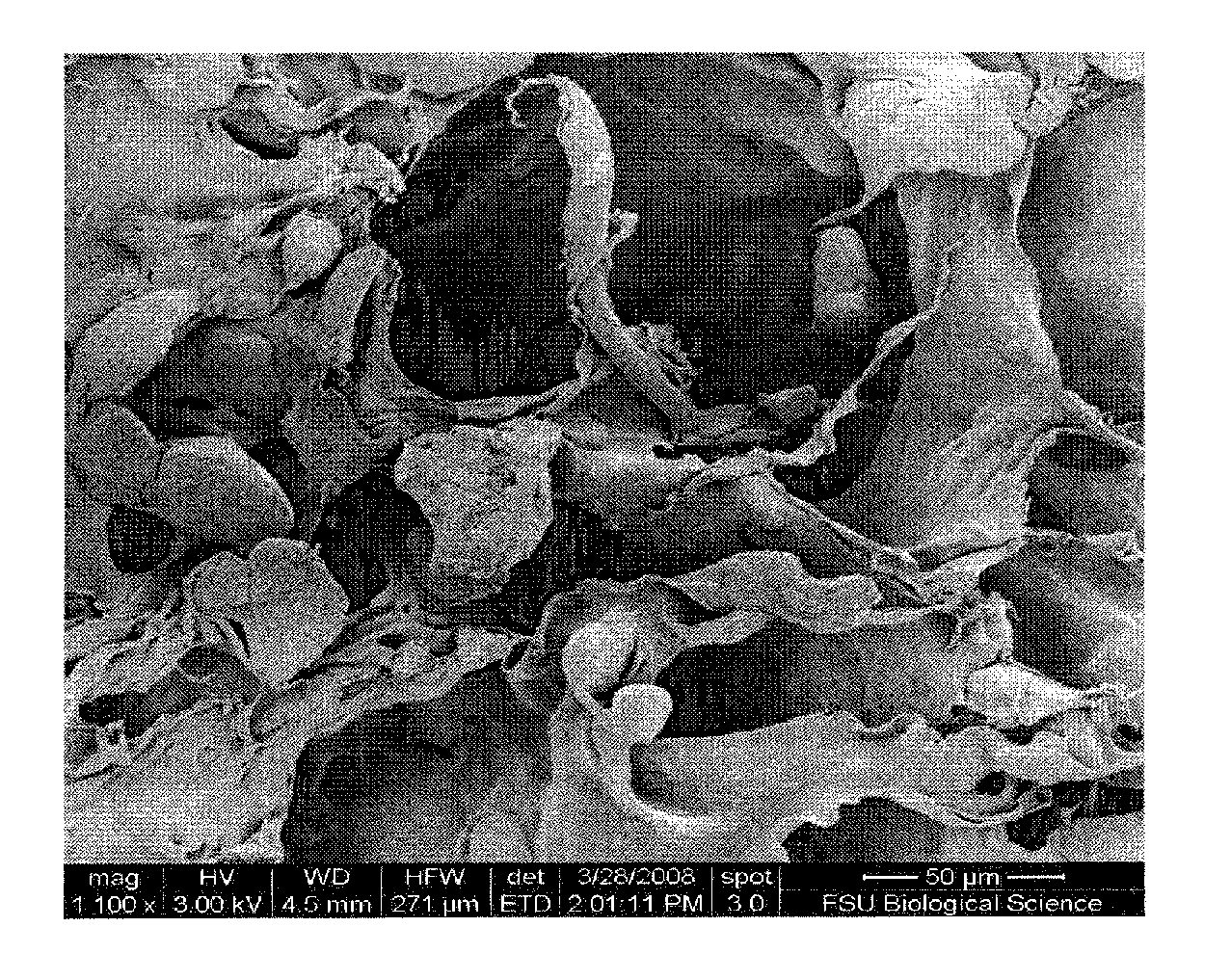
[0060]The 3D HCG scaffolds were prepared by lyophilization, following an established procedure reported in our early studies (Zhao et al., 2002, 2006). An inverse correlation between pore size and freezing temperature was found to be in agreement with previous reports, indicating that the pore structure of the HCG scaffolds can be readily adjusted by regulating the freezing temperature (O'Brien et al., 2005; Zhao et al., 2002). The surface texture of the scaffolds under all freezing conditions appeared rough, with pore sizes serving as suitable templates for cell adhesion, migration and growth, varying from 99±24 μm for HCG80 scaffolds to 158±23 μm in the HCG20 scaffold (FIG. 10).

[0061]Table 2 lists the pore size of each fabricated scaffold, detailing the effect of freezing temperature on final construct pore size.

TABLE 2Freezing TemperaturePore Size−80° C. (HCG80) 99 ± 24 μm−50° C. (HCG50)128 ± 38 μm−20° C. (HCG20)158 ± 23 μm

example 3

Cell Seeding and Preconditioning

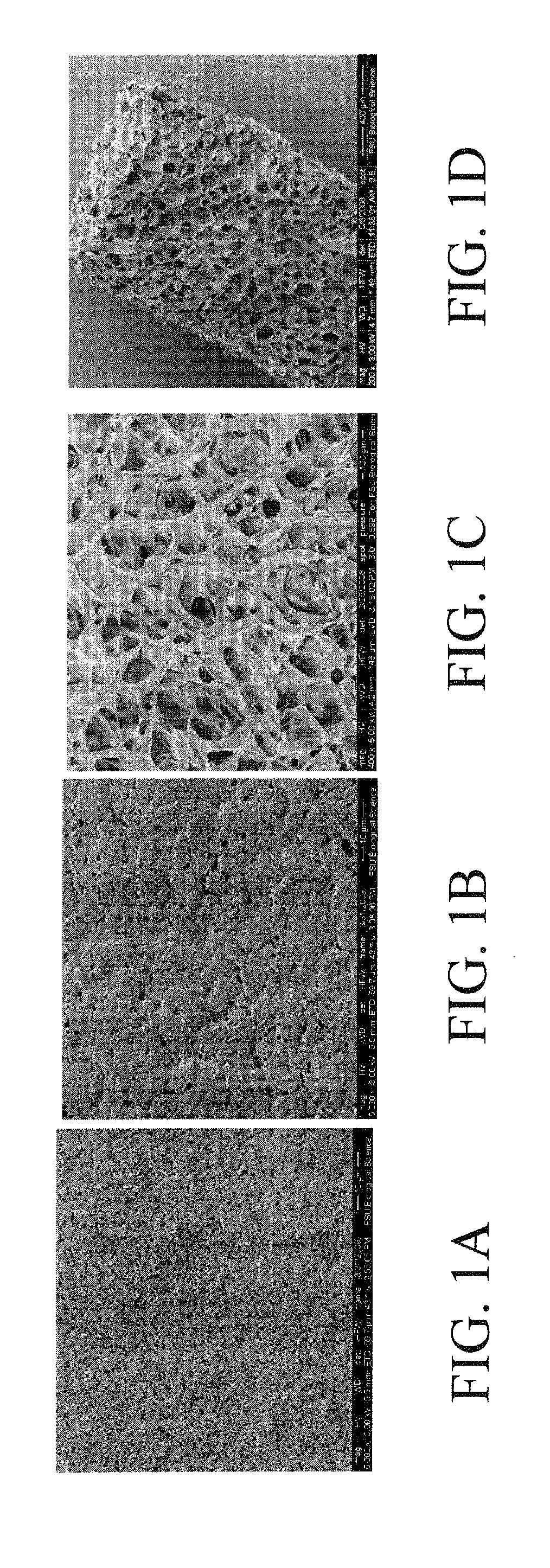
[0062]Seeding efficiency had an inverse relationship to both flow rate and pore size, as shown in FIGS. 11A and 11B. However, preconditioning of the HCG scaffolds in serum containing media had no effect on cell-seeding efficiency at 1.0 ml / min (FIG. 11C). DAPI images obtained from depth filtration 6 h after seeding at 1.0 ml / min show good cell penetration and distribution throughout the 1 mm thickness of the scaffolds (FIGS. 11D and 11E). SEM images of preconditioned samples (FIGS. 12A-12C) displayed a more fibrous surface texture after 7 days of complete medium preconditioning, giving it a less rough appearance as compared to samples stored in PBS (FIGS. 12D-12F) over the same time period. Preconditioning also induced the appearance of nanopores on the surface of the scaffold (FIG. 12C) when imaged at ×30,000. Preconditioning did not appear to effect the overall pore structure of the HCG scaffold (FIGS. 12A and 12D).

[0063]To examine the impact of per...


PUM

PropertyMeasurementUnit
Diameteraaaaaaaaaa
Diameteraaaaaaaaaa
Lengthaaaaaaaaaa

Abstract

The subject invention concerns a perfusion bioreactor device and methods of using the same. A bioreactor device of the invention can be used to grow cells and tissue in a controlled in vitro environment. A perfusion bioreactor device of the invention can have multiple perfusion chambers that can be controlled individually. Transverse or parallel flow of a fluid can be provided to each chamber. Cells can be seeded on a hydrogel and/or 3D scaffold to provide a 3D environment in the bioreactor device where the cells can adhere, proliferate, migrate, secrete growth and/or differentiation factors, and/or undergo differentiation, etc. The subject invention also concerns hydrogels and 3D scaffolds that can be used to grow and/or differentiate cells thereon.

Description

CROSS-REFERENCE TO RELATED APPLICATION[0001]The present application claims the benefit of U.S. Application Ser. No. 61 / 311,007, filed Mar. 5, 2010, which is hereby incorporated by reference herein in its entirety, including any figures, tables, nucleic acid sequences, amino acid sequences, and drawings.GOVERNMENT SUPPORT[0002]This invention was made with government support under grant number W81XWH-07-1-0363 awarded by the Department of Defense Peer Reviewed Medical Research Program. The government has certain rights in the invention.BACKGROUND OF THE INVENTION[0003]Osteoporosis and bone-related diseases and injuries are major public health threats for over 44 million Americans. Osteoporosis is characterized by excessive loss of bone and micro-architectural deterioration of bone tissue leading to bone fragility and increase susceptibility to fractures of hip, spine, and wrist. An overall imbalance between osteoblast-mediated bone formation and osteoclast-mediated bone resorption is ...

Claims


Application Information

Patent Timeline
IPC IPC(8): C12N15/85C12N5/071C12N5/0775C12M3/00B82Y5/00
CPCA61L27/26A61L27/38C12N2533/72C12N2533/54C12N2533/18C12N5/0654C12M29/10C12M25/14A61L2430/02A61L27/46A61L27/52A61L27/56A61L2300/258A61L2300/624C08L89/06C08L5/08
Inventor MA, TENG
Owner FLORIDA STATE UNIV RES FOUND INC
Why Eureka
Try Eureka