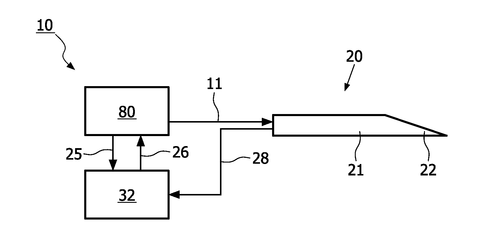
[0010]This object is solved by an optical examination device adapted to be at least partially inserted into a turbid medium according to claim 1. The optical examination device comprises: a shaft portion adapted to be inserted into the turbid medium. The shaft portion comprises a tip portion adapted to be the foremost portion during
insertion into the turbid medium. At least one
light source device adapted to emit a beam of broad-band light is provided in a region adapted to be inserted into the turbid medium of the shaft portion. The beam of broad-band light comprises different
wavelength bands which are differently modulated. At least one
photodetector for detecting broad-band light is provided in the region adapted to be inserted into the turbid medium of the shaft portion. Since the optical examination device is provided with the at least one
light source in the region of the shank portion which is adapted to be inserted into the turbid medium, the beam of broad-band light can be reliably emitted towards and scattered in the
region of interest of the turbid medium, such as tissue located at a specific position in the case of medical applications. Since the beam of broad-band light comprises different
wavelength bands which are differently modulated, spectroscopic information can be acquired with a simple photodetector in combination with a
demodulation unit. The
demodulation unit can be realized as a compact
electronic circuit or can be implemented in
software on a suitable processor. Thus, sophisticated and expensive spectrometers can be dispensed with. In this context, broad-band light comprising different wavelength bands means light which comprises a large number of wavelengths with continuous wavelength spectra in at least one
wavelength band. Broad-band means that a wide range of wavelengths is covered. The plurality of wavelength bands can be modulated at different frequencies and / or timing sequences. Since the at least one photodetector is provided in the region which is adapted to be inserted into the turbid medium,
scattered light can directly be detected inside the turbid medium with the at least one photodetector. Thus, the
scattered light does not have to be coupled into optical fibers which would lead to the problem of only small numerical apertures available. Further, in a case in which a plurality of detection positions is provided, instead of an additional
optical fiber for each detection position (which would be required if the scattered light would have to be guided to a
spectrometer located outside the turbid medium such as a
mammal body) only electrical connections from the photodetectors to the outside of the turbid medium (e.g. to the outside of the
mammal body) are required. This comes along with a considerable cost reduction and results in a less complicated
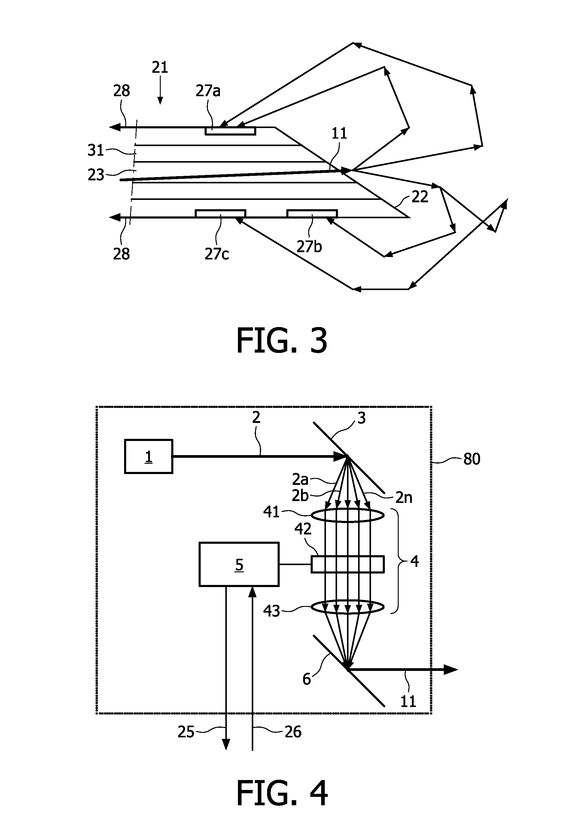
system. In particular, the at least one photodetector (or a plurality of photodetectors) can be arranged in a side region of the shaft portion.
[0012]According to one aspect, the at least one photodetector is a
photodiode. Photodiodes can be conveniently fabricated with high detection efficiency and at low costs. Further, they can be realized in a very compact fashion such that integration into the shaft portion, compact arrangement on an inner or outer surface of the shaft portion, or compact arrangement on a core element to be placed in a hollow channel inside the shaft portion (such as a mandrin in the case of a
biopsy needle) is possible.
[0013]According to an aspect, the shaft portion is provided with a plurality of photodetectors arranged at different positions relative to the shaft portion. In this case, spectroscopic information contained in the scattered light can be acquired at different spatial positions. As a consequence, spatial resolution of the properties of the region of the turbid medium (e.g. tissue) which is located in front of the tip portion becomes possible.
[0014]According to an aspect, the optical examination device comprises a
demodulation and analysis unit adapted to perform a
spectral analysis of a
signal received from the at least one photodetector. In this case, information about the region of the turbid medium in front of the tip portion is analyzed with regard to the distribution of different wavelength bands. As a consequence, information about the scattering properties and / or
chromophore concentration in this region of the turbid medium can be reliably acquired.
[0017]According to an aspect, the shaft portion forms at least a part of a
biopsy needle. In this case, inadvertently
puncturing tissue which should not be punctured, such as nerves or blood vessels, can be prevented. In an alternative, the shaft portion forms at least a part of a
catheter or of an
endoscope.

