

Apparatus and method for devices for imaging structures in or at one or more luminal organs
- Summary
- Abstract
- Description
- Claims
- Application Information
AI Technical Summary
Benefits of technology
Problems solved by technology
Method used
Image

Examples
Embodiment Construction
[0011]t is therefore one of the objects of the present disclosure to reduce or address the deficiencies and / or limitations of such prior art approaches, procedures, methods, systems, apparatus and computer-accessible medium.
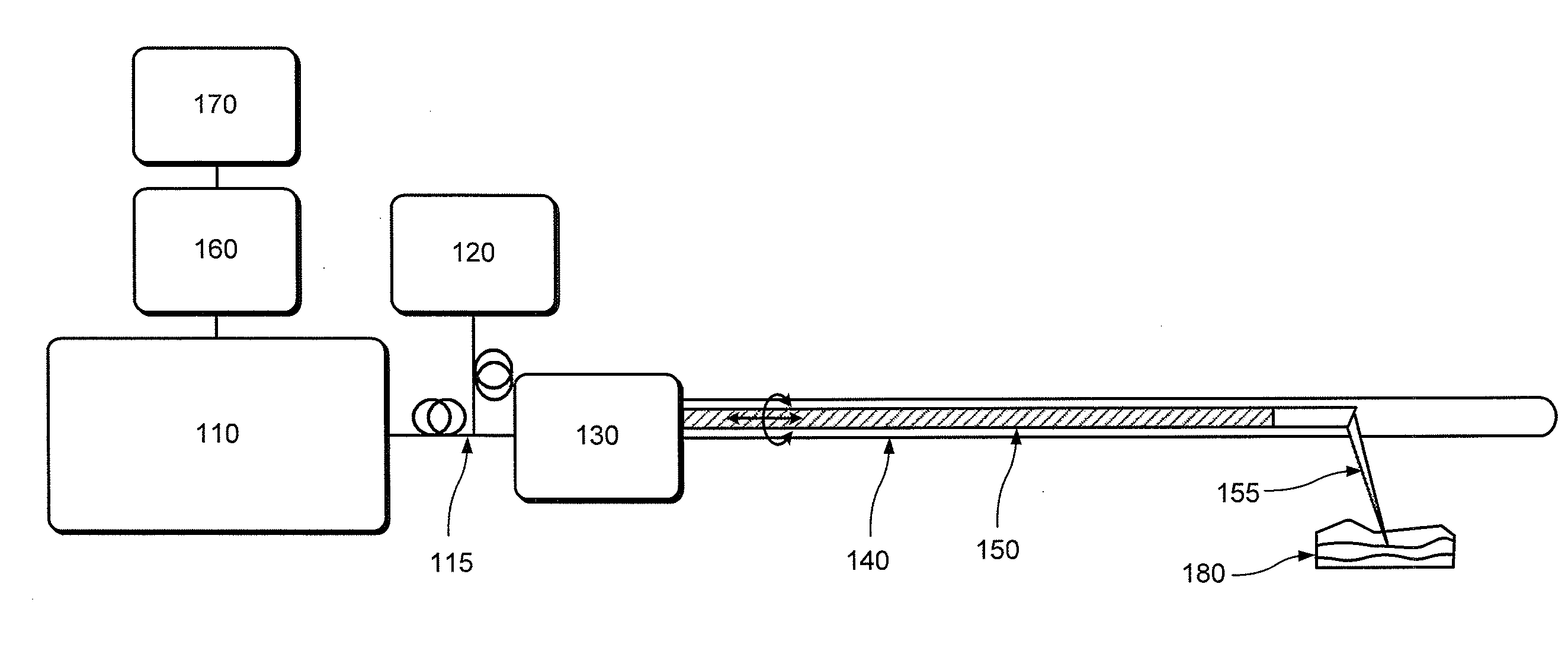
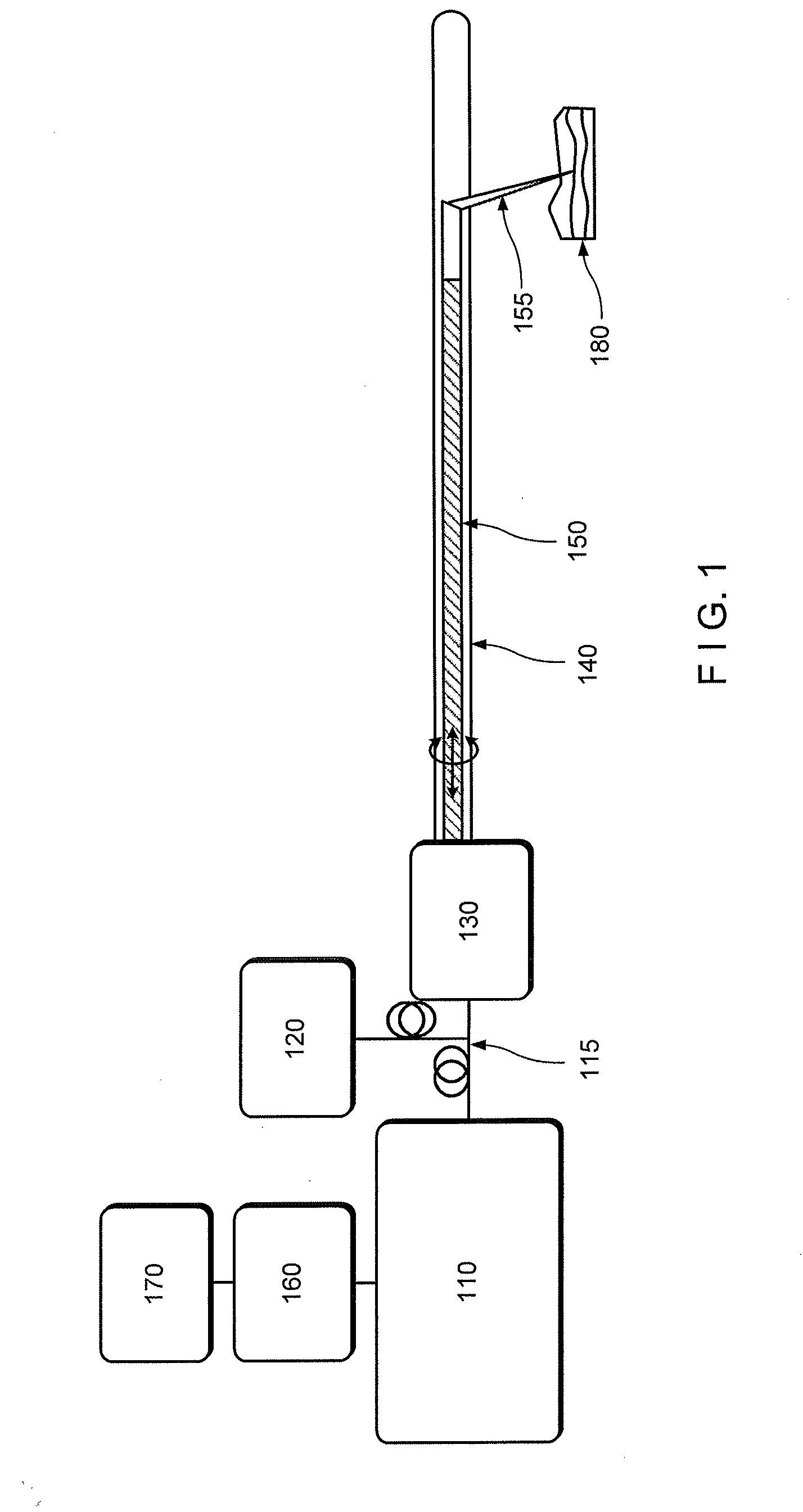
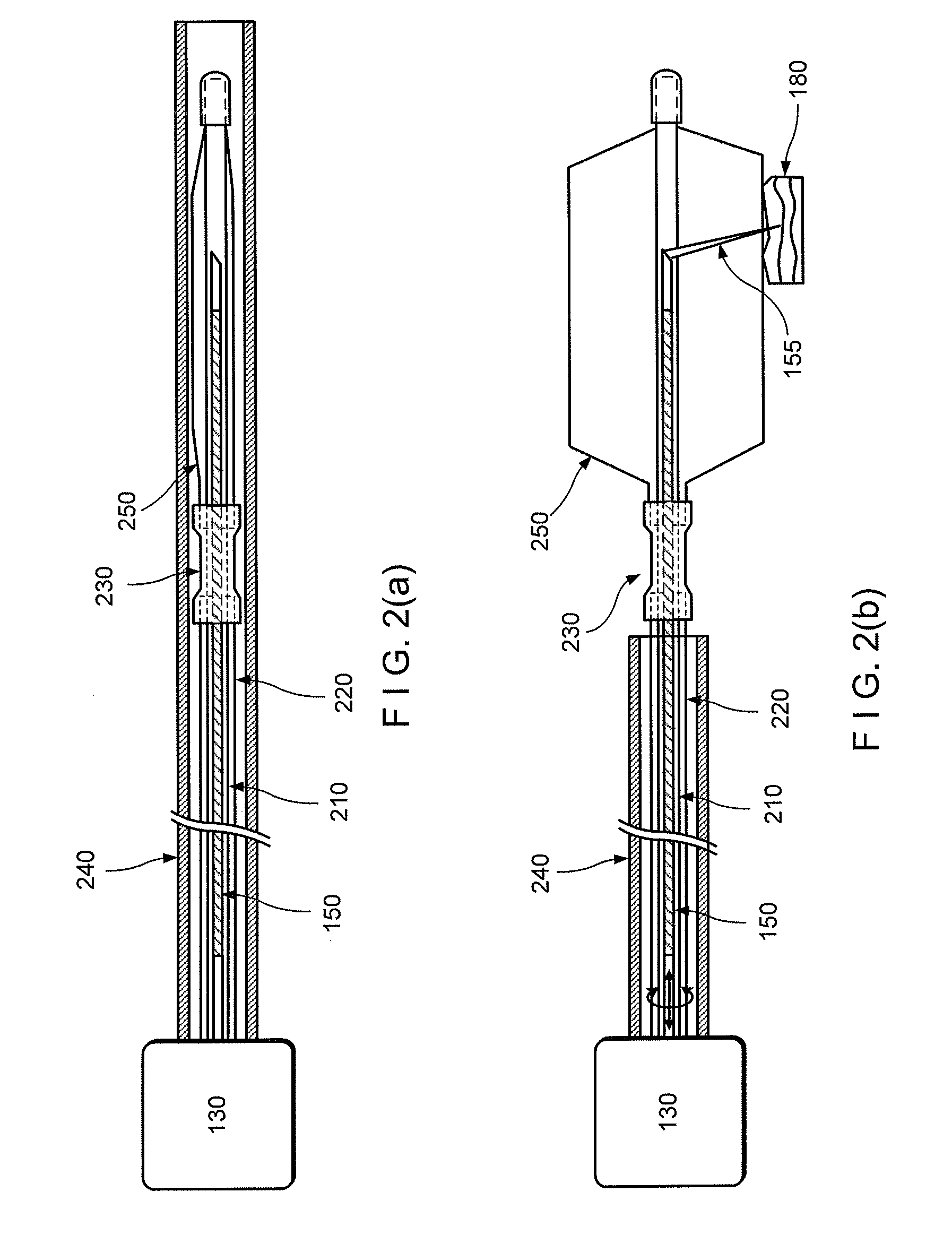
[0012]For example, one object of the present disclosure is to provide an exemplary microscopic imaging device according to an exemplary embodiment for approximately centering an imaging probe within a luminal organ to enable comprehensive microscopy of the majority of the luminal organ. A further object of the present disclosure is to provide a device which can be configured to perform luminal organ microscopy that can operate in standalone mode, thus at least partially obviating the need for conscious sedation. Another object of the present disclosure is to utilize one or more in vivo microscopy technologies through one or more tethered capsule. It is yet object of the present disclosure to provide a balloon catheter that centers the optics while exerting a mini...
PUM
 Abstract
Description
Claims
Application Information