Rapid fret-based diagnosis of bacterial pathogens
a bacterial pathogen and rapid fret technology, applied in the field of diagnostics, can solve the problems of high case-fatality rate of inhalational disease, system inability to differentiate between virulent and avirulent strains of i>bacillus anthracis, and high skill level of the technician performing the assay, so as to achieve quick and efficient results
- Summary
- Abstract
- Description
- Claims
- Application Information
AI Technical Summary
Benefits of technology
Problems solved by technology
Method used
Image
Examples
example 1
Materials and Methods
[0077]Bacteria
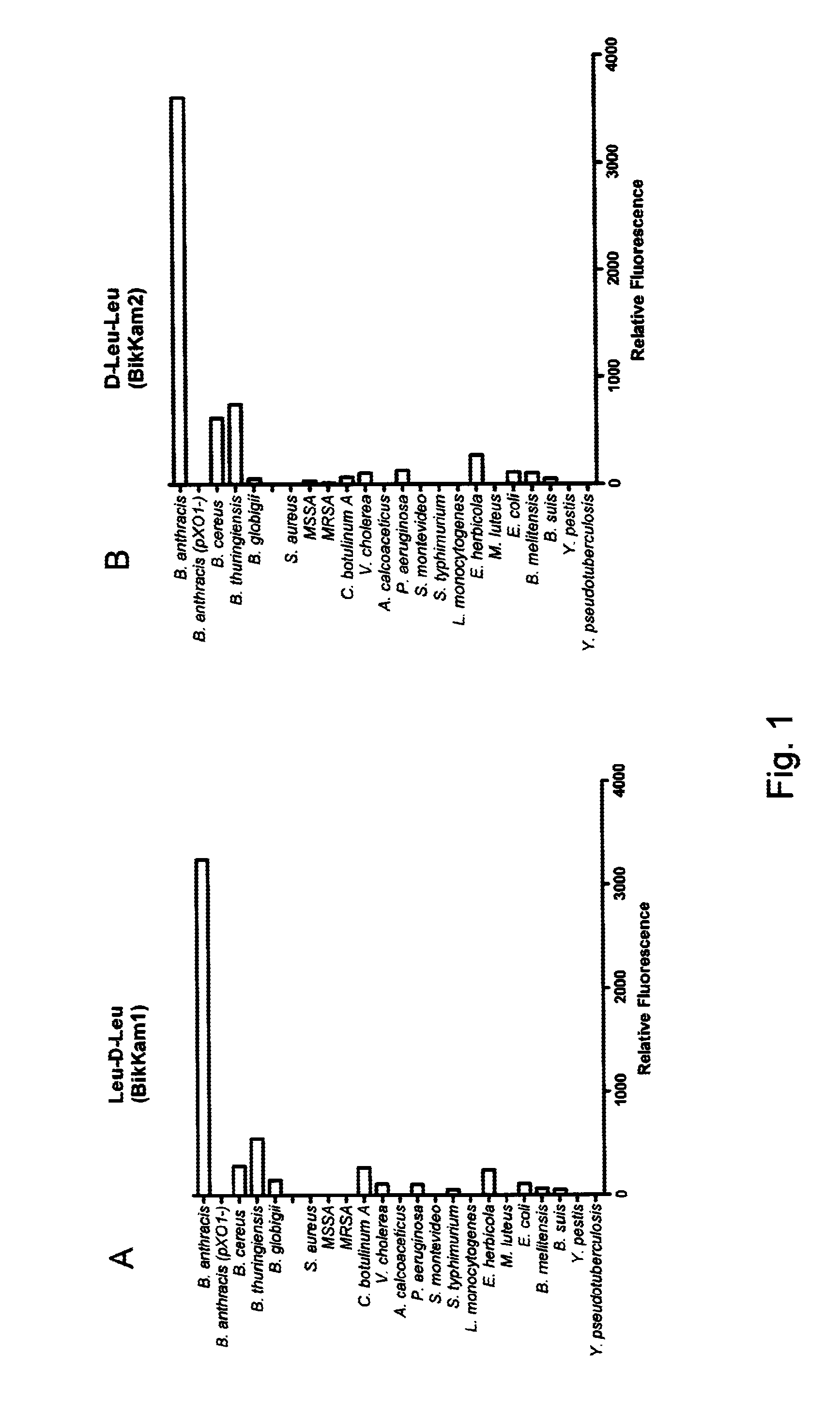
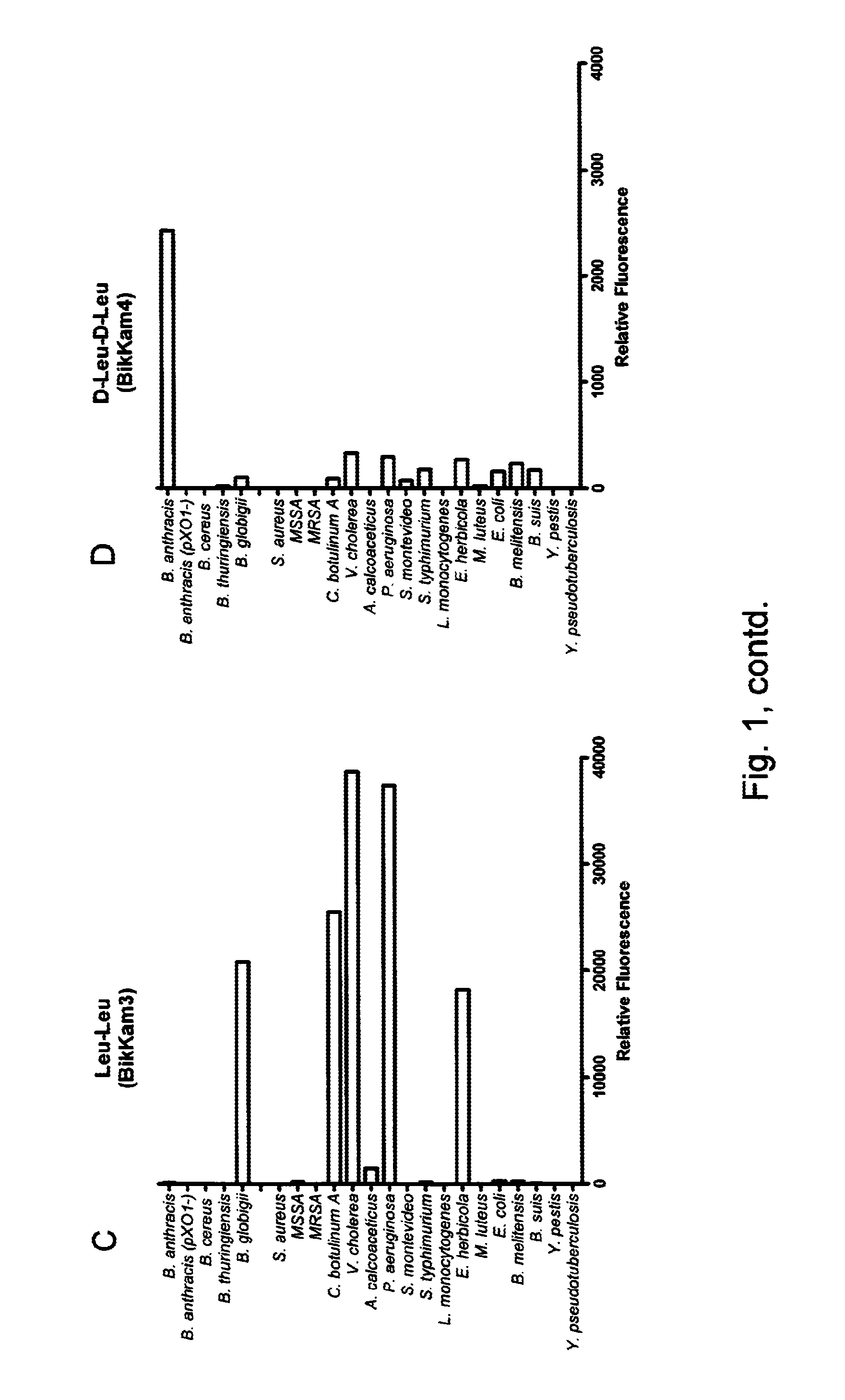
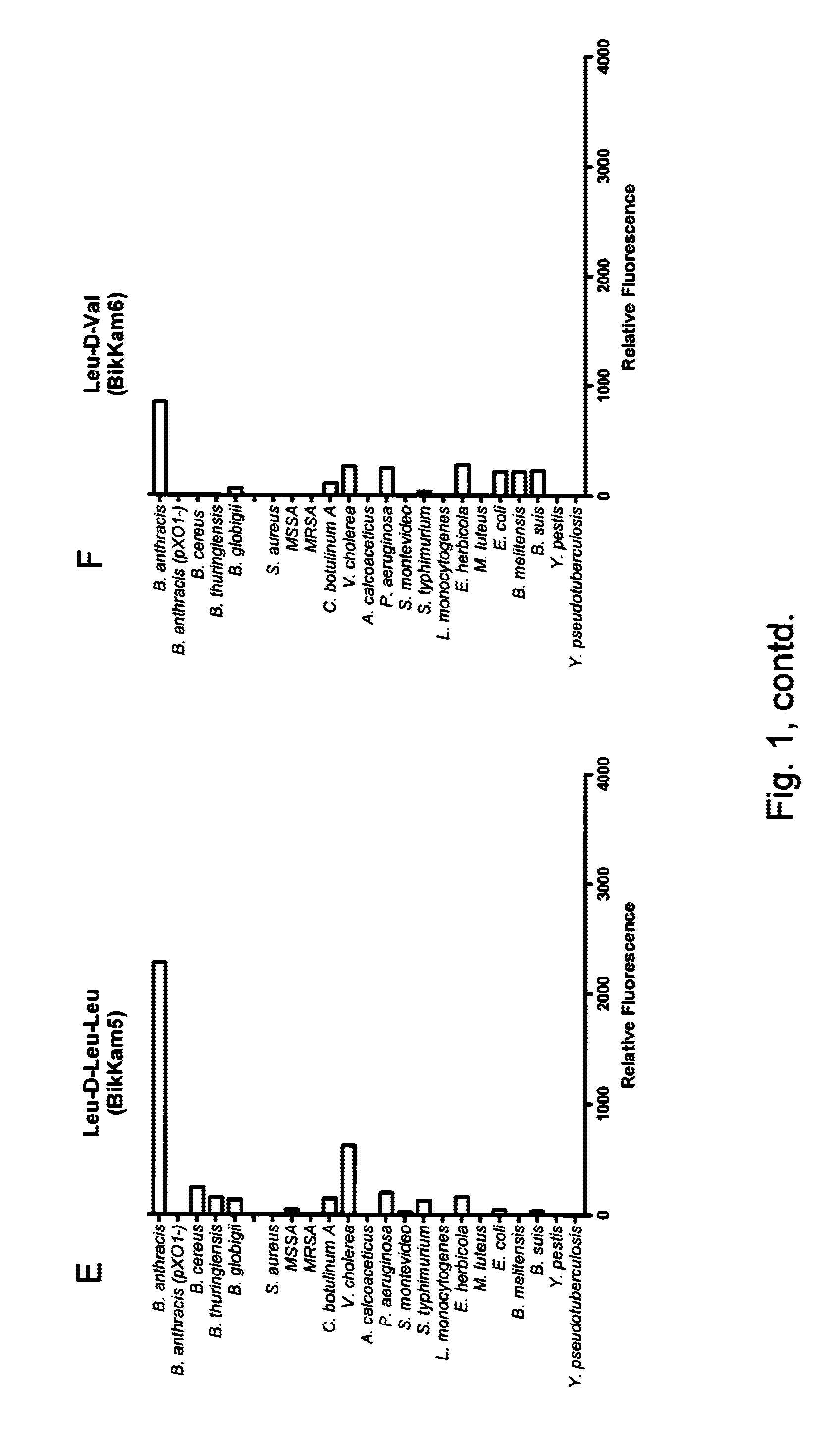
[0078]Bacteria were grown overnight in Brain Heart Infusion (BHI) medium or 70% Human serum (HuS) at the appropriate growth temperature. Next day the bacteria were pelleted by centrifugation for 10 min at 10 000 rpm. Supernatant was sterilized by filtration through an 0.22 uM filter (MilliPore). For mass spectrometric analysis and confirmation of the in vitro and ex vivo results sera of mice that were suffering a B. anthrax infection was filtrated on a Waters Sep-Pak classic cartridge WAT051910 C18 column.
[0079]Reagents
[0080]FRET peptides were synthesized by PepScan (The Netherlands). Identity and purity were verified by mass spectrometry and reverse phase HPLC. Anthrax Lethal Factor (LF) was purchased from List Laboratories. Thermolysin, Collagenase Clostridium and lysostaphin were purchased from Sigma. Collagenase Vibrio / Dispase was purchase from Roche. LF inhibitor 12155 was synthesized by Pyxis (The Netherlands). Nine experimental peptides were...
example 2
Material and Methods
[0090]B. cereus and B. globigii cells were grown overnight in 5 ml BHI medium at 37° C. Next day the bacteria were diluted 1:10 in BHI and grown in presence of 0.01 M BikKam1 (PepScan, The Netherlands). During growth, cell samples were taken with 1 hour time intervals. The cell samples were washed twice with PBS, reconstituted with 20 uL PBS and spotted onto glass slides. As a negative control bacteria grown for 16 hr without BikKam1 were used. Fluorescence microscopy was performed with a Leitz Orthoplan microscope using a 1000 fold magnification.
[0091]Results
[0092]After 2 hr incubation, fluorescent BikKam1 fragments could be detected in the cytoplasm of B. cereus (FIG. 5B). At 4 hr, the fluorescent BikKam1 fragments were besides in the cytoplasm also present in the cell wall at the new division sides (the side wall) (FIG. 5C). Finally, after 16 hr incubation, all BikKam1 fragments were moved from the cytoplasm to the side walls of B. cereus (FIG. 5D). The bacter...
PUM
| Property | Measurement | Unit |
|---|---|---|
| Fraction | aaaaa | aaaaa |
| Volume | aaaaa | aaaaa |
| Fluorescence | aaaaa | aaaaa |
Abstract
Description
Claims
Application Information
 - R&D
- Intellectual Property
- Life Sciences
- Materials
- Tech Scout
- Unparalleled Data Quality
- Higher Quality Content
- 60% Fewer Hallucinations
Browse by: Latest US Patents, China's latest patents, Technical Efficacy Thesaurus, Application Domain, Technology Topic, Popular Technical Reports.
© 2025 PatSnap. All rights reserved.Legal|Privacy policy|Modern Slavery Act Transparency Statement|Sitemap|About US| Contact US: help@patsnap.com


