Inverse-fluorescence correlation spectroscopy

a technology of inverse fluorescence and correlation spectroscopy, which is applied in the field of inverse fluorescence correlation spectroscopy, can solve the problem of the necessity of labeling biomolecules with small fluorescent markers, and achieve the effect of high brightness

Inactive Publication Date: 2012-03-01
WENNMALM STEFAN +1
View PDF1 Cites 10 Cited by
  • Summary
  • Abstract
  • Description
  • Claims
  • Application Information

AI Technical Summary

Benefits of technology

[0028]The medium can comprise, or consist of, conventional organic fluorescent molecules, but is not limited to those. Since the only requirement is a high total signal from the medium, i

Problems solved by technology

A drawback of FCS as described above, as with all fluorescence based methods for analyzing biom

Method used


Image

  • Inverse-fluorescence correlation spectroscopy
  • Inverse-fluorescence correlation spectroscopy
  • Inverse-fluorescence correlation spectroscopy

Examples


exemplary embodiment 1

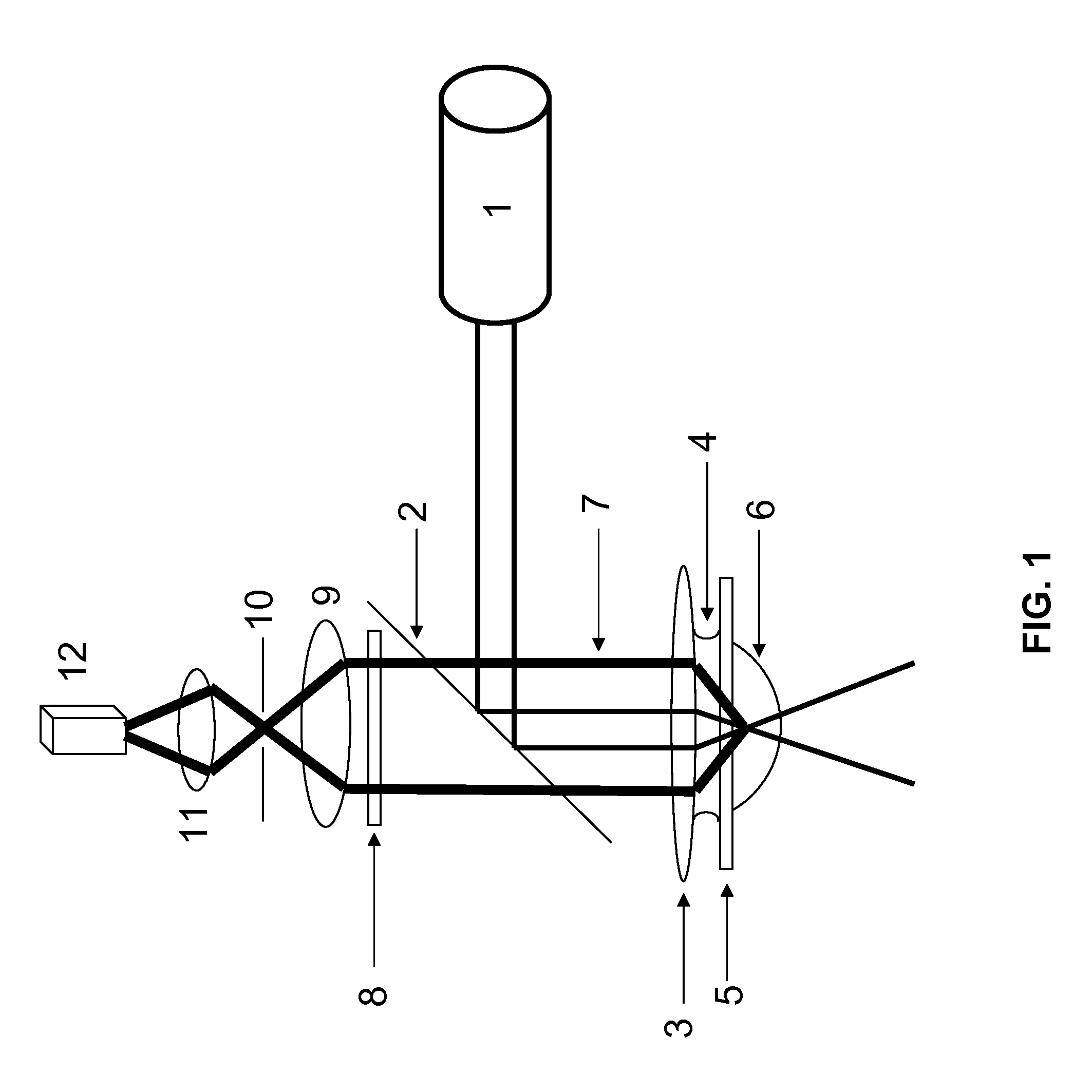
A Fluorescence Correlation Spectroscopy System of the Present Disclosure

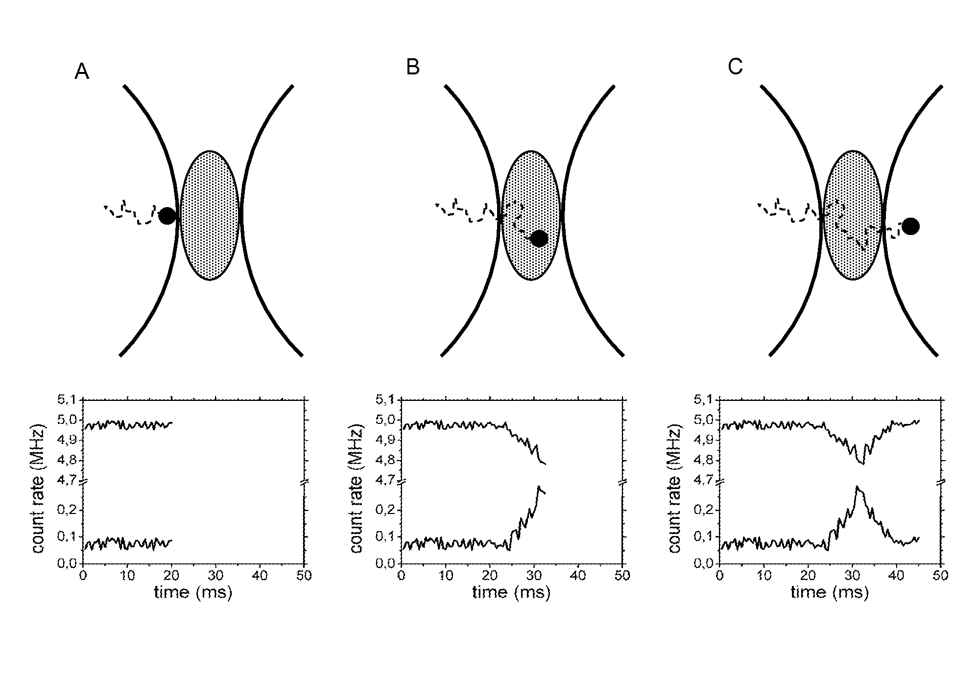
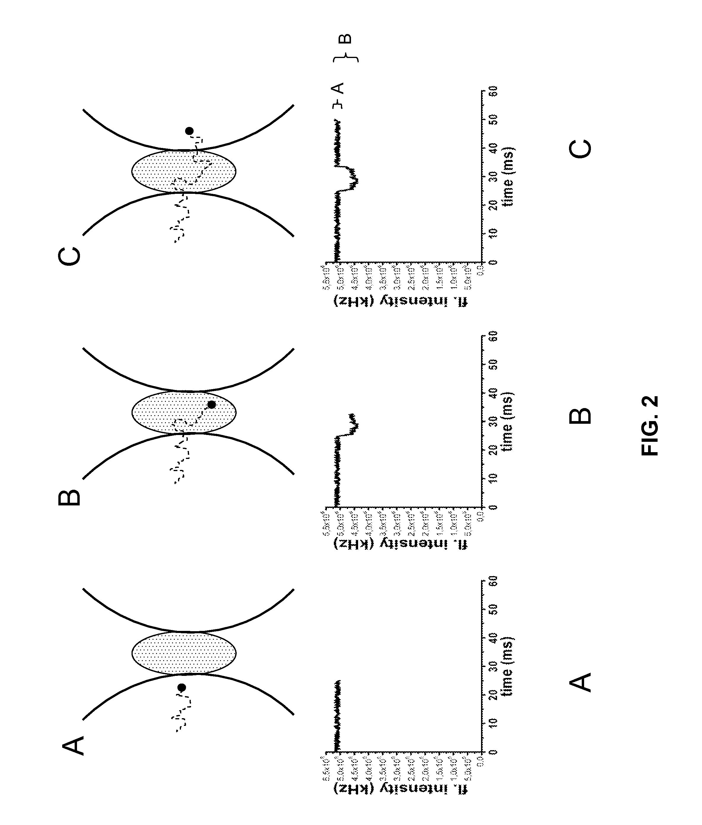
[0207]Generally, if proteins or other biomolecules are analyzed by iFCS or iFCCS, zero-mode waveguides (ZMWs) can be used in combination with a standard FCS-microscope. The detection volumes in ZMWs are 1000-10 000 times smaller than diffraction-limited detection volumes, which allows proteins and similar sized biomolecules to generate negative spikes of magnitude larger than the noise in the medium-signal. If the FCS-microscope in addition is equipped with alternative photo-detectors that are capable of detecting considerably higher count rates than what APDs are capable of, for example PMTs in DC-mode or photo-diodes, which both give a current as output, then the relative photon-noise in the signal from the medium can be significantly reduced. This in turn allows even smaller proteins and biomolecules to be detected.

exemplary embodiment 2

Analyzing the Concentration of and Size of Unlabeled Proteins by iFCS

[0208]Using iFCS, protein molecules can be analyzed without being fluorescently labeled. iFCS gives an estimate of the concentration and diffusion coefficient of the proteins. The size-estimate of particles / proteins possible from iFCS is the same as in standard FCS in that the size is estimated from the diffusion coefficient. This is in contrast to iFCCS, which allows a direct estimate of the volume of particles / proteins. However, by using FIDA / PCH (Chen et al., 1999; Kask et al., 1999) as described above, the volume of particles / proteins can be estimated in iFCS, i.e. also for unlabeled particles / proteins.

[0209]An experiment can be carried out as follows:

[0210]The protein-sample is dissolved in the signal-generating medium (for example 1 mM alexa 488 carboxylic acid dissolved in a buffer of pH 8.5), and analyzed in the detection volume created by the ZMW on the iFCS-microscope. The automatically generated autocorr...

exemplary embodiment 3

Analyzing the Binding of a Labeled Protein to an Unlabeled Protein by iFCCS

[0212]In standard FCS, the binding of a labeled biomolecule to an unlabeled one can be detected only if the resulting complex has a diffusion time at least 1.6 times longer than the unbound labeled protein (Meseth et al., 1999). For spherical molecules, this corresponds to a volume at least ˜4 times that of the unbound labeled protein. iFCCS is more sensitive and should allow identification of complexes whose size differs less from that of unbound labeled proteins; with the ability to identify complexes with a volume 1.6 times larger than that of unbound labeled proteins, complexes can be 2.5 times smaller than the resolution-limit in standard FCS and still be identified by iFCCS

[0213]An experiment can be carried out as follows:

[0214]Fluorescently labeled protein-molecules, e.g. a labeled antibody (protein A), is mixed with another, unlabeled protein (protein B), whose volume can be smaller or larger than tha...


PUM


Abstract

A method is disclosed for analyzing particles or biomolecules in a liquid sample, including: detecting a signal and fluctuations in the signal from a detection volume in the sample; wherein the signal is generated from signal-generating molecules in the medium surrounding the particles or biomolecules and the fluctuations are transient reductions in the signal as the particles or biomolecules transit through the detection volume; and analyzing the detected fluctuations to obtain information about the particles or biomolecules in the liquid sample. At least one example embodiment of the present invention relates to a fluorescence correlation spectroscopy system including a laser, a zero-mode waveguide, guiding device for guiding the laser into the zero-mode waveguide, device for collecting fluorescence emission from excited molecules within the waveguide, a detector for detecting the fluorescence emission and means for autocorrelating the detected fluorescence signal, wherein the detector comprises a photomultiplier tube. Moreover, at least one embodiment relates to the use of a fluorescence correlation spectroscopy system for analyzing molecules of interest in a sample by detecting and analyzing fluctuations in a fluorescence signal that is generated from sample molecules surrounding the molecules of interest, wherein the fluctuations are transient reductions in the detected fluorescence signal.

Description

TECHNICAL FIELD OF THE INVENTION[0001]The invention relates to analysis of diffusing particles and biomolecules in solution or in cells. In biophysics, biochemistry, and cell biology, methods are needed for analyzing the interaction of biomolecules. A particular requirement on such methods is the possibility to measure interactions even at low concentrations, down to nano-molar and lower concentrations.[0002]The invention also relates to the field of analyzing particles that may not be biological, for example particles in solutions and emulsions. Examples are the need to determine the concentration and size of particles in engine-fuels, for environmental and health purposes, or the need for analyzing aggregation of particles in for example cosmetic products such as skin lotions.[0003]It may also be of interest to analyze particles or biomolecules in a solid. How the invention may be applied to such analyses will be clarified below. The description of the invention below will however...

Claims


Application Information

Patent Timeline
IPC IPC(8): G01J3/44G01N21/64G01J3/30
CPCG01N21/6408G01N21/658G01N21/65G01N21/6458G01N15/0205G01N2015/0038
Inventor WENNMALM, STEFANWIDENGREN, JERKER
Owner WENNMALM STEFAN
Why Eureka
Try Eureka