
Method for treatment of emphysema

a technology for emphysema and pulmonary artery disease, applied in the field of emphysema treatment, can solve the problems of difficult to confirm in each case which one is satisfactory, inhibit satisfactory expiration, lung occlusion, etc., and achieve the effect of less-invasive reduction of the volume of pulmonary artery or alveolar sac suffering

Inactive Publication Date: 2012-03-01
TERUMO KK
View PDF2 Cites 9 Cited by
  • Summary
  • Abstract
  • Description
  • Claims
  • Application Information

AI Technical Summary

Benefits of technology

[0009]It is an object of the present invention to provide a method for reducing the volume of the pulmonary alveoli or alveolar sac suffering from emphysema.
[0011]According to this method pertaining to the present invention, it is possible to less-invasively reduce the volume of pulmonary alveoli or alveolar sacs suffering from emphysema.

Problems solved by technology

COPD frequently involves these symptoms at the same time and makes it difficult to confirm in each case which one of the diseases is actually the cause of the lung occlusion.
This inhibits satisfactory expiration.
Moreover, emphysema decreases the effective area and vascular bed (capillary vessels running point to point on the surface of the pulmonary alveoli) of the pulmonary alveoli, thereby reducing the gas exchange capacity of the lung as a whole.
In addition, emphysema involves inflammation that destroys elastin and collagen, thereby causing the lung to decrease elasticity.
This result is that the lung cannot keep stretching and expanding the respiratory tract, and this permits easy deformation of the bronchia.
However, it merely relieves the patient's condition but is not an effective method of therapy.
All of these pharmacotherapies help control emphysema and alleviate its symptom, but they are not necessarily effective.
In addition, there are several methods of surgical treatment which include lung reductive surgery in which removal of damaged parts from the lung is made to promote expansion of the normal parts of the lung and lung transplantation; however, these surgical methods have disadvantages of a heavy burden on the patient and difficulties in securing the lung for substitution (see Ware J H, et al., “Cost effectiveness of Lung-Volume-Reduction Surgery for Patients with Severe Emphysema”, The New England Journal of Medicine 2003; 348:2092-2102; National Emphysema Treatment Trial Research Group, “A Randomized Trial Comparing Lung-Volume-Reduction Surgery with Medical Therapy for Severe Emphysema”, The New England Journal of Medicine 2003; 348:2059-2073).
However, the current noninvasive therapy is limited in possibilities of success.
However, once left indwelling in the lung, it prevents access to any place beyond its indwelling point (see Mark L. Mathis, PneumRx, Inc., U.S. Pat. No. 7,549,984 B1).
In this method, however, it needs a certain length of time for the lung parenchyma to be destroyed by the reaction of the living body.
Thus, at present, no effective method for treatment of emphysema exists in the relevant field of art.

Method used


Image

  • Method for treatment of emphysema
  • Method for treatment of emphysema
  • Method for treatment of emphysema

Examples


example 1

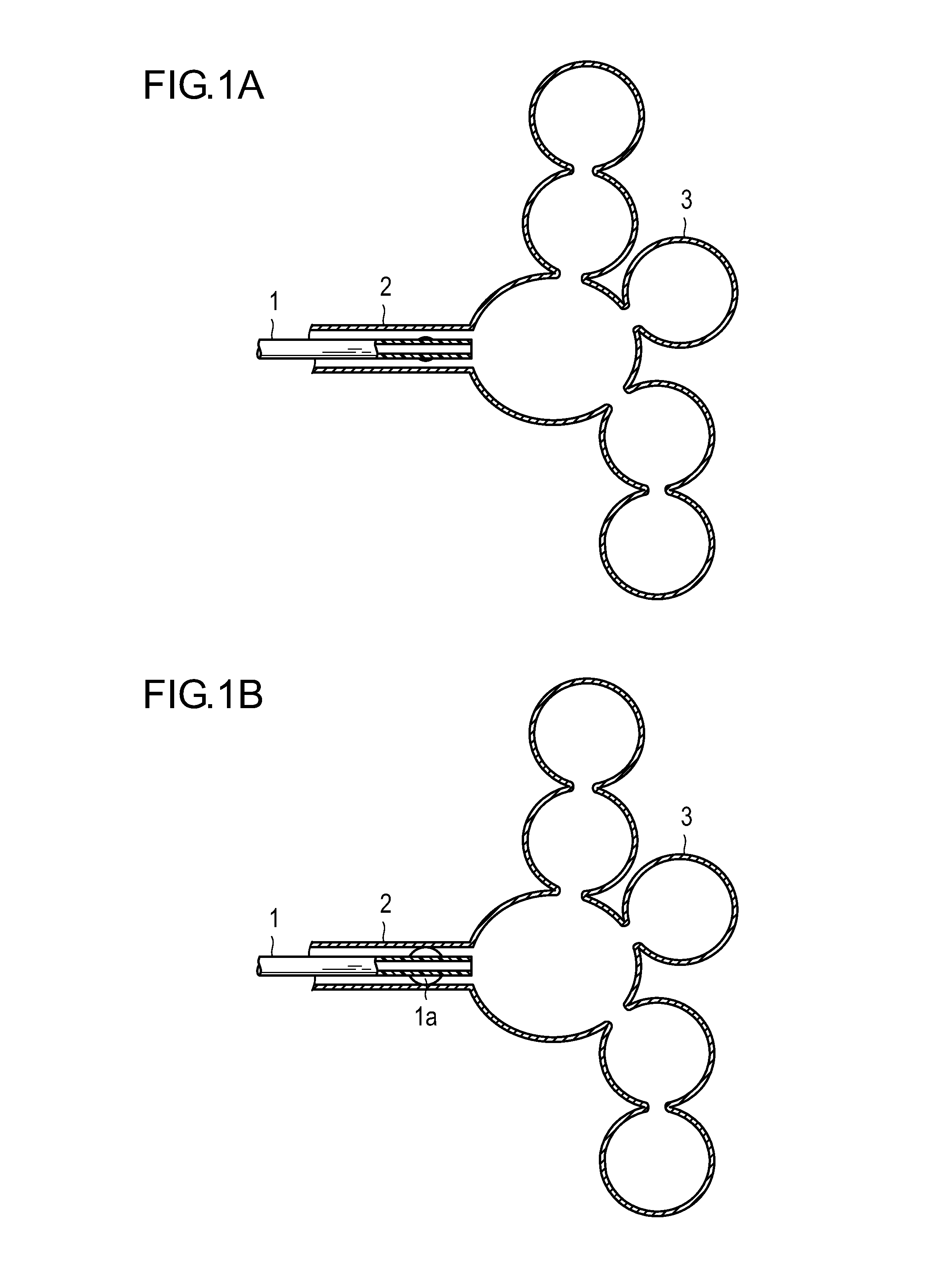
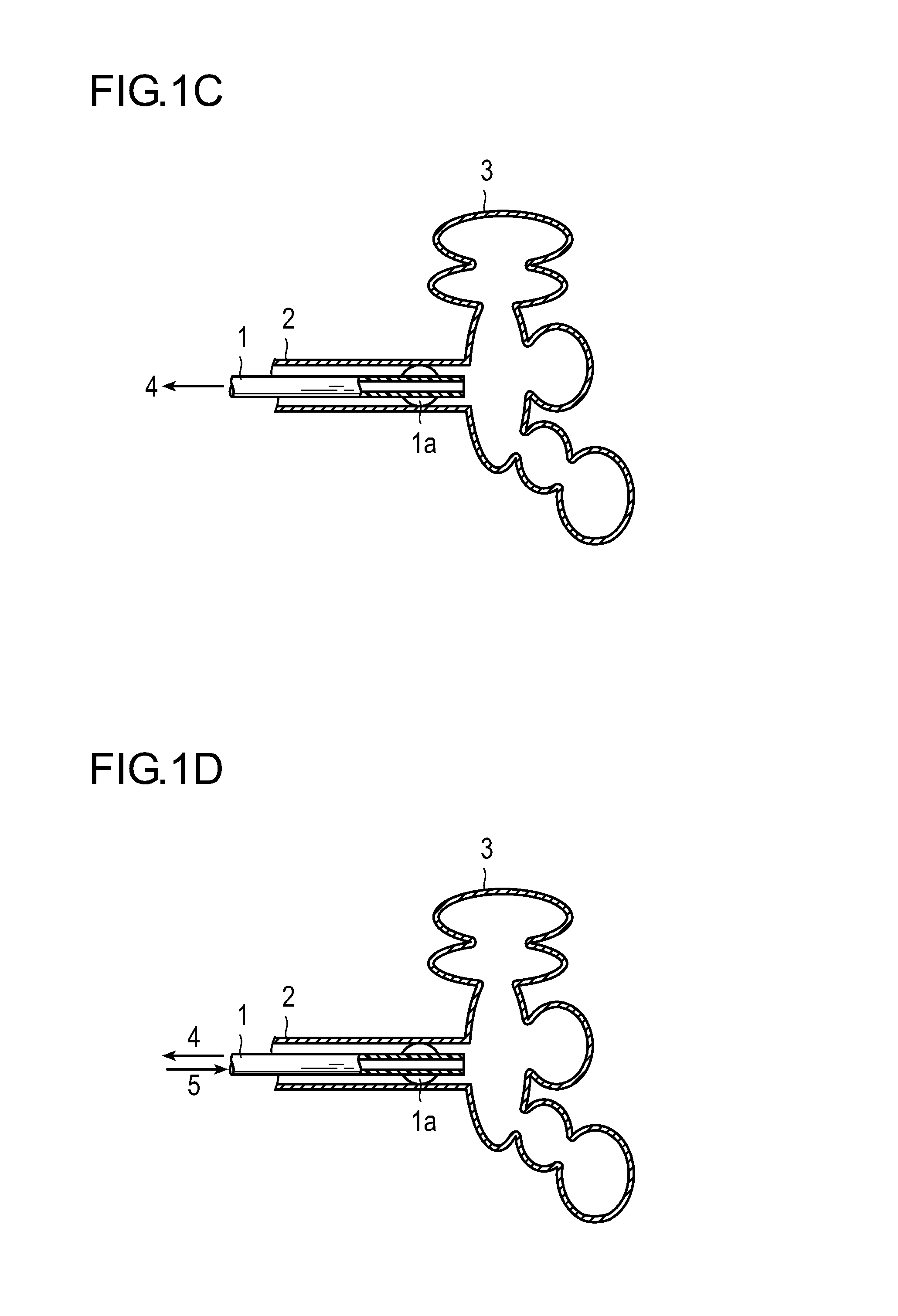
[0076]As shown in FIG. 1A, an OTW-type PTCA balloon catheter 1 [Ryujin Plus OTW (registered trademark); Medical Instrument Approval Number: 21600BZZ00035; made by TERUMO CORPORATION] for use in treatment of stenosis of blood vessel lumen in a cardiovascular region was inserted through a working lumen (not shown) of a bronchoscope into the lumen of a bronchiole 2. Here, a guide wire [Runthrough (registered trademark), made by TERUMO CORPORATION] (outside diameter: 0.014 inch) was preliminarily inserted into the working lumen of the bronchoscope. The distal end of the guide wire was advanced into the vicinity of a desired alveolar parenchyma 3 suffering from emphysema under X-ray fluoroscopy. Next, a catheter was advanced through the function of the guide wire into the vicinity to the desired alveolar parenchyma 3 suffering from emphysema under X-ray fluoroscopy, followed by pulling out the guide wire.

[0077]Subsequently, as shown in FIG. 1B, using a syringe connected to a balloon part...

example 2

[0082]As shown in FIG. 2A, a microcatheter (for example, FINECROSS (registered trademark), made by TERUMO CORPORATION) 1 was inserted through a working lumen (not shown) of a bronchoscope into the lumen of a bronchiole 2. Here, a guide wire [Runthrough (registered trademark), made by TERUMO CORPORATION] (outside diameter: 0.014 inch) was preliminarily inserted in the working lumen of the bronchoscope. The distal end of the guide wire was advanced into the vicinity of a desired alveolar parenchyma 3 suffering from emphysema under X-ray fluoroscopy. Next, a catheter was advanced through the function of the guide wire into the vicinity of the desired alveolar parenchyma 3 suffering from emphysema under X-ray fluoroscopy, followed by pulling out the guide wire.

[0083]Subsequently, as shown in FIG. 2B, using an indeflator connected to a balloon part dilating lumen disposed at a proximal portion of the catheter 1, a balloon 1a was dilated with air, thereby closing a bronchiole 2. As shown ...


PUM


Abstract

Disclosed is a method for treatment of emphysema by which it is possible to reduce the volume of pulmonary alveoli or alveolar sacs abnormally enlarged with destruction by emphysema. The method for treatment of emphysema includes: (a) inserting a catheter having a balloon into a bronchus or bronchiole; (b) dilating the balloon to occlude the bronchus or bronchiole; (c) shrinking pulmonary alveoli or alveolar sacs on the downstream side of the bronchus or bronchiole occluded in the step (b); and (d) injecting a hardening agent through the catheter into the respiratory region and hardening the hardening agent.

Description

BACKGROUND[0001]1. Technical Field[0002]The present invention relates to a method for treatment of emphysema. More particularly, the invention relates to a method for treatment of emphysema by reducing the volume of the pulmonary alveoli or alveolar sac which have been anomalously expanded with destruction by emphysema.[0003]2. Description of the Related Art[0004]Chronic obstructive pulmonary disease (COPD) means a large group of pulmonary diseases that hinder normal respiration, and is a disease which brings about lung occlusion arising from the presence of at least one selected from asthma, emphysema and chronic bronchitis. COPD frequently involves these symptoms at the same time and makes it difficult to confirm in each case which one of the diseases is actually the cause of the lung occlusion. A case is clinically diagnosed as COPD by constant decrease in expiration flow from the lung over several months, which continues for two years or more in the case of chronic bronchitis. T...

Claims


Application Information

Patent Timeline
IPC IPC(8): A61M25/10A61F2/958
CPCA61B17/00491A61B17/12104A61B2019/5466A61B2017/00495A61B2017/22067A61B17/12186A61B2090/3966
Inventor TADA, YUICHIANZAI, TAKAONOGAWA, ATSUHIKO
Owner TERUMO KK
Learn More