
Convergent parameter instrument

Inactive Publication Date: 2012-03-29
POINT OF CONTACT
View PDF11 Cites 87 Cited by
  • Summary
  • Abstract
  • Description
  • Claims
  • Application Information

AI Technical Summary

Benefits of technology

[0009]The present invention provides a skin imaging system that is high tech, low-cost, robust, portable, and accessible at the point of care. The instrument integrates some or preferably all of high resolution color imaging, surface mapping, perfusion imaging, thermal imaging, and Near Infrared (“NIR”) spectral imaging. Digital color photography is employed for color evaluation of skin disorders. Use of surface mapping to accurately measure body surface area and reliably identify wound areas has been proven. Perfusion mapping has been employed to evaluate burn wounds and trauma sites. Thermal imaging is an accepted and efficient technique for studying skin temperature as a tool for medical assessment and diagnosis. NIR spectral imaging may be used to measure skin hydration, an indicator of skin health and an important clue for a wide variety of medical conditions such as kidney disease or diabetes. Visualization of images acquired by the different modalities are be controlled through a common control set, such as user-friendly touch screen controls, graphically displayed as 2D and 3D images, separately or integrated, and enhanced using image processing to highlight and extract features. All skin parameter instruments are non-contact which means no additional risk of contamination, infection or discomfort. All scanning modalities may be referenced to the 3D surface acquired by the 3D surface mapping instrument. Combining the technologies creates a convergent parameter system with capability to assess injury to and diseases of the skin.

Problems solved by technology

No standard of care regarding skin imaging exists.
However, no comparable standard of care exists for imaging skin.
Such an assessment does not take advantage of the remarkable advances in nontraditional surface imaging, and lacks the ability to quantify the skin's condition, restricting the clinician's ability to diagnose and monitor skin-related ailments.
Pressure ulcers are a skin condition with severe patient repercussions and enormous facility costs.
Pressure ulcers cost medical establishments in the United States billions of dollars annually.
The pressure ulcer, a serious secondary complication for patients with impaired mobility and sensation, develops when a patient stays in one position for too long without shifting their weight.
Constant pressure reduces blood flow to the skin, compromising the tissue.
A pressure ulcer can develop quickly after a surgery, often starting as a reddened area, but progressing to an open sore and ultimately, a crater in the skin.
Management of patients with severe burns and other trauma is affected by the location, depth, and size of the areas burned, and also affects prediction of mortality, need for isolation, monitoring of clinical performance, comparison of treatments, clinical coding, insurance billing, and medicolegal issues.
Accurate initial determination of burn depth is difficult even for the experienced observer and nearly impossible for the occasional observer.
Total Burn Surface Area (“TBSA”) measurements require human input of burn location, severity, extent, and arithmetical calculations, with the obvious risk of human error.
MRI, ultrasonography, and angiograms are used to visualize these malformations, but are costly and sometimes require anesthesia and dye injections for the patient.

Method used


Image

  • Convergent parameter instrument
  • Convergent parameter instrument
  • Convergent parameter instrument

Examples


Embodiment Construction

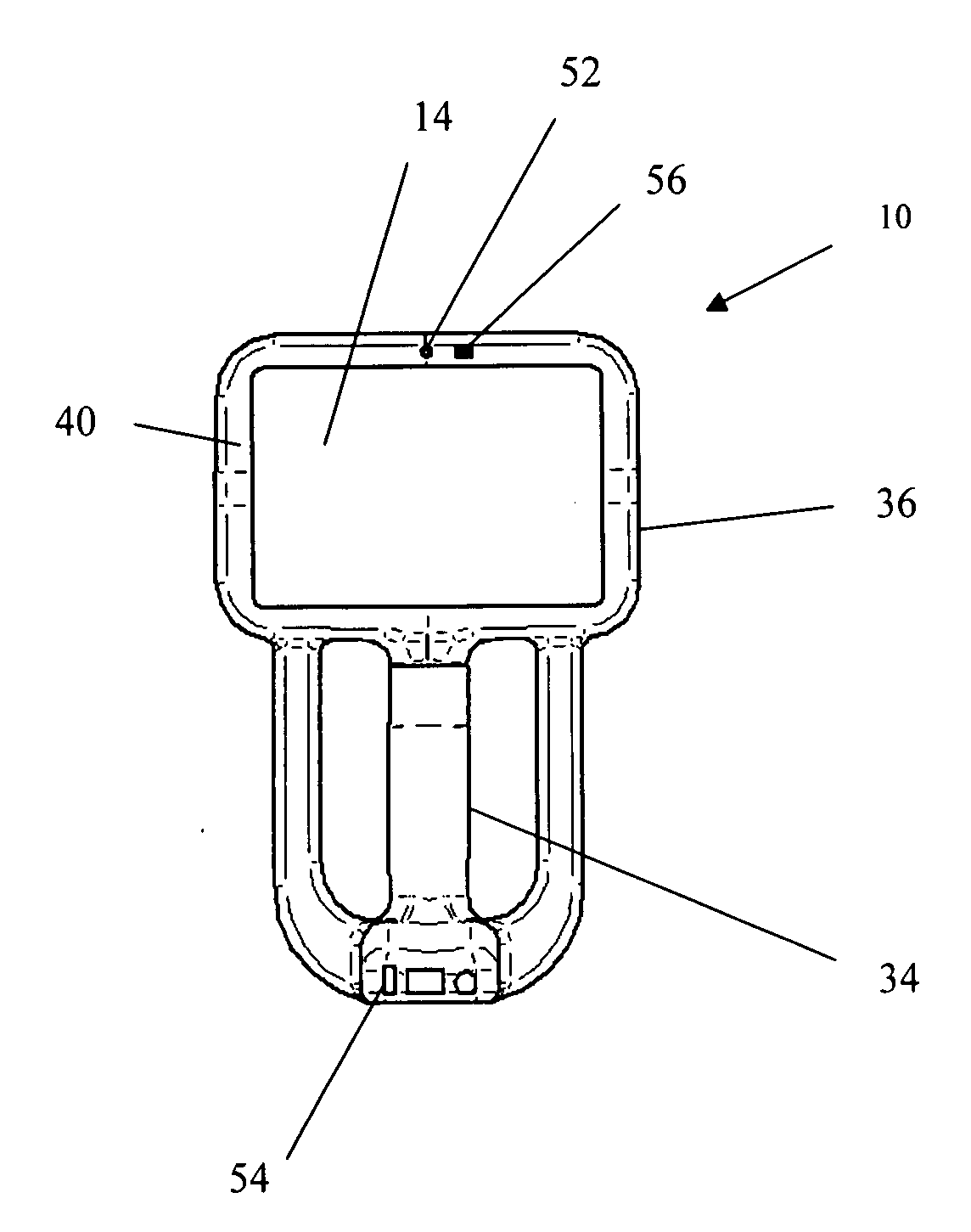
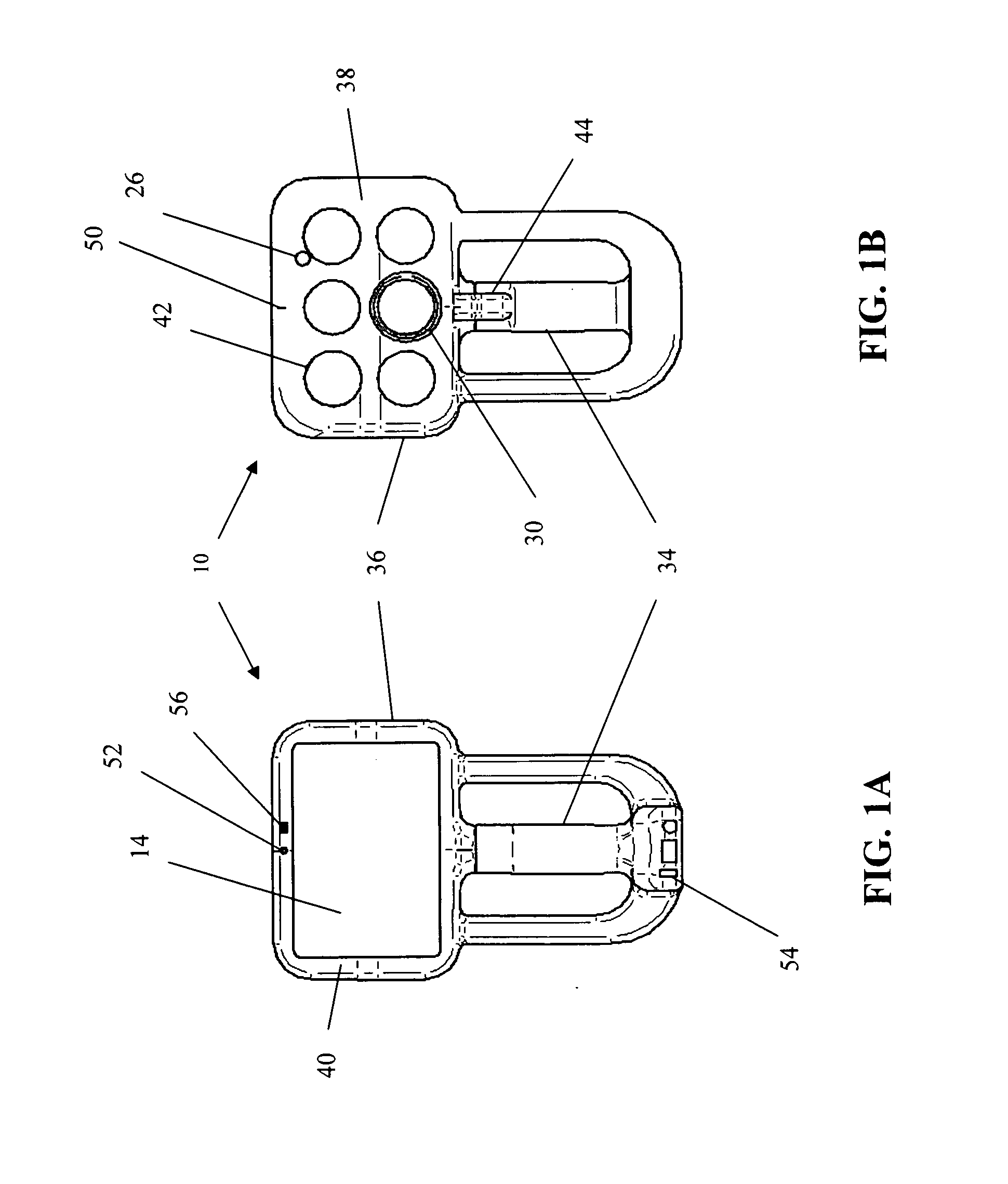
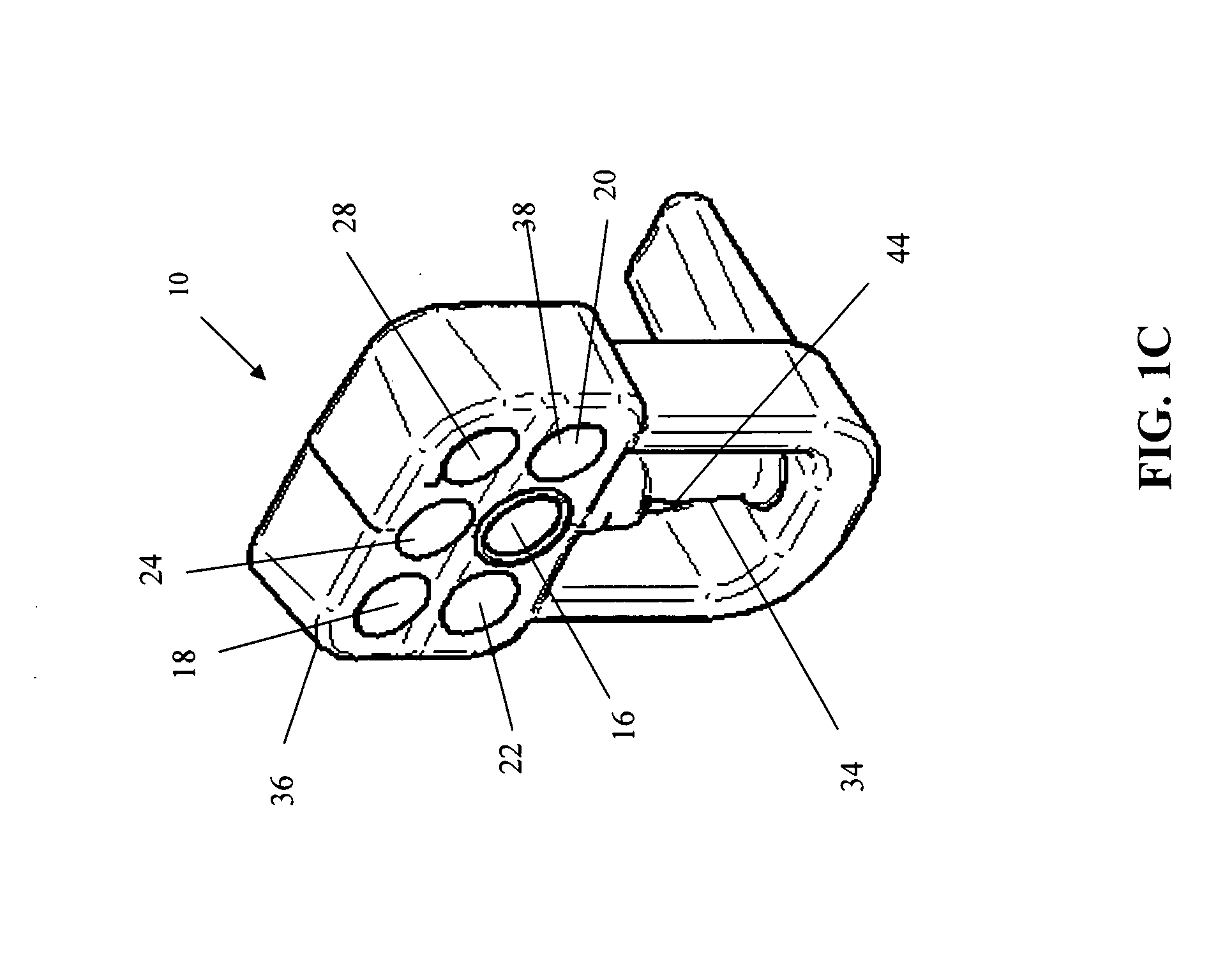
[0020]The present invention involves the integration of up to five imaging modules into a handheld convergent parameter instrument 10. Each imaging module utilizes a common control set 12 and a common display 14.

[0021]The first imaging technique is high resolution color digital photography, used for the purpose of medical noninvasive optical diagnostics and monitoring of diseases. Digital photography, when combined with controlled solid state lighting, polarization filtering, and coordinated with appropriate image processing techniques, derives more information that the naked eye can discern. Clinically inspecting visible skin color changes by eye is subject to inter- and intraexaminer variability. The use of computerized image analysis has therefore been introduced in several fields of medicine in which objective and quantitative measurements of visible changes are required. Applications range from follow-up of dermatological lesions to diagnostic aids and clinical classifications ...


PUM


Abstract

The present invention relates to a convergent parameter instrument and method for performing real-time imaging using multiple imaging techniques. More particularly, the present invention relates to a handheld convergent parameter instrument providing some or preferably all of real-time imaging, including surface mapping, color imaging, perfusion imaging, thermal imaging, and near infrared spectroscopy, and including a common control set and a common display.

Description

BACKGROUND OF THE INVENTION[0001](a) Field of the Invention[0002]The present invention relates to a convergent parameter instrument and method for performing real-time imaging using multiple imaging techniques. More particularly, the present invention relates to a handheld convergent parameter instrument providing some or preferably all of real-time imaging, including surface mapping, color imaging, perfusion imaging, thermal imaging, and near infrared spectroscopy, and including a common control set and a common display.[0003](b) Background of the Invention[0004]Skin, the largest organ of the body, has been essentially ignored in medical imaging. No standard of care regarding skin imaging exists. Computerized Tomography (“CT”), Magnetic Resonance Imaging (“MRI”), and ultrasound are routinely used to image within the body for signs of disease and injury. Researchers and commercial developers continue to advance these imaging technologies to produce improved pictures of internal orga...

Claims


Application Information

Patent Timeline
IPC IPC(8): A61B6/00
CPCA61B5/0077A61B5/441A61B5/055A61B6/5247A61B6/5229A61B5/0261A61B5/70A61B5/0082A61B5/015A61B2505/01A61B2560/0431A61B2560/0443A61B2576/02A61B5/0035A61B5/004G16H30/40
Inventor WHITESTONE, JENNIFER J.MERSCH, STEVEN H.
Owner POINT OF CONTACT
Learn More