

Microfluidic system for measuring cell barrier function
a microfluidic system and cell barrier technology, applied in the field of microfluidic systems for measuring cell barrier function, can solve the problems of not considering bbb transport at all in drug development programs, 1% of brain volume reached, and inability to effectively penetrate the brain parenchyma
- Summary
- Abstract
- Description
- Claims
- Application Information
AI Technical Summary
Benefits of technology
Problems solved by technology
Method used
Image

Examples
example 1
A. Materials and Methods
[0068]Materials and Reagents. Poly(dimethysiloxane) (PDMS) (Sylgard 184) was purchased from Dow Corning (Midland, Mich.). SU-8 2150 for two-step positive photoresist features was purchased from MicroChem. Co. (Newton, Mass.). Polytester membranes were purchased from Corning Inc. (Corning, N.Y.). All cell lines were purchased from ATCC (Manassas, Va.). Ag / AgCl electrodes of 500 μm diameter were purchased from World Precision Instruments (Sarasota, Fla.).
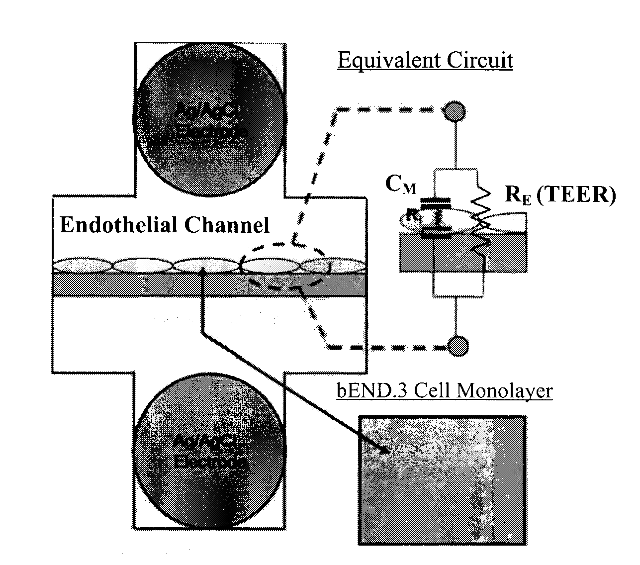
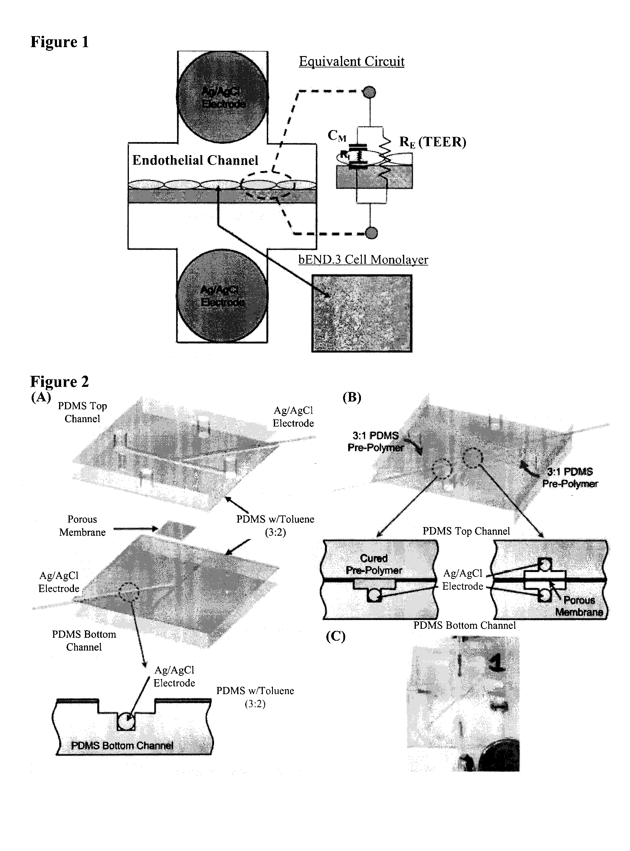
[0069]Device Design and Fabrication. The device is composed of two layers of PDMS microfluidic channels, which are designed with integrated microgrooves that permit the physical registration of two electrodes on opposite sides but in immediate proximity to a porous membrane on which cells are cultured for impedance measurement (Tung et al., J. Sens. Actuators, B 2004, 98, 356-367). The porous membrane on which cells attach and grow (initial diameter 24 mm and with a thickness of 10 μm) was first cut from the Tr...
example 2
Fabrication of Layered Microfluidic Channels and the Establishment of Cell Culture Protocol
[0083]Layered microfluidic channels were fabricated using soft lithography (Duffy et al., Analytical Chemistry 70, 4974-4984 (1998)). Briefly, PDMS prepolymer was mixed with curing agent at a weight ratio of 10 (prepolymer):1 (curing agent) and was cast onto two 4 inch silicon wafer containing a positive relief pattern that was 200 μm thick, one for the top layer and one for the bottom layer. The mixture was cured at 60° C. for 2 hours, then the cured PDMS layer was peeled from the silicon wafer. Access holes were punched with a 16 gauge blunt syringe (1.65 mm outer diameter) forming the inlet and outlet holes for each channel. In order to glue the top and the bottom layers together, PDMS mortar layers were created using PDMS and toluene mixed at 3 (PDMS):2 (toluene) weight ratio. The mixture was well mixed using a vortex mixer and it was allowed to sit for 5 minutes to remove bubbles. The tol...
example 3
[0097]Static Microfluidic Model with b.End3 Cell Line
Permeability of [14C]-Mannitol, [14]-Inulin, and [14C]-Dextran Across b.End3 Monolayer in Static Microfluidic Model
[0098]In order to examine the size selectivity of the tight junctions formed by the endothelial cells in the static model, three paracellular transport markers with different molecular weight (MW) are selected: [14C]-mannitol (MW=182.17), [14C]-inulin (MW=6179), and [14C]-dextran (MW=105000) (Wishart, D. S. et al. Nucleic Acids Res., D901-906 (2008)). If endothelial monolayers grown in this model possess size selectivity, it is expected that differences in permeability for these three marker molecules is observed. Specifically, mannitol is predicted to demonstrate the highest permeability across cell monolayer through paracellular transport due to it being the smallest among the selected molecules; while dextran is predicted to display the lowest permeability due to its large molecular weight and size. The permeabilit...
PUM
| Property | Measurement | Unit |
|---|---|---|
| Speed | aaaaa | aaaaa |
| Transport properties | aaaaa | aaaaa |
Abstract
Description
Claims
Application Information