
Method and compositions for detecting botulinum neurotoxin

a botulinum neurotoxin and composition technology, applied in the field of botulinum neurotoxin detection methods and compositions, can solve the problems of inability to study the catalytic kinetics of toxins, complicated and expensive amplification process, etc., to reduce the fret ratio of the entire population of cells, and significantly reduce the fluorescence signal of yfp

Active Publication Date: 2012-08-30
WISCONSIN ALUMNI RES FOUND
View PDF3 Cites 3 Cited by
  • Summary
  • Abstract
  • Description
  • Claims
  • Application Information

AI Technical Summary

Benefits of technology

Enables the sensitive and rapid detection of botulinum neurotoxins at picomolar levels within hours, allowing for real-time monitoring of toxin enzymatic kinetics and high-throughput screening of inhibitors, suitable for use in living cells and neurons.

Problems solved by technology

These toxins are a well-recognized source of food poisoning, often resulting in serious harm or even death of the victims.
Botulinum neurotoxins are listed as a bioterror threat due to their extreme potency and the lack of immunity in the population.
This method requires large numbers of mice, is time-consuming and cannot be used to study toxin catalytic kinetics.
A number of amplified immunoassay systems based on using antibodies against toxins have also been developed, but most of these systems require complicated and expensive amplification process, and cannot be used to study toxin catalytic activity either.
Although HPLC and immunoassay can be used to detect cleaved substrate molecules and measure enzymatic activities of these toxins, these methods are generally time-consuming and complicated, some of them require specialized antibodies, making them inapplicable for large scale screening.
However, it requires synthesis of peptides with modified amino acid derivatives and is not suitable for use in living cells.

Method used


Image

  • Method and compositions for detecting botulinum neurotoxin
  • Method and compositions for detecting botulinum neurotoxin
  • Method and compositions for detecting botulinum neurotoxin

Examples


example 1

Bio-Sensors Based on CFP-YFP FRET Pair and Botulinum Neurotoxin Protease Activity

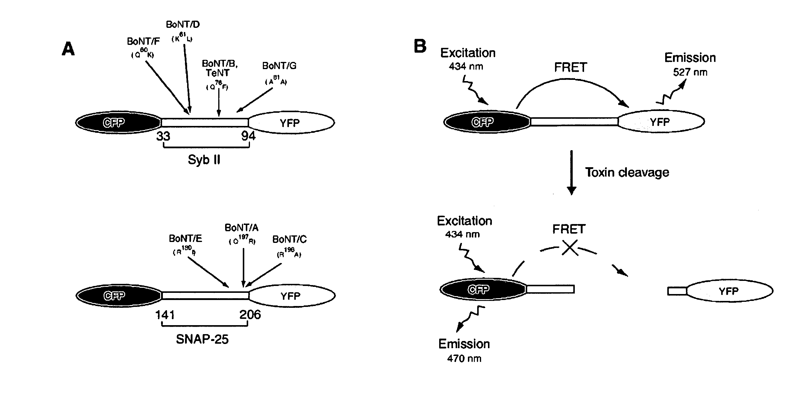
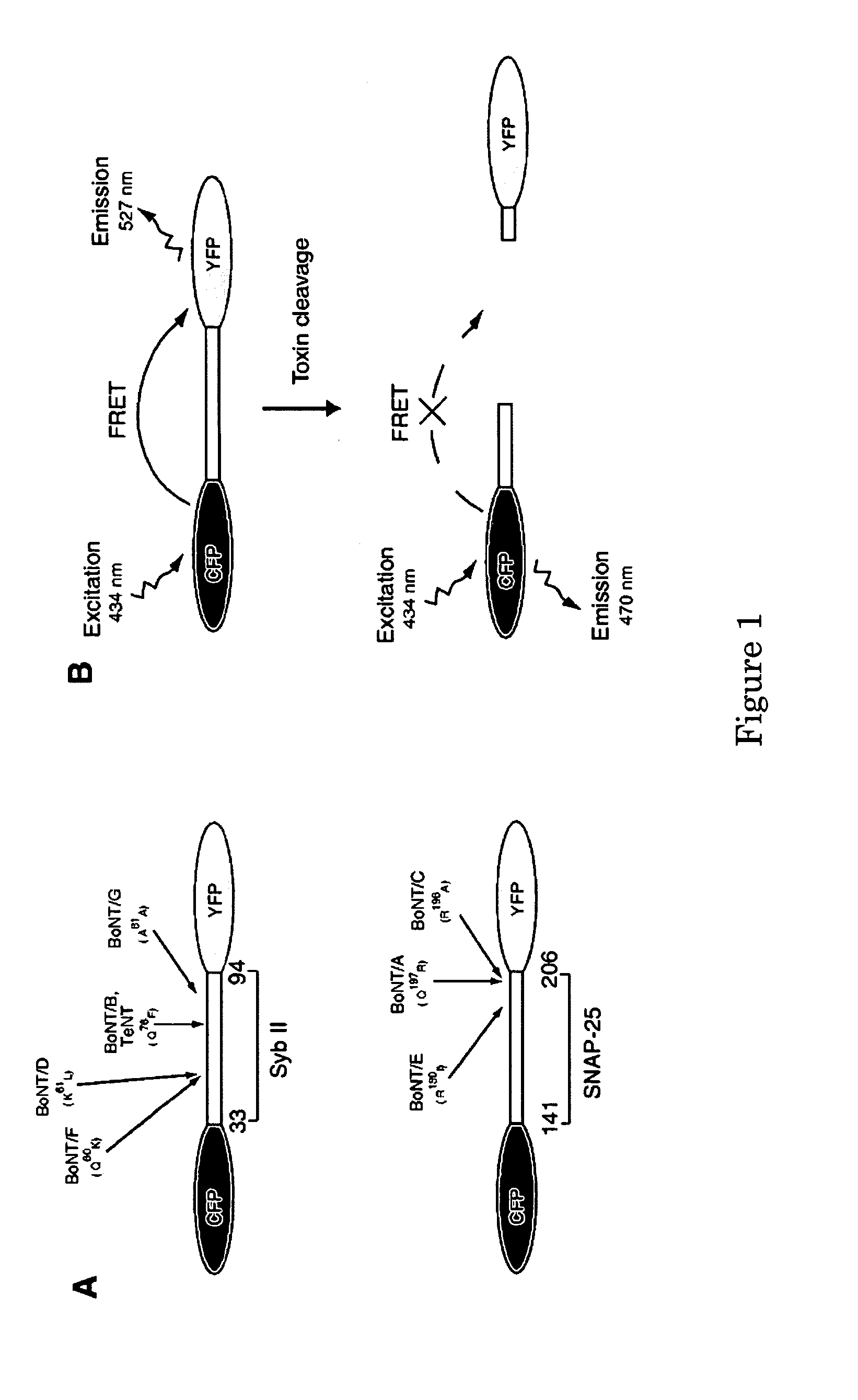
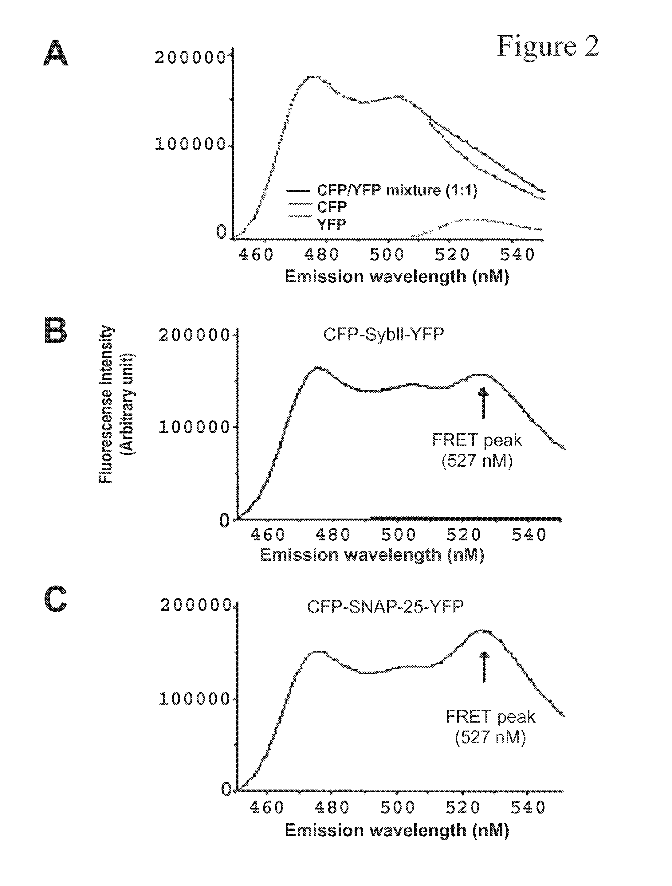
[0093]In order to monitor botulinum neurotoxin protease activity using FRET method, CFP and YFP protein are connected via syb II or SNAP-25 fragment, denoted as CFP-Syb11-YFP and CFP-SNAP-25-YFP, respectively (FIG. 1A). Short fragments of toxin substrates were used instead of the full-length protein to optimize the CFP-YFP energy transfer efficiency, which falls exponentially as the distance increases. However, the cleavage efficiency by BoNTs decreases significantly as the target protein fragments get too short. Therefore, the region that contain amino acid 33-96 of synaptobrevin sequence was used because it has been reported to retain the same cleavage rate by BoNT / B, F, and TeNT as the full length synaptobrevin protein does. Similarly, residues 141-206 of SNAP-25 were selected to ensure that the construct can still be recognized and cleaved by BoNT / A and E.

[0094]The FRET assay is depicted in FIG. 1B....

example 2

Monitoring the Cleavage of Bio-Sensor Proteins by Botulinum Neurotoxins in Vitro

[0096]300 nM chimera protein CFP-Syb11-YFP was mixed with 50 nM prereduced BoNT / B holotoxin in a cuvette. The emission spectra were collected at different time points after adding BoNT / B (0, 2, 5, 10, 30, 60 minutes, etc). At the end of each scan, a small volume of sample (30 μl) was taken out from the cuvette and mixed with SDS-loading buffer. These samples later were subjected to SDS-page gels and the cleavage of chimera proteins were visualized using an antibody against the his6 tag in the recombinant chimera protein. As shown in FIG. 3A, the incubation of bio-sensor protein with BoNT / B resulted in a decrease of YFP emission and increase of CFP emission. The decrease of FRET ratio is consistent with the degree of cleavage of the chimera protein by BoNT / B (FIG. 3A, low panel). This result demonstrates the cleavage of the bio-sensor protein can be monitored in real time by recording the change in its FR...

example 3

Monitoring Botulinum Neurotoxin Protease Activity in Real Time Using a Microplate Spectrofluorometer

[0098]The above experiments demonstrated that the activity of botulinum neurotoxin can be detected in vitro by monitoring the changes of the emission spectra of their target bio-sensor proteins. We then determined if we could monitor the cleavage of bio-sensor proteins in real time using a microplate reader—this will demonstrate the feasibility to adapt this assay for future high-throughput screening. As shown in FIG. 4A, 300 nM CFP-SNAP-25-YFP chimera protein was mixed with 10 nM BoNT / A in a 96-well plate. CFP was excited at 436 nm and the fluorescence of the CFP channel (470 nM) and YFP channel (527 nM) were recorded over 90 minutes at 30 second intervals. Addition of BoNT / A resulted in the decrease of YFP channel emission and the increase of CFP channel emission. This result enabled us to trace the kinetics of botulinum neurotoxin enzymatic activity in multiple samples in real time...


PUM


Abstract

The present invention provides a molecular construct capable of fluorescent resonance energy transfer (FRET), comprising a linker peptide, and donor and acceptor fluorophore moieties, where the linker peptide is a substrate of a botulinum neurotoxin selected from the group consisting of synaptobrevin, syntaxin and SNAP-25, or a fragment thereof capable being cleaved by the botulinum neurotoxin, and separates the donor and acceptor fluorophores by a distance of not more than 10 nm, and where emission spectrum of the donor fluorophore moiety overlaps with the excitation spectrum of the acceptor fluorophore moiety; or where the emission spectra of the fluorophores are detectably different. Also provided are isolated nucleic acid expressing the construct, kits comprising said construct and cell lines comprising said nucleic acid. Further provided are methods of detecting a BoNT using the above described construct via FRET, and methods for detecting a BoNT using surface plasmon resonance imaging.

Description

CROSS-REFERENCES TO RELATED APPLICATIONS[0001]This is a divisional application of U.S. patent application Ser. No. 11 / 014,845, filed Dec. 20, 2004, which claims the benefit of U.S. Provisional Application Ser. No. 60 / 530,645, filed Dec. 19, 2003, and U.S. Provisional Application Ser. No. 60 / 579,254, filed Jun. 15, 2004, the contents of which are incorporated herein by reference in their entirety.STATEMENT REGARDING FEDERALLY SPONSORED RESEARCH[0002]This invention was made with United States government support awarded by the National Institutes of Health under the grant numbers NIH GM56827 and MH061876. The government has certain rights in this invention.BACKGROUND OF THE INVENTION[0003]Botulinum neurotoxins (BoNTs) are produced by Clostridium botulinum and are the most potent toxins known. These toxins are a well-recognized source of food poisoning, often resulting in serious harm or even death of the victims. There are seven structurally related botulinum neurotoxins or serotypes (...

Claims


Application Information

Patent Timeline
Patent Type & Authority Applications(United States)
IPC IPC(8): C40B30/08C07K14/00C07K2/00C07K7/08G01N21/64G01N21/55C07K14/435G01N33/542G01N33/569
CPCC07K14/43595C07K14/705C07K2319/50C07K14/435G01N33/542G01N33/56911G01N2333/33C07K2319/60C12Y304/24069A61P25/00A61P31/04A61P39/02A61P43/00Y02A50/30G01N2333/952G01N33/573
Inventor CHAPMAN, EDWIN R.DONG, MIN
Owner WISCONSIN ALUMNI RES FOUND
Learn More