Optical imaging contrast agents and uses thereof

a contrast agent and optical imaging technology, applied in the field of optical imaging contrast agents, can solve the problems of describing contrast agents, long dwell time, and time required for clearance of unbound contrast, and can introduce significant inconvenience in imaging workflow

Inactive Publication Date: 2012-09-27
TARON
View PDF4 Cites 24 Cited by
  • Summary
  • Abstract
  • Description
  • Claims
  • Application Information

AI Technical Summary

Benefits of technology

[0020]In another aspect of the invention, we describe a method of optical imaging of a subject comprising, administering a microbubble modified to incorporate an optically active probe and a targeting ligand to the subject and thereafter imaging the subject with an optical imaging scanner. After imaging, the microbubbles within the targeted tissue are destroyed by application of low-frequency ultrasound pulses intended to eliminate the microbubbles by outward rectified gas diffusion and subsequent deflation of the microbubble. A second population of microbubbles bearing identical or different optically active probe and identical or different targeting ligands is subsequently administered, and the process is repeated.
[0021]In another embodiment of the invention, we describe a method for delivering an optically active probe into one or several targeted cells. Said optically active probe is incorporated into or immobilized onto the shell of the microbubble, in addition to a targeting ligand. Microbubbles maintain proximity with the targeted cell(s) by the bond between the targeting ligand and molecular target on the cell(s). The optically active probe is transferred from the microbubble to the adjacent targeted cells by the process of acoustically-activated delivery. Said acoustic activation should consist of high-amplitude ultrasound of sufficient energy to induce nonlinear oscillation and subsequent violent destruction of the microbubble. Cells that receive the optically active probe may be subsequently imaged and quantified using optical imaging.
[0022]In another aspect of the invention, we describe therapeutic uses of the described optically active modified microbubbles. In one embodiment of the invention, the therapeutic method comprises, administering a therapeutically effective amount of microbubbles modified by the addition of an optically active probe and a therapeutic substance to a subject, imaging the subject for a period of time during which accumulation of the microbubbles at a desired loca...

Problems solved by technology

A significant difficulty with previously described contrast agents relates to the time required for clearance of unbound contrast.
A long dwell time can introduce significant inconvenience in imaging workflow, especially in a high-throughput research setting.
Additionally, a long dwell period can negate the possibility of administering multiple contrast agent doses within the same imaging setting,...

Method used


Image

  • Optical imaging contrast agents and uses thereof
  • Optical imaging contrast agents and uses thereof
  • Optical imaging contrast agents and uses thereof

Examples


specific examples

[0048]The following examples illustrate, but in way are intended to limit the invention.

example 1

Synthesis of Microbubbles by Sonication

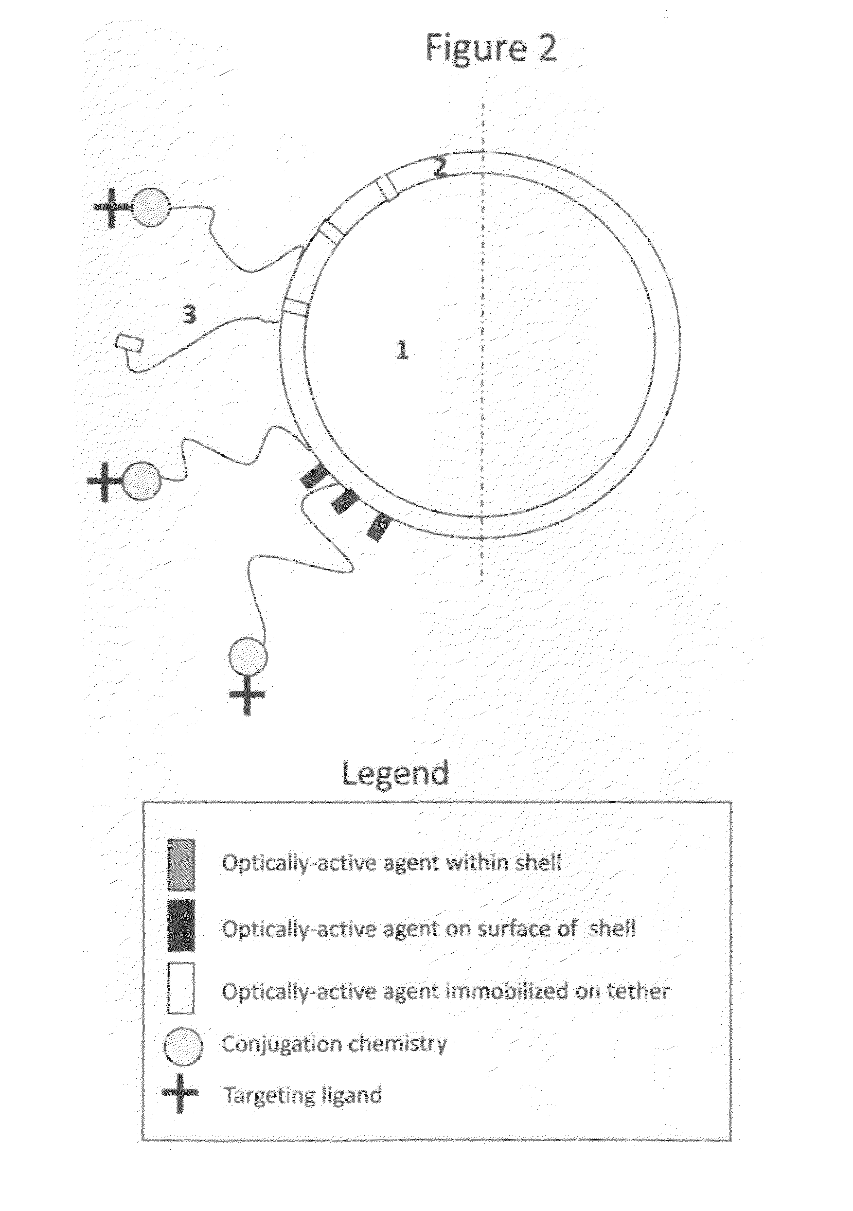
[0049]Microbubbles consisting of a lipid monolayer encapsulating decafluorobutane gas (SynQuest) are prepared by mixing 40 mg of distearoyl phosphatidylcholine (Avanti Polar Lipids) and 20 mg polyoxyethylene-40 stearate (Sigma-Aldrich). This mixture is added to 20 mL of sterile normal saline and sonicated to clarity using a probe-type sonicator. Decafluorobutane gas is dispersed through the aqueous phase via a thin capillary tube, and sonication continued until the formation of microbubbles, sizes of 1-30 um (mean size 1-5 um). The resulting microbubbles are stabilized with a lipid monolayer and augmented with polyoxyethylene as an emulsifier. Microbubbles so prepared are stored refrigerated under a perfluorocarbon atmosphere until further use.

[0050]Microbubbles are also prepared with different shell compositions, including denatured proteins, sugars, and non-biological polymers. For example, decafluorobutane microbubbles stabilized by a shell ...

example 2

Synthesis of Microbubbles with Reactive Groups

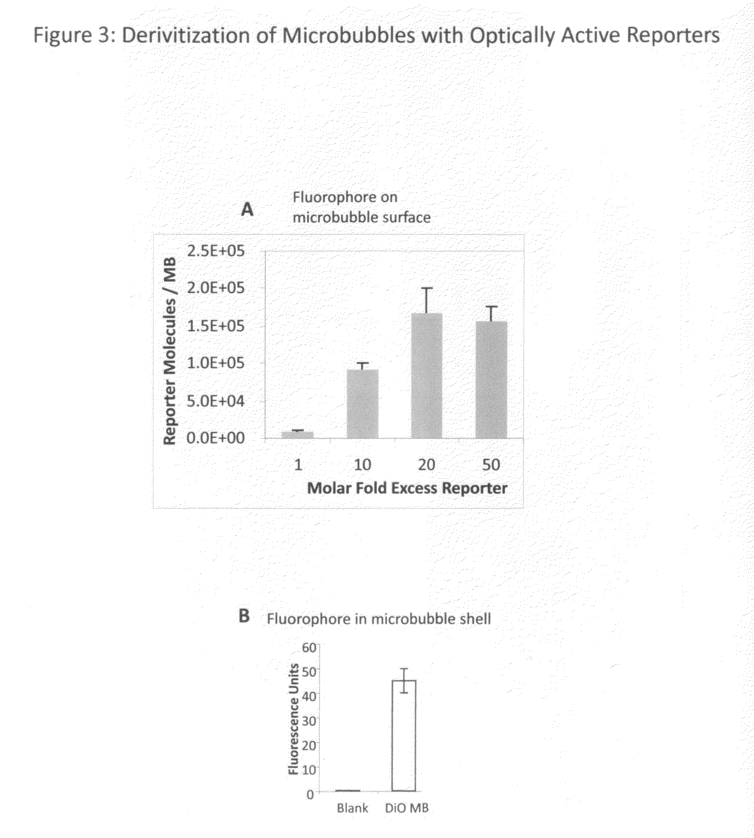
[0051]Microbubbles bearing the reactive group 2-pyridyl disulfide (PDP), to which targeting ligands or optically active probes may be immobilized, are prepared as follows. Microbubbles consisting of a lipid monolayer encapsulating decafluorobutane are prepared by mixing 40 mg of phosphatidylcholine, 20 mg polyoxyethylene 40 stearate, and 5 mg of PDP-PEG(2000)-disteroylphosphatidylethanolamine (Avanti). This mixture is added to 20 mL of sterile normal saline (Baxter) and sonicated to clarity using a probe-type sonicator. Decafluorobutane gas is dispersed through the aqueous phase via a thin capillary tubing and sonication continued with the formation of microbubbles, sizes of 1-30 um (mean size 1-5 um). The resulting microbubbles are stabilized with a lipid monolayer and augmented with a polyoxyethylene brush for improved stability and PDP residues on a long polymer tether for the subsequent ligand conjugation. Microbubble material is sto...


PUM


Abstract

The invention relates to microbubble contrast agent compositions comprising a gas core encapsulated by a monolayer shell, said shell comprising a first surfactant, a second surfactant having a higher water solubility than said first surfactant, an anchor molecule attached to said shell at the gas-shell interface and an optically-active probe, said probe being detectable by fluorescence, near-infrared, bioluminescence, or other optical imaging methods. Uses of the described compositions for imaging and therapeutic applications are contemplated.

Description

CROSS-REFERENCES TO RELATED APPLICATIONS[0001]This application claims the benefit of priority to U.S. Provisional Application No. 61 / 277,192 filed on Sep. 22, 2009, the contents of which is incorporated herein by reference.TECHNICAL FIELD[0002]The present invention relates to the field of contrast agents for imaging in the setting of research in vitro and in vivo in animal models, and for diagnostic imaging of veterinary animals and humans. More specifically, the invention relates to microparticle contrast agents that can be used for bioluminescence, fluorescence, and other optical imaging modalities. Use of the described agent for functional imaging (for example, visualizing and measuring blood flow and blood volume) and molecular imaging (for example, visualizing and measuring molecular receptors within the body) are contemplated. Additionally, therapeutic applications are contemplated.BACKGROUND OF THE INVENTION[0003]Optical imaging is a molecular and functional imaging technique...

Claims


Application Information

Patent Timeline
IPC IPC(8): A61K49/00
CPCA61K49/0002A61K49/223A61K49/0091A61K49/0032A61B8/06A61B8/065A61B8/0883A61B8/0891A61B8/481A61B8/5223A61N7/00G16H50/30
Inventor RYCHAK, JOSHUA J.
Owner TARON
Why Eureka
Try Eureka