
Carotid-artery-plaque ultrasound-imaging method and evaluating device

a carotid artery and ultrasound imaging technology, applied in ultrasonic/sonic/infrasonic diagnostics, instruments, applications, etc., can solve the problems of large-scale devices, poor resolution, and difficulty in repeating intravascular ultrasound

Inactive Publication Date: 2013-02-21
UNIVERSITY OF TOKUSHIMA
View PDF7 Cites 5 Cited by
  • Summary
  • Abstract
  • Description
  • Claims
  • Application Information

AI Technical Summary

Benefits of technology

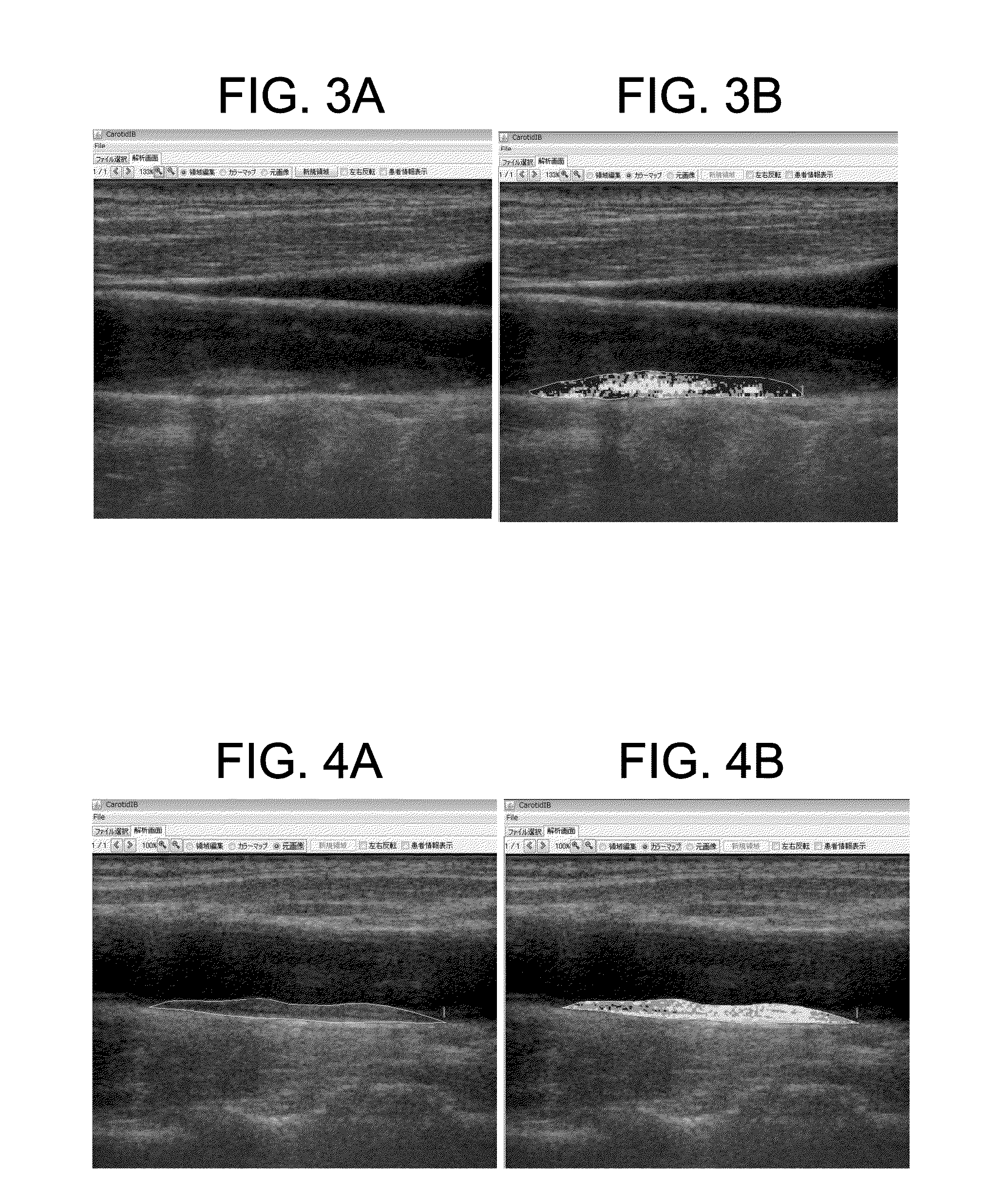
The present invention is about a device that can evaluate carotid artery plaque using echo signals obtained by a special method. The device includes an obtaining portion, a memory portion, a coloring portion, and a display portion. The obtaining portion gets the raw signals of echo signals from the carotid artery plaque. The memory portion stores the relationship between color ranges and colors. The coloring portion uses the raw signals to classify the reference areas of the echo image into different color ranges and colors those reference areas. The display portion displays the colored echo image. This invention makes it easy to distinguish and early detect vascular disorder based on the carotid artery echo image analysis. Another feature is that the device can display divided areas that show both uncolored and colored echo images simultaneously, making the display more visible and allowing for comparison between the two images.

Problems solved by technology

As a result, it is difficult to repeatedly conduct the intravascular ultrasound.
However, there is a problem with radiation exposure in the case of CT, while there is a problem that the resolution is poor in the case of MRI.
In addition, CT and MRI require a large-scale device.
For this reason, these noninvasive diagnosis methods cannot be conducted in a small clinic.
Also, outpatients cannot freely have these noninvasive diagnosis methods.
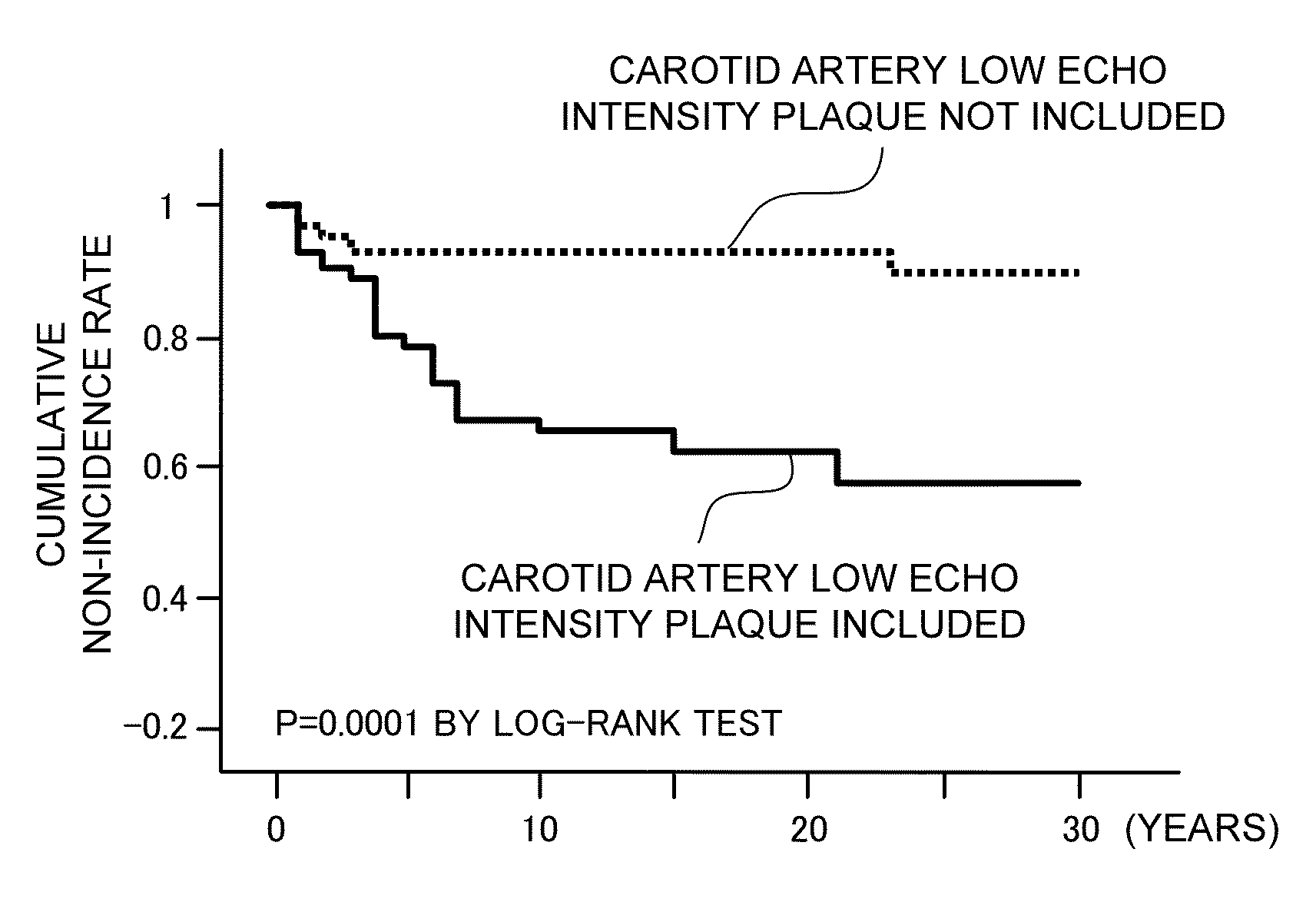
That is, it is said that, if a large amount of low echo intensity plaque is included in carotid artery, this means vulnerability or instability of coronary artery (Non-patent Literature 4).
However, the carotid artery echo test is not a quantitative test but a qualitative rest.
Accordingly, even the same plaque may result in different diagnosis results depending on evaluator or testing devices.
As a result, there is a problem that it is difficult to properly evaluate tissue characteristics of the plaque (Non-patent Literature 1).
However, when plaque properties are classified with the naked eye based on the echo intensity levels displayed in gray scale, since the criterion of intensity level discrimination is blurry, it is difficult to quantitatively evaluate plaque properties.
For this reason, it is difficult to compare images with each other.
As a result, there is problems that time-variation of a patient cannot be properly checked, and that patients' cases cannot be properly compared with each other.
In addition, it is difficult to evaluate plaque properties in severely calcified lesion, high grade lesion such as high grade internal carotid artery lesion, tortuous vessel, or the like.
It can be understood that difficult to quantitatively evaluate plaque properties based on echo intensity.
Accordingly, there is a problem that even the same RF signal will be represented by different levels in gray scale in the produced echo image.
However, these improvements are not succeeded up to now.
However, if these methods are adapted to noninvasive carotid artery analysis, an echo reflection wave passes through skin or tissue.
Since the echo reflection wave is analyzed, it is difficult to simply adopt these methods to noninvasive carotid artery analysis.

Method used


Image

  • Carotid-artery-plaque ultrasound-imaging method and evaluating device
  • Carotid-artery-plaque ultrasound-imaging method and evaluating device
  • Carotid-artery-plaque ultrasound-imaging method and evaluating device

Examples


example 1

Validity Confirmation of Statin Group Drug

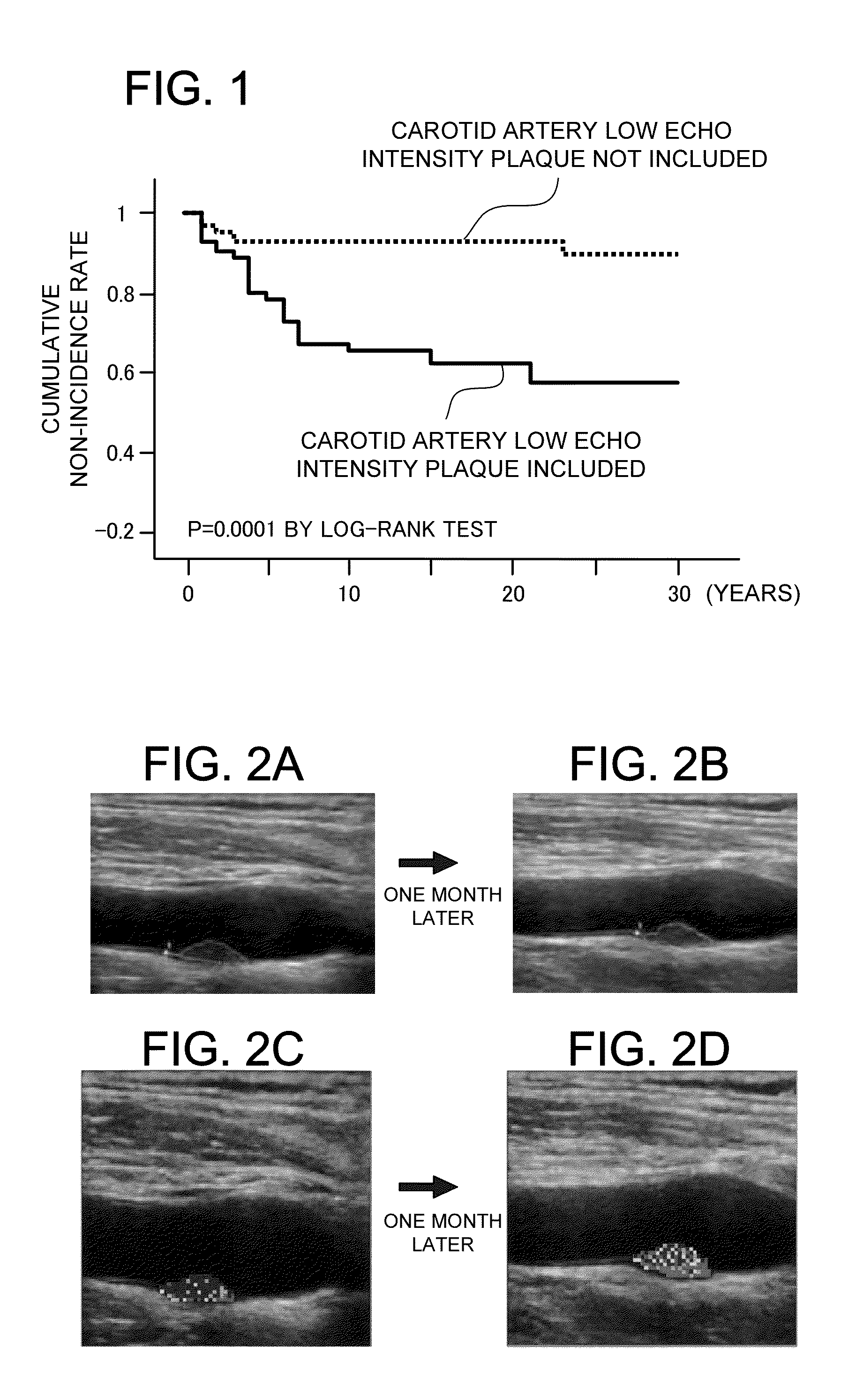
[0120]Examples of the present invention will be described. First, the following description describes an example according to the present invention that confirms of the validity of a statin group drug. In this example, time variation of the properties of a carotid artery plaque is evaluated before and after medication of a statin group drug. Thus, the confirmation of the validity of the medicine according to the present invention is evaluated. Specifically, a statin is given to a patient who has carotid artery plaque. The shape change is observed between at the medication and at the end of medication for one month.

[0121]In this specification, the “medicine” is not specifically limited as long as it can improve the properties of carotid artery plaque. That is, the “medicine” is not specifically limited as long as it can be used as hyperlipemia drugs. For example, the “medicine” can be hyperlipemia drugs such as statin drugs, fibrate drugs, EP...

example 2

Property Evaluation of Carotid Artery Plaque

[0130]The following description describes an example that evaluates the properties of carotid artery plaque by using the determination method of the present invention. In this example, three types of carotid artery echoes with different plaque intensity distributions are measured, and are compared with each other by using the method according to the present invention. In this example, carotid artery echo images of patients are obtained. The patients are a patient who has the hypoechoic plaque in the carotid artery (57-years-old female), a patient who has isoechoic plaque (51-years-old male), and a patient who has hyperechoic plaque (73-years-old male). The carotid artery echo images are evaluated by the determination method according to the present invention. FIGS. 6 and 7 show the results. FIGS. 6A to 6C show colored images and specific data of three types of plaque that have intensity distributions corresponding to so-called hypoechoic p...

example 3

Comparison between Analysis Result Based on Colored Image, and Actual Tissue Clinical Image

[0131]To verify whether the analysis result of the plaque properties obtained by the determination method of the present invention agrees with actual histopathological tissue image or not, the plaque property analysis of a CEA patient obtained by the method according to the present invention is compared with actual removed tissue. In this embodiment, the data of CEA patients with high degrees of stenosis in carotid artery (61-years-old male, and 79-years-old male) before the CEA obtained by the carotid artery echo determining method of the present invention is compared with the removed specimen that corresponds to the echo part of carotid artery and is obtained after CEA (HE stained). FIG. 8 show the result of the 61-years-old male. FIG. 9 show the result of the 79-years-old male. FIG. 8A shows a colored image of CEA case. FIG. 8B shows an image corresponding to FIG. 8A with a part to be remov...


PUM


Abstract

The carotid artery plaque properties are quantitatively determined by a carotid artery echo whereby predicting incidence of cerebral infarction or myocardial infarct. A method includes obtaining, coloring, and displaying steps. In the obtaining step, the integrated amounts per unit time of echo signals corresponding to pixels of an echo image of carotid artery plaque are obtained. The signals are obtained when the image is obtained by an IBS method. In the coloring step, the relationship between ranges and colors is referenced. The available full range of the integrated amount is previously divided into the ranges. The colors are assigned to the ranges. The pixels are classified into the ranges in accordance with the integrated amounts corresponding to the pixels. The pixels of the image are colored in colors corresponding to the ranges of the pixels. In the displaying step, the colored echo image is displayed on a display portion 30.

Description

BACKGROUND OF THE INVENTION[0001]1. Field of the Invention[0002]The present invention relates to a carotid-artery-plaque ultrasound-imaging method, a carotid-artery-plaque evaluating device, a carotid-artery-plaque evaluating program, and a computer-readable storage medium that can noninvasively evaluate the properties of plaque in carotid artery by using ultrasound examination (ultrasonic pulse).[0003]2. Description of the Related Art[0004]Diagnosis of carotid artery plaque tissue characteristics can be used not only to evaluate stroke risk but also to predict ischemic heart disease events or to estimate therapeutic effect of lipid lowering drug for dyslipidemia. In particular, for diabetes mellitus, this diagnosis is important to estimate the prognosis or QOL of a patient.[0005]Presently, intravascular ultrasound examination (IVUS) is used as invasive diagnosis of carotid artery plaque properties. However, the intravascular ultrasound requires catheterization in which a catheter t...

Claims


Application Information

Patent Timeline
IPC IPC(8): G06K9/00
CPCA61B8/5223A61B8/0891G16H50/30
Inventor SATA, MASATAKAYAMADA, HIROTSUGU
Owner UNIVERSITY OF TOKUSHIMA
Learn More