
Early detection and staging of colorectal cancer using a panel of micro rnas

a colorectal cancer and micro-rnas technology, applied in combinational chemistry, biochemistry apparatus and processes, library screening, etc., can solve the problems of not detecting early malignancy, low detection efficiency, and fatal malignant tumors, and achieve high sensitivity and specificity, and simple assays.

Inactive Publication Date: 2013-05-30
MOR RES APPL LTD
View PDF4 Cites 5 Cited by
  • Summary
  • Abstract
  • Description
  • Claims
  • Application Information

AI Technical Summary

Benefits of technology

The present invention provides a way to diagnose colorectal cancer (CRC) by using a specific panel of microRNAs (tiny RNA molecules) as biomarkers. This method has high sensitivity and specificity for CRC, and can even detect small amounts of cancer cells in the body. The invention is based on the unexpected results obtained when testing the expression level of selected miRNAs in tumor samples obtained from CRC patients and control samples. The CRC-specific miRNA panel was found effective in diagnosing and staging CRC with significantly increased sensitivity and specificity, especially for detecting CRC metastasis and differentiating between various lymphatic stages of CRC metastasis.

Problems solved by technology

Malignant tumors are fatal, mostly due to their capacity to invade neighboring tissues and metastasize through the lymphatic system and bloodstream to nearby or distant organs.
Unfortunately, in addition to its high false-positive rate, the sensitivity of the FOBT remains around 30%-50% and may not detect early malignancy, since not all carcinomas shed blood in the early phase of their development.
However, despite its accuracy, fiber-optic colonoscopy is currently used to screen high-risk populations (e.g. individuals with genetic predisposition and / or family history) and its application to large population screening has failed mainly due to low compliance, high-costs and complications associated with the procedure.
The variety of molecular events resulting in a phenotype of colonic adenoma or carcinoma limits these assays to detection of tumors harboring the mutations or hypermethylated genes examined while other tumors may go undetected.
Numerous serum markers, such as carcinoembryonic antigen (CEA), carbohydrate antigen 19-9, and lipid-associated sialic acid, have been investigated in colorectal cancer, but their low sensitivity has limited their role, as reflected by the American Society of Clinical Oncology (ASCO) guidelines, to monitoring therapy and for post-therapy surveillance.
In particular, no set of miRNAs has been developed for successfully diagnosing pre-cancerous polyps.

Method used


Image

  • Early detection and staging of colorectal cancer using a panel of micro rnas
  • Early detection and staging of colorectal cancer using a panel of micro rnas
  • Early detection and staging of colorectal cancer using a panel of micro rnas

Examples


example 1

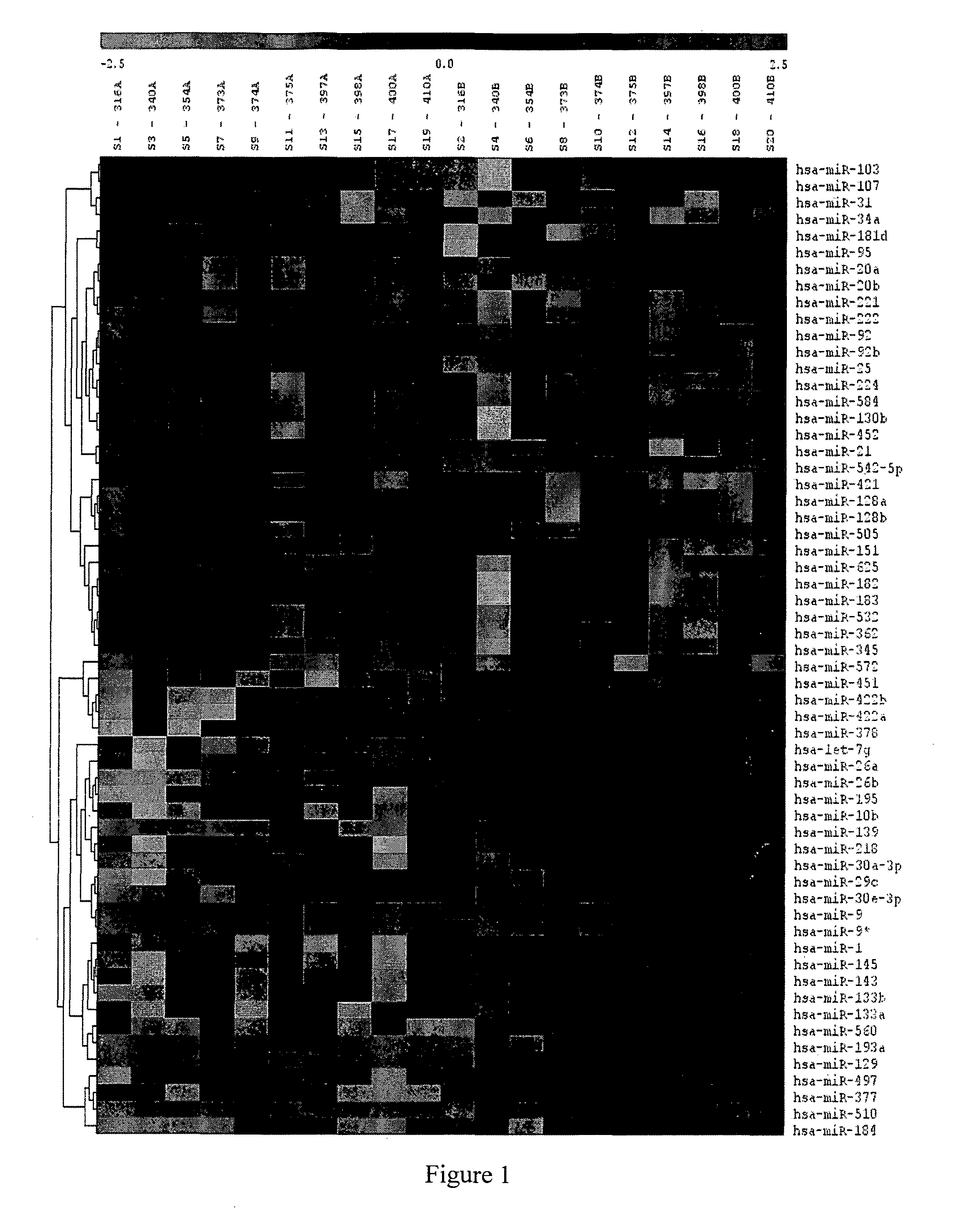
Identification and Selection of CRC-Specific miRNA

[0186]Samples from CRC patient were tested on a miRNA microarray (LC sciences, Houston, USA). The samples included RNA obtained from the tumor and from tissue adjacent to the tumor, and two normal colon samples obtained from non cancer patients. The selection criteria were up-regulation of the miRNA expression in most of the patients and a significantly higher expression in tumors versus adjacent tumor tissue.

[0187]Other microRNAs, shown to be upregulated in CRC, were selected from a pool of miRNAs (˜900) found in publicly available sources (e.g. WO2009 / 140670, WO2009 / 059026, WO2010 / 004562 and WO2009 / 111643) and electronic databases (e.g. www.mirbase.org, www.genecards.org).

[0188]Candidate miRNAs, from both sources, were checked in-silico for their expression profile in normal lymphatic tissue. Only miRNA which showed very low expression in normal lymphatic tissue and lymphocytes were selected. The selected miRNA were subjected to fu...

example 2

Validation of miRNA Expression in Tumor Tissue, Adjacent Normal Tissue, PBMC and Normal Colon Tissue

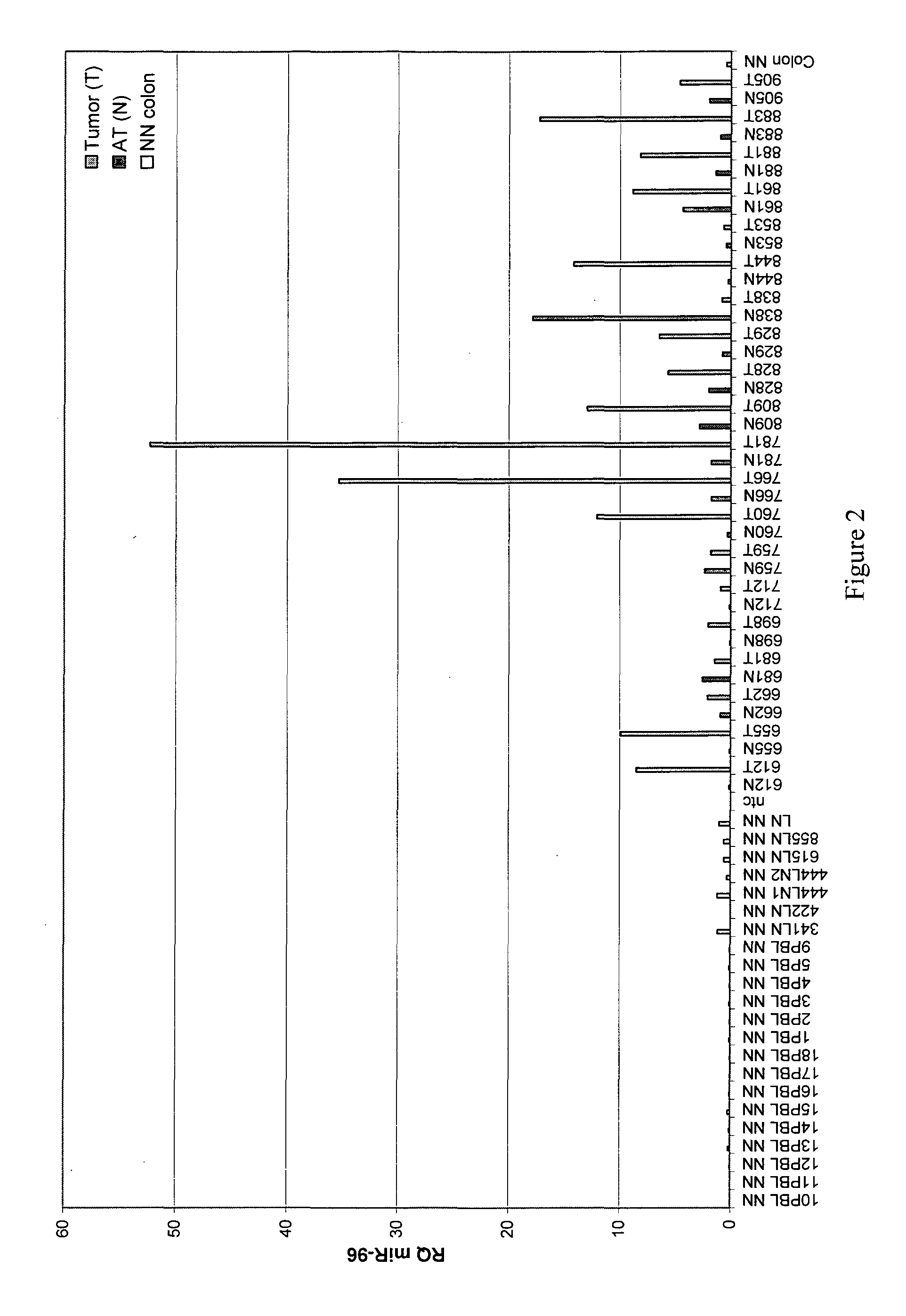
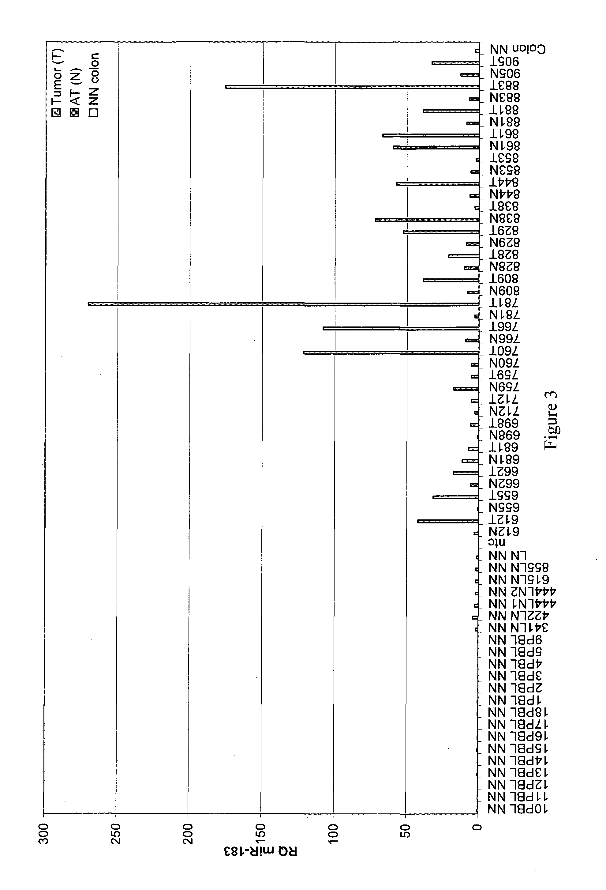
[0190]In order to create a diagnostic panel, the expression levels of each miRNA identified (in Example 1) as being over-expressed in colon tumor samples (T) was tested and compared to a panel of (i) paired adjacent normal (AT) tissues obtained from patients with adenocarcinoma of the colon, (ii) PBMCs of healthy individuals (designated PBL) and (iii) RNA extracted from normal colonic tissues (Ambion®) (FIGS. 2-9). Only miRNAs that were exclusively expressed in tumor tissues as validated by real-time PCR were selected for the panel. This selection process resulted in the identification of 8 miRNAs suitable for the diagnostic panel.

[0191]FIG. 2 shows the expression profile of hsa-mir-96. Significantly higher expression is seen in tumor samples (T) as compared to the adjacent normal tissue (AT) obtained from patients with adenocarcinoma of the colon, or the PBMC of healthy individuals. ...

example 3

Ultrastaging of Sentinel Lymph Nodes of CRC Patients Using the miRNA Panel

[0200]In order to assess lymphatic staging, 86 sentinel lymph nodes (SLNs) obtained from 20 CRC patients were studied. Each SLN was cut into two fragments. One half was subjected to enhanced pathological examination using H&E and immunohistochemistry staining for cytokeratin (CK). The other half was snap frozen in liquid nitrogen, RNA was then extracted and the expression of the miRNA panel was studied. Expression of at least two miRNAs from the miRNA panel indicated a CRC positive lymph node. The detailed analysis of SLN ultrastaging of CRC patients is shown in Table 3 below.

TABLE 3Analysis of SLN ultrastaging of 20 CRC patientsPathologymiRNA panelPanelsampleH&ECK2096183194200a200b200c203429screen612sln1neg.neg.+−−++++++sln2neg.neg.−−−−−−−−−sln3neg.neg.−−−−−−−−−sln4pos.−++++++++655sln1neg.neg.+++++++++sln2pos.−−−−−−−−−sln3neg.neg.−−−−−−−−−sln4pos.++−−−−+−+662sln1neg.pos++−−−−−−+sln2neg.pos−−−−−−−−−sln3neg.pos...


PUM

PropertyMeasurementUnit
diameteraaaaaaaaaa
diameteraaaaaaaaaa
diameteraaaaaaaaaa

Abstract

The present invention provides compositions, methods and kits for diagnosing cancer, specifically the diagnosis of colorectal cancer (CRC). More specifically, the invention provides simple assays, with high sensitivity and specificity for CRC, wherein a panel of microRNA (miRNA) are used as biomarkers.

Description

FIELD OF THE INVENTION[0001]The present invention is directed to the field of cancer diagnosis and staging, specifically of colorectal cancer. In particular, the invention provides compositions, methods and diagnostic kits using a panel of microRNA molecules.BACKGROUND OF THE INVENTION[0002]Colorectal cancer (CRC) is the fourth most common cancer and second leading cause of cancer-related death in the US, with more than 140,000 new cases diagnosed annually (Jemal A et al. CA Cancer J Clin 2009: 59; 225-249). In Western countries, adenocarcinoma of the colon and rectum accounts for more new cases of cancer per year than any anatomic site except the lung.[0003]Features of malignant adenocarcinoma, distinct from benign tumors, include invasion and metastasis. Malignant tumors are fatal, mostly due to their capacity to invade neighboring tissues and metastasize through the lymphatic system and bloodstream to nearby or distant organs.[0004]The survival and prognosis of CRC patients depen...

Claims


Application Information

Patent Timeline
Patent Type & Authority Applications(United States)
IPC IPC(8): C12Q1/68
CPCC12Q1/6886C12Q2600/178C12Q2600/112
Inventor NISSAN, AVIRAMROSENBAUM, STELLA MITRANIILYAYEV, NADIAHALLE, DAVID
Owner MOR RES APPL LTD
Learn More