
Quantitative protein assay using single affinity capture agent for identification and detection

Inactive Publication Date: 2005-07-28
KIM RAYMOND A
View PDF2 Cites 4 Cited by
  • Summary
  • Abstract
  • Description
  • Claims
  • Application Information

AI Technical Summary

Benefits of technology

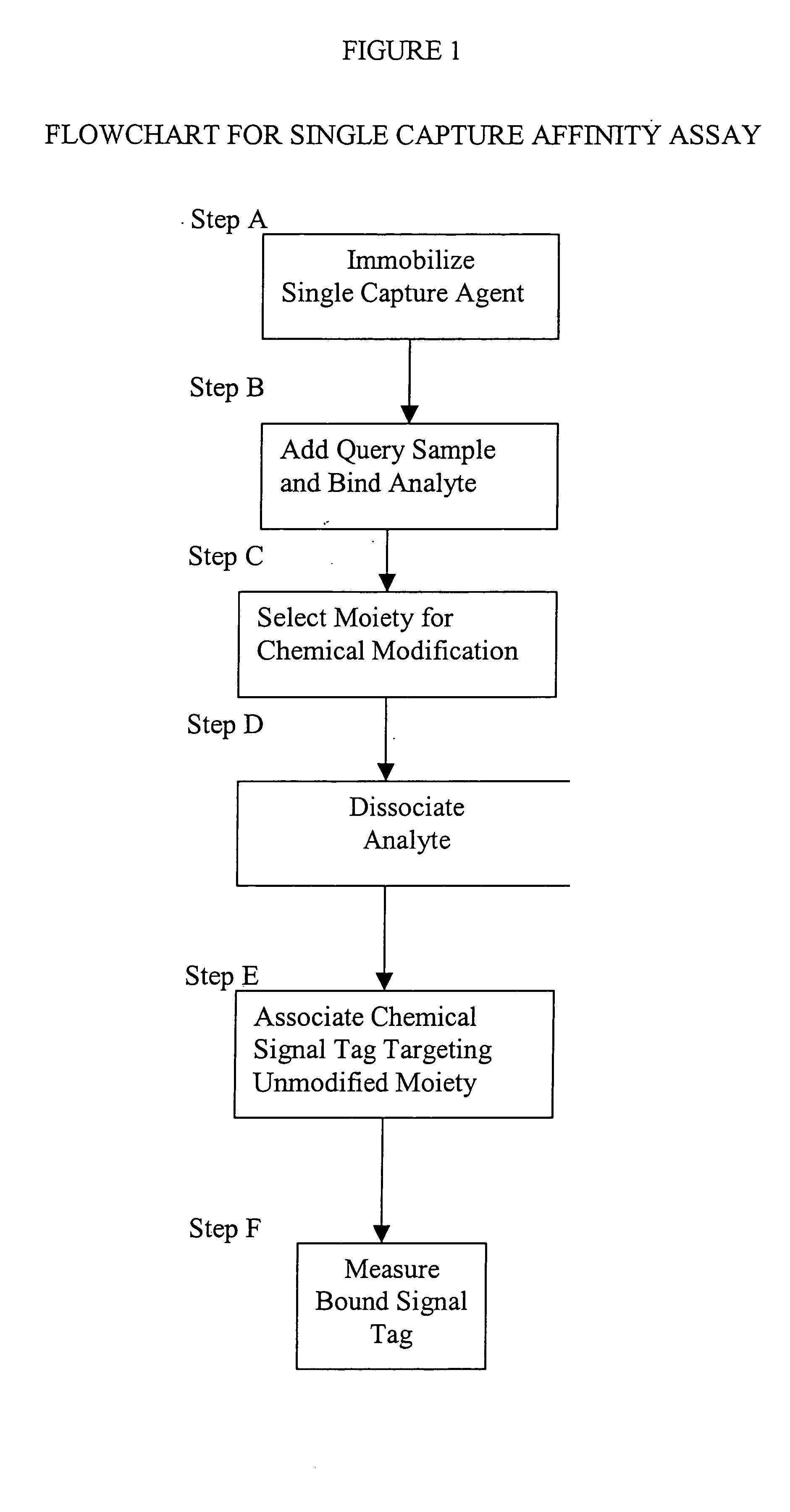
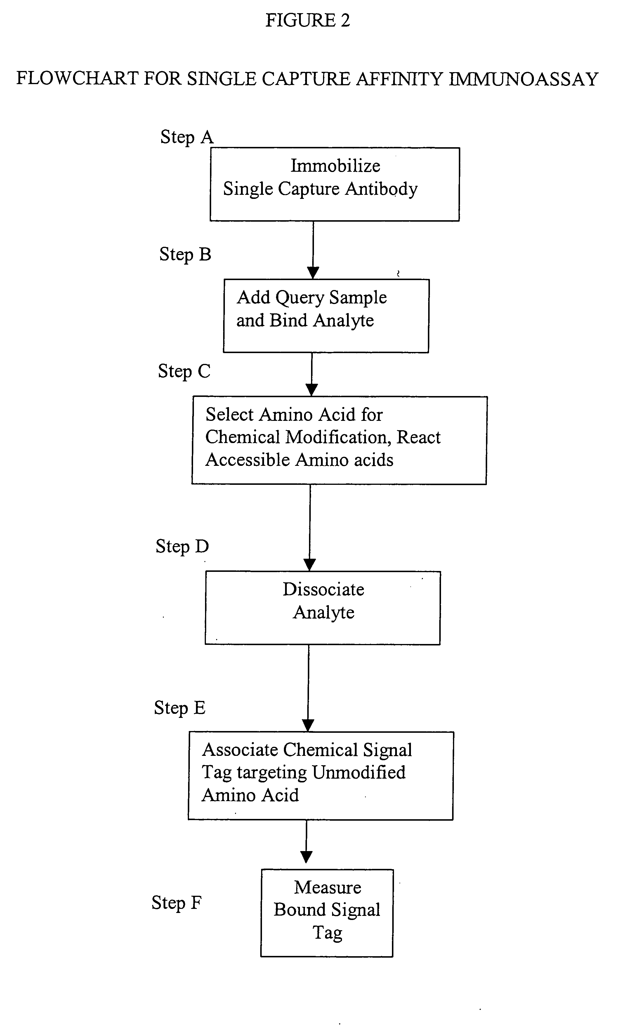
[0014] This invention provides a method that permits identification and quantification of analyte using only one affinity agent, thus significantly simplifying the assay development process. This is achieved by using one affinity agent as both a capture agent to effect both specific binding to the target analyte (i.e. the protein identification) coupled with chemical modification steps, and to detect and quantify the binary binding between the capture agent and the analyte (i.e. the protein quantification). The requirement of a second detection agent is eliminated in this invention through the use of a selective chemical modification process.
[0018] This invention may be applied to the detection and distinguishing of various types of post-translation modification that are known to occur for proteins. For example, assay of phosphorylated proteins is greatly simplified by the use of only one antibody to quantify each phosphorylation state. This capability permits inclusion of a greater number of antibody specificities in multiplexed assay format for parallel measurement of multiple signal transduction pathways. This invention allows the mechanistic analysis of drug leads for target validation, potential off-target activities, the emergence of compensatory drug resistance mechanisms, and secondary or concurrent target assessment for combinatorial therapy.
[0019] This invention may be applied to comprehensive surveillance of pathogens and toxins, which is a critical component of bio-defense strategy. Over 50 biological agents including bacteria and spores (Gram positive and negative), viruses (DNA and RNA, enveloped and non-enveloped), protozoa, and toxins have been selected by the NIH as high priority select agents in order to spur therapeutic and diagnostic product development. Many of these select agents are also blood-borne pathogens transmissible by blood transfusions or tissue transplantations, a significant concern for the safety of our blood and organ supplies. Routine surveillance of these agents requires advanced screening technologies capable of assaying multiple pathogens and bio-toxins in a single sample. Currently, these tests are performed in uniplex assay format using immunological, serological, microbiological, or nucleic-acid based (i.e. PCR-based) methods. These tests, although suitable for single analyte measurement, are difficult to scale up for multi-analyte measurement, and even if feasible, the high cost of large scale assay production makes this approach impractical for routine screening. On the other hand, multiplexed assays using microarrays or beads can be developed for nucleic acids or proteins measurement, provided the chemical compositions of the analytes are compatible within the assay. For instance, RT PCR-based RNA detection has been multiplexed for diagnosis of viral pathogens, e.g. HIV and HCV. However, the difficulty of combining different PCR amplification strategies for different types of nucleic acids (for instance RNA and DNA molecules from a mixture of RNA and DNA viruses) makes large-scale PCR multiplexing technical challenging. In contrast, protein-based multiplexed assays, e.g. antibody microarrays, provide more versatile assay platform in that the antibodies specific for multiple types of pathogens (bacteria, viruses, and protozoa), bio-toxins (anthrax, botulism), and prions can be all multiplexed in a single assay for parallel detection. In addition to direct analyte measurement, the host's response to pathogens, e.g. protective antibodies, can also be profiled to detect the prior exposure or latent infection, thus adding additional dimension to the pathogen coverage. This invention provides an enabling technology with which to develop such multiplexed assays.

Problems solved by technology

The remarkable progress that has been made in genomic science using nucleic acid microarrays unfortunately has not been equally applicable to protein microarrays.
While useful for analysis of individual proteins, or uniplex analysis, many methods of protein detection are not easily amenable to the increased complexities of multi-analyte analysis.
Currently, significant challenges exist in the development of antibody reagents for protein microarrays, especially ELISA immunoassays.
One of the bottlenecks in the development of ELISA is the requirement of two validated antibodies that bind to an analyte without competing for the same binding site (or epitope).
The process of identifying antibodies that can work together as a pair in multiplexed sandwich-ELISA format, however, has proven to be an extremely challenging task when assembling large panels of microarray specificities (Haab B B, et al., Genome Biology.
The difficulty lies with the technical and logistic nature of having to screen through many combinations of antibodies in order to match up a pair that binds to a cognate antigen with high affinity and specificity, but without competing for the same epitope.
A second difficulty in the development of ELISA is the selection of pairing status of antibodies as either capture or detection agents.
These difficulties in antibody reagent development in ELISA are largely responsible for the extremely limited choices of multiplexable contents, which are currently limited to cytokines, chemokines, and growth factors.
In addition to the difficulties of antibody pair-matching constraint and orientation considerations, multi-analyte assays generally do not provide adequate detection sensitivity because of their inability to use signal amplification schemes commonly available to uniplex ELISA systems.
The lack of signal amplification renders multi-analyte assays generally inadequate for applications requiring measurement of low abundance analytes.
Both of these approaches also require the binding of a hapten to the analyte prior to affinity capture, which potentially may alter or interfere with native protein interactions for the analyte.
Further, unbound hapten can be difficult to remove from the testing system, thereby increasing the background noise and decreasing detection sensitivity to low abundance proteins.

Method used


Image

  • Quantitative protein assay using single affinity capture agent for identification and detection
  • Quantitative protein assay using single affinity capture agent for identification and detection
  • Quantitative protein assay using single affinity capture agent for identification and detection

Examples


Embodiment Construction

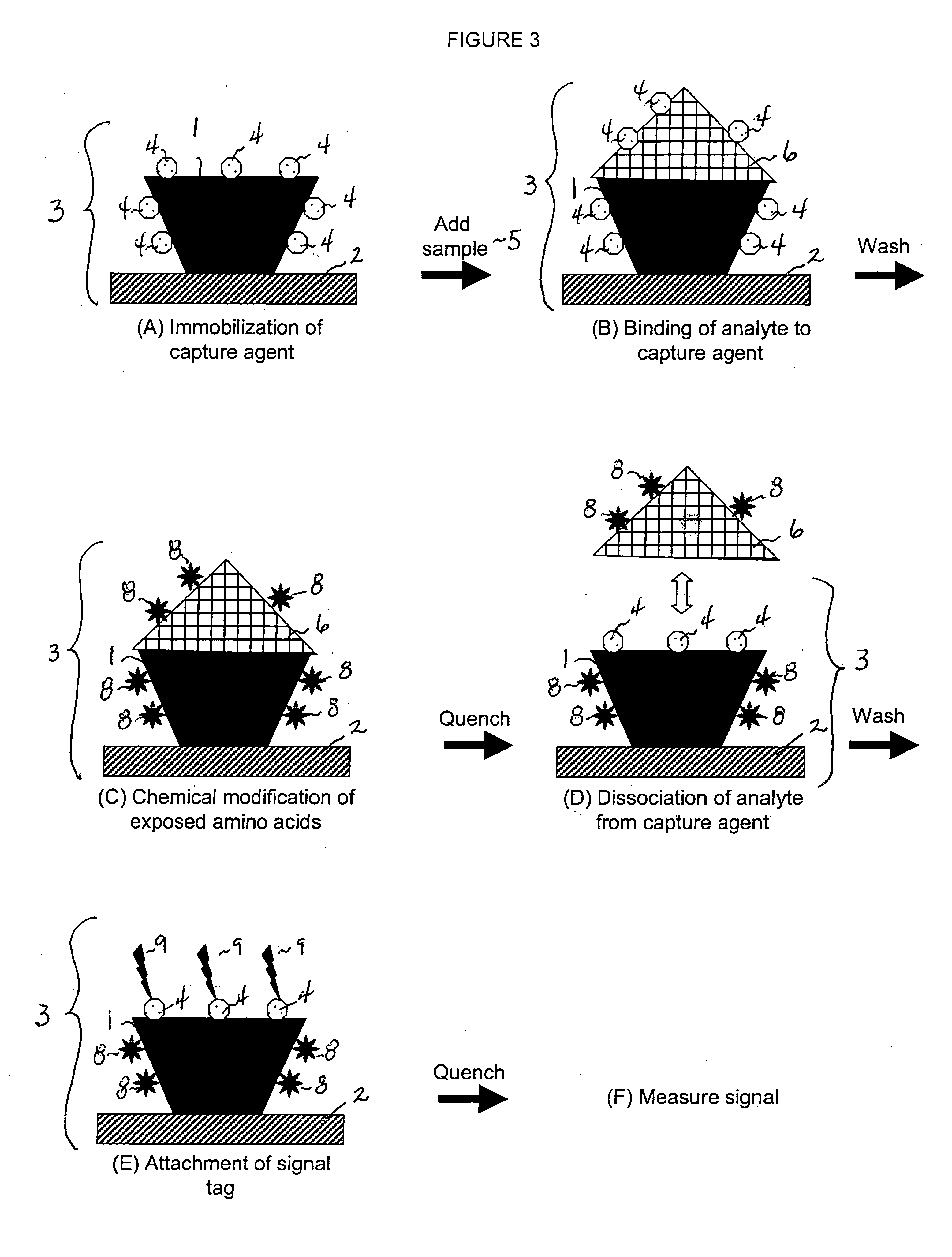
[0082] Antibody Immobilization (Corresponding to Step A of FIGS. 1-3). A murine IgG monoclonal antibody (US Biologicals; Swampscott, Mass.; # 11903-60) specific for human IgG Fc fragment was immobilized onto a solid support, the bottom of 96-well microplates by incubating 50 μl of 100 ng / ml antibody in phosphate-buffered saline (PBS; USB Corp., Cleveland, Ohio; #75889) for 1 hr. A consideration was the selection of microplate substrate compatible with the single affinity capture assay. Several microplate types, e.g. treated, untreated, and coated polystyrene as well as polypropylene, were tested. The results showed that different types of plastics performed differently in the assay, and even the same type of plastic from different vendors gave widely varying performance. The factors considered in the choice of solid support included capability of protein binding and amount of inherent background signal. Of the solid supports tested, the γ-irradiated polystyrene plates (E&K Scientifi...


PUM

PropertyMeasurementUnit
Areaaaaaaaaaaa
Affinityaaaaaaaaaa

Abstract

This invention discloses a single capture affinity assay and apparatus for the identification and quantification of an analyte in a biological sample. The assay uses an array of affinity capture agents specific for an analyte wherein the analyte is removed prior to detection. After the analyte is bound to the capture agent, a moiety of the complex is chemically modified, then the analyte is dissociated from the capture agent, revealing unmodified moiety located in the analyte-capture agent contact interface, which is detected by binding with a signal tag and the signal is quantitated. Analyte detection is achieved without using labeled secondary detection agents. The method is especially applicable to proteins, with antibodies as capture agents and selected amino acids as lysine being modified. The method may be used for uniplex or multiplexed analysis, and is applicable to high-throughput protein measurement.

Description

RELATED INFORMATION [0001] This application claims benefit of and priority to U.S. provisional patent application Ser. No. 60 / 527,838 filed Dec. 09. 2003.FIELD OF INVENTION [0002] The invention is directed to a method of identification and quantification of biomolecules, especially proteins, by a single capture agent assay technology. The invention is analyte specific, quantitative, and is applicable to antibody microarrays for uniplex or multiplexed high-throughput protein measurement. BACKGROUND OF THE INVENTION [0003] Proteins are a major functional component of biological cells. The analysis of proteins is vital, both in basic biomedical research and in the biotechnology industry, especially in the discovery of new therapeutic and diagnostic entities. While accurate identification and quantitative measurement of small amounts of proteins has long been an endeavor in protein biochemistry, it has become even more important given the demands of proteomic research. The remarkable pr...

Claims


Application Information

Patent Timeline
IPC IPC(8): C12Q1/68G01N33/543
CPCG01N33/54306
Inventor KIM, RAYMOND A.
Owner KIM RAYMOND A
Learn More