In vivo imaging method for cancer
a cancer and in vivo imaging technology, applied in the field of in vivo imaging, can solve the problems of low brain uptake, inconsistent imaging, and low relative ability to detect cancer cells
- Summary
- Abstract
- Description
- Claims
- Application Information
AI Technical Summary
Benefits of technology
Problems solved by technology
Method used
Image
Examples
example 1
Synthesis of 9-(2-[18F]Fluoro-ethyl)-5-methoxy-2,3,4,9-tetrahydro-1H-carbazole-4-carboxylic acid diethylamide (imaging agent 5
Example 1(a)
Benzyloxy Acetyl Chloride (1)
[0133]To benzyloxyacetic acid (10.0 g, 60.0 mmol, 8.6 mL) in dichloromethane (50 mL) was added oxalyl chloride (9.1 g, 72.0 mmol, 6.0 mL) and DMF (30.0 mg, 0.4 mmol, 32.0 μL) and stirred at RT for 3 h. There was initially a rapid evolution of gas as the reaction proceeded but evolution ceased as the reaction was complete. The dichloromethane solution was concentrated in vacuo to give a gum. This gum was treated with more oxalyl chloride (4.5 g, 35.7 mmol, 3.0 mL), dichloromethane (50 mL), and one drop of DMF. There was a rapid evolution of gas and the reaction was stirred for a further 2 h. The reaction was then concentrated in vacuo to afford 11.0 g (quantitative) of Benzyloxy acetyl chloride (1) as a gum. The structure was confirmed by 13C NMR (75 MHz, CDCl3) bc 73.6, 74.8, 128.1, 128.4, 128.6, 130.0, and 171.9.
Examp...
example 1 (
Example 1(m)
9-(2-[18F]Fluoro-ethyl)-5-methoxy-2,3,4,9-tetrahydro-1H-carbazole-4-carboxylic acid diethylamide (imaging agent 5)
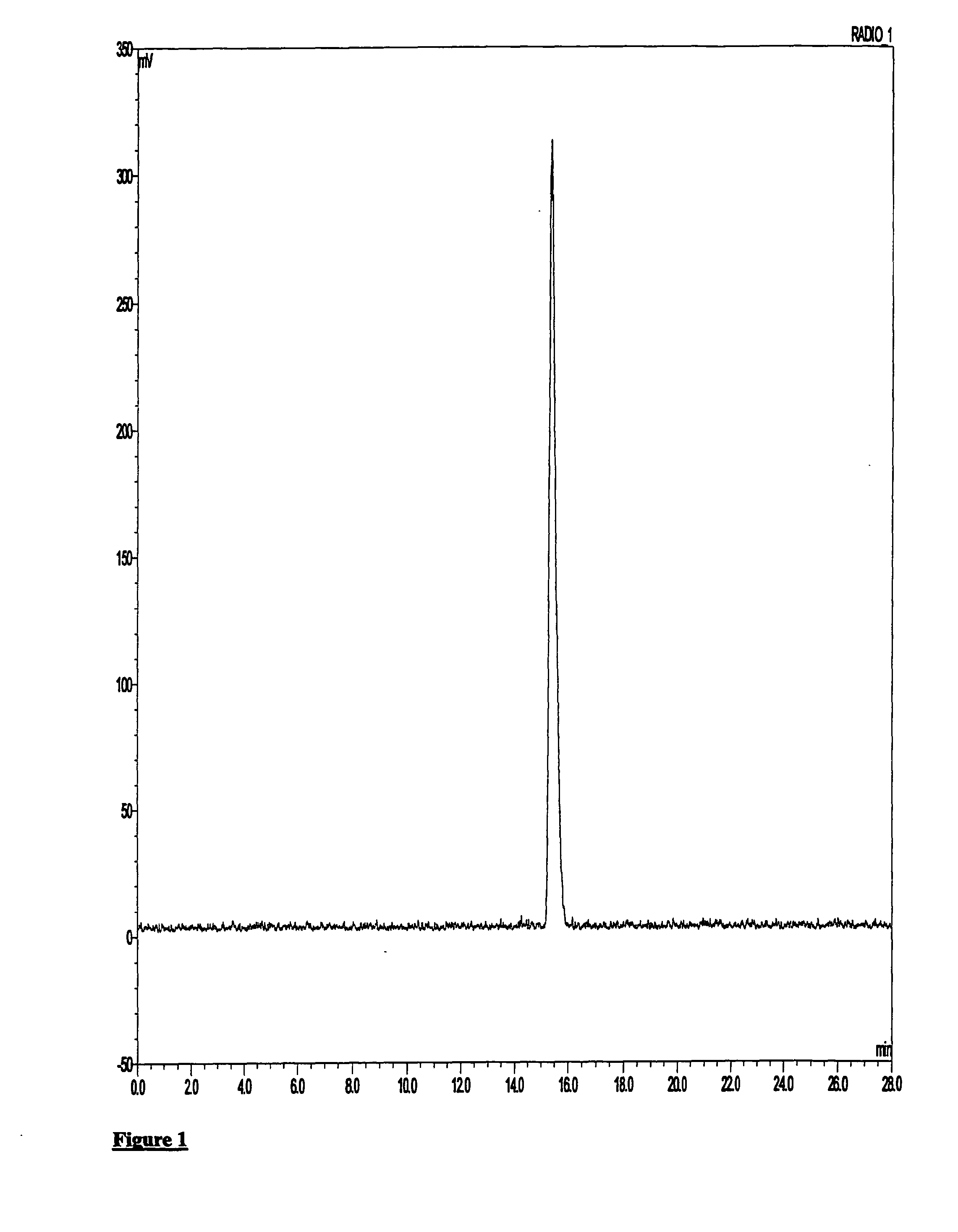
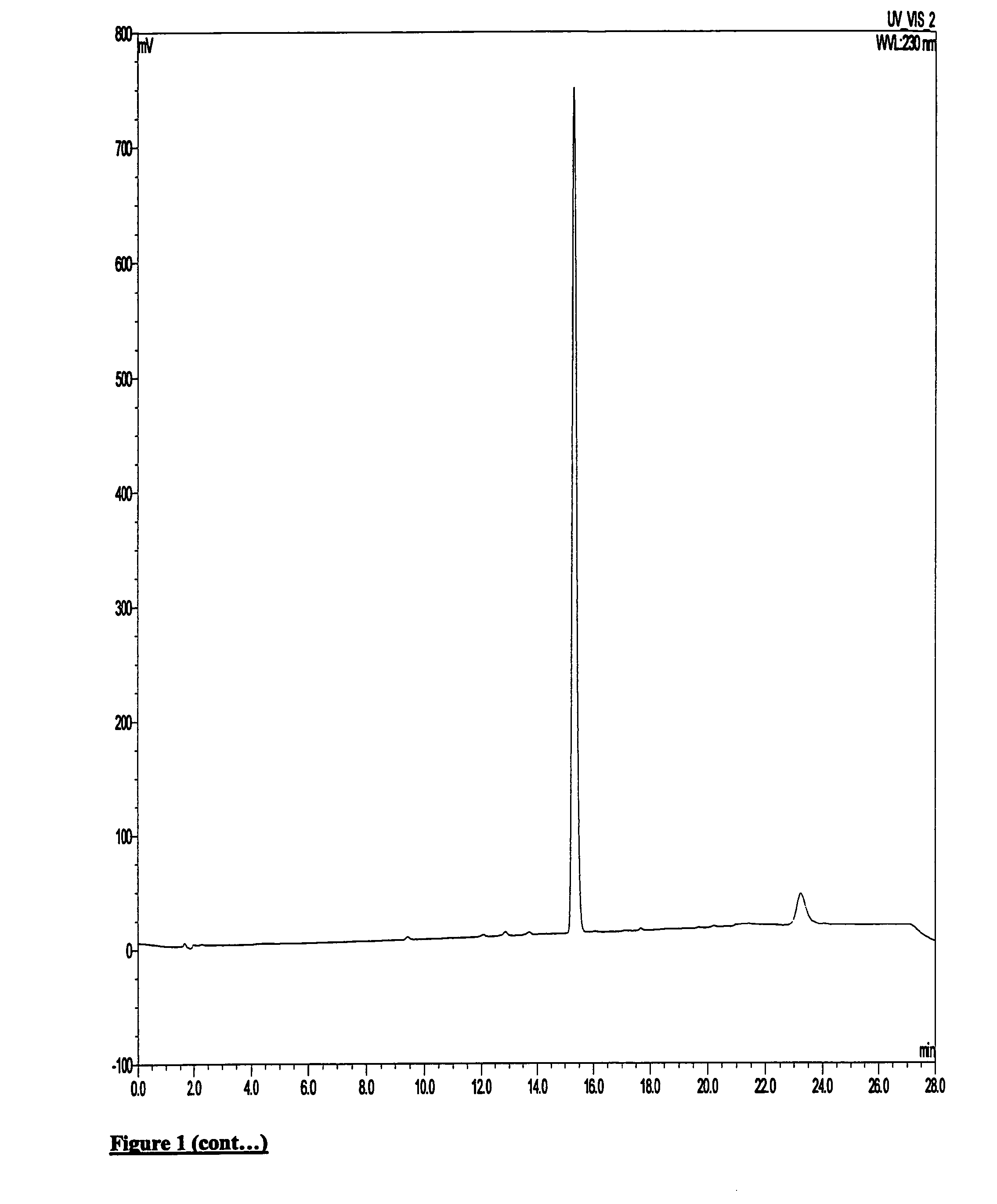
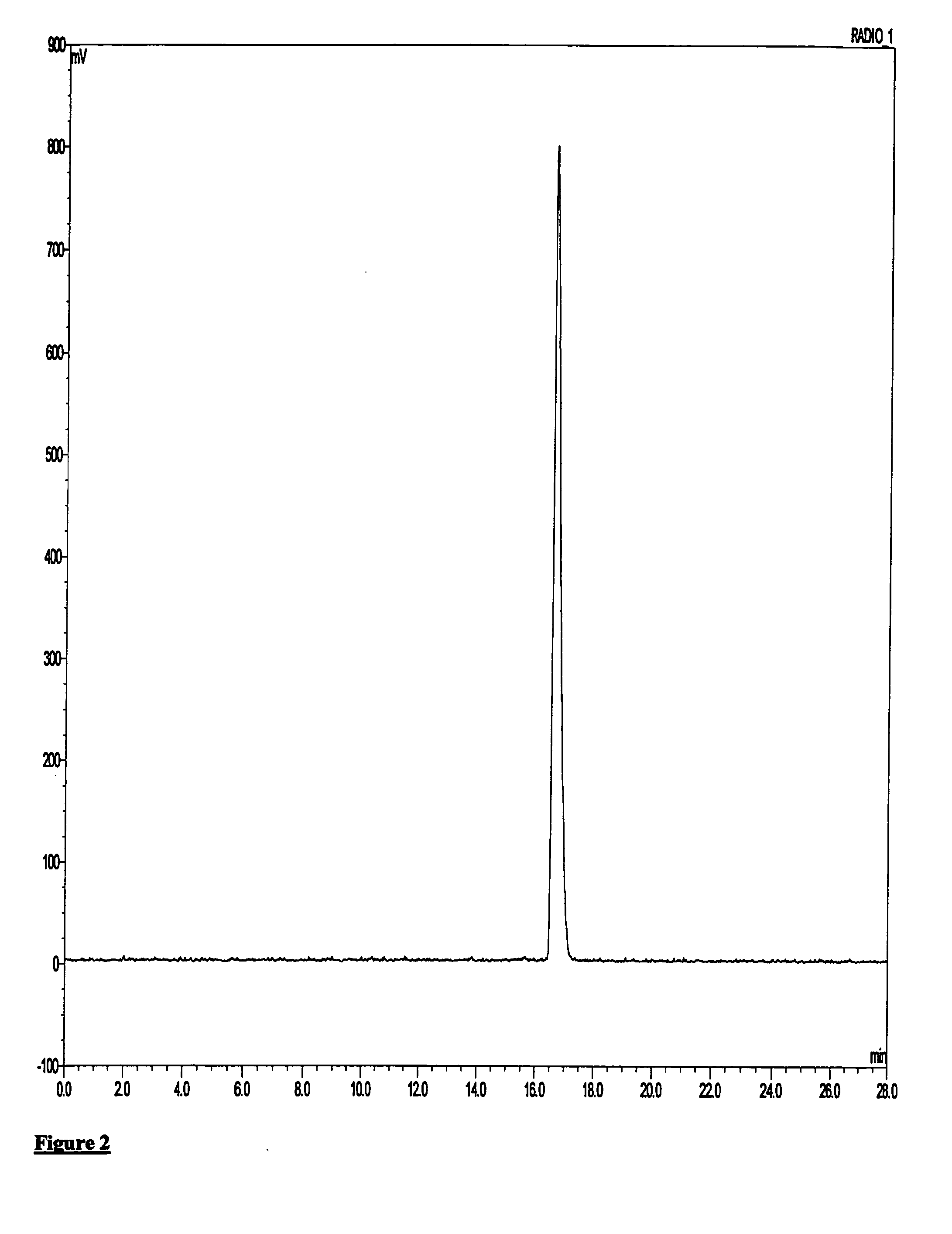
[0145][18F]Fluoride was supplied from GE Healthcare on a GE PETrace cylcotron. Kryptofix 2.2.2 (2 mg, 5 μmol), potassium bicarbonate (0.1 mol dm−3, 0.1 ml, 5 mg, 5 mol) and acetonitrile (0.5 ml) was added to [18F]F− / H20 (ca. 400 MBq, 0.1-0.3 ml) in a COC reaction vessel. The mixture was dried by heating at 100° C. under a stream of nitrogen for 20-25 mins. After drying and without cooling, methanesulphonic acid 2-(4-diethylcarbamyl-5-methoxy-1,2,3,4-tetrahydro-carbazol-9-yl)ethyl ester (0.5-1 mg, 1.2-2.4 μmol) in acetonitrile (1 ml) was added to the COC reaction vessel and heated at 100° C. for 10 mins. After cooling, the reaction mixture was removed and the COC reaction vessel rinsed with water (1.5 ml) and added to the main crude reaction.
[0146]Following this, the crude product was applied to semi-preparative HPLC: HICHROM ACE 5 C18 column (100×10 mm i.d.),...
example 2
Synthesis of 9-(2-Fluoro-ethyl)-5-methoxy-2,3,4,9-tetrahydro-1H-carbazole-4-carboxylic acid diethylamide (non-radioactive imaging agent 5)
Example 2(a)
Fluoroethyl Tosylate (12)
[0148]2-Fluoroethanol (640 mg, 10 mmol, 0.6 mL) was dissolved in pyridine (10 mL) under nitrogen. The solution was stirred at 0° C. and tosyl chloride (4.2 g, 21.8 mmol) added portionwise to the solution over a period of 30 min, keeping the temperature below 5° C. The reaction was stirred at 0° C. for 3 h. Ice was slowly added followed by water (20 mL). The reaction mixture was extracted into ethyl acetate and washed with water. Excess pyridine was removed by washing with 1 N HCl solution until the aqueous layer became acidic. Excess tosyl chloride was removed by washing with 1 M aqueous sodium carbonate. The organic layer was washed with brine, dried over magnesium sulfate and concentrated in vacuo to give 2.1 g (98%) of fluoroethyl tosylate (12) as a colourless oil. The structure was confirmed by 13C NMR (75 ...
PUM
| Property | Measurement | Unit |
|---|---|---|
| Electric charge | aaaaa | aaaaa |
| Capacitance | aaaaa | aaaaa |
| Volume | aaaaa | aaaaa |
Abstract
Description
Claims
Application Information
 - R&D
- Intellectual Property
- Life Sciences
- Materials
- Tech Scout
- Unparalleled Data Quality
- Higher Quality Content
- 60% Fewer Hallucinations
Browse by: Latest US Patents, China's latest patents, Technical Efficacy Thesaurus, Application Domain, Technology Topic, Popular Technical Reports.
© 2025 PatSnap. All rights reserved.Legal|Privacy policy|Modern Slavery Act Transparency Statement|Sitemap|About US| Contact US: help@patsnap.com


