Utilizing lipopolysaccharide in exhaled breath condensate to diagnose gram negative pneumonia

a technology of gram negative bacteria and exhaled breath, which is applied in the field of gram negative bacterial pneumonia diagnosis methods and devices, can solve the problems of sterile blood, high mortality rate, and difficulty in obtaining useful sputum samples from humans with pneumonia, and achieve the effect of promoting condensation

Inactive Publication Date: 2013-07-25
CHARLOTTE MECKLENBURG HOSPITAL AUTHORITY
View PDF9 Cites 3 Cited by
  • Summary
  • Abstract
  • Description
  • Claims
  • Application Information

AI Technical Summary

Benefits of technology

[0013]Alternative devices for collecting exhaled breath condensate are also known. These devices include those disclosed in Gaston et al., U.S. Pat. Nos. 6,033,368 and 6,419,634; Hunt et al., U.S. Pat. No. 6,585,661; Baddour, U.S. application Ser. No. 10 / 257,912; and EU Patent No. 0,759,169 B1 to Winsel et al., all of which are incorporated by reference herein in their entireties. In addition, Kline, U.S. application Ser. Nos. 10 / 742,721 and 10 / 778,477, two commonly-assigned non-provisional patent applications, disclose devices for collecting exhaled breath and are incorporated by reference herein in their entireties.
[0014]Exhaled condensate is known to contain many molecules that can serve as markers of many lung diseases, as reviewed by Kharitinov et al. in 2001 (Biomarkers 7 (1):1-32, 2002.). However, the concept of measuring lipopolysaccharide in exhaled breath condensate for the purpose of diagnosing Gram negative pneumonia or other Gram negative bacterial infections has not been disclosed previously.
[0015]The present invention comprises a method for determining whether a subject has Gram negative bacterial pneumonia based on the presence of lipopolysaccharide in exhaled breath condensate collected from the subject. The present invention further comprises the collection devices utilized to collect exhaled breath condensate from both spontaneously breathing and mechanically ventilated subjects and the devices utilized to determine whether lipopolysaccharide is present in the collected exhaled breath condensate.
[0016]Broadly defined, the present invention, according to one aspect, is a method for diagnosing and monitoring intrapulmonary Gram negative bacterial infection in an air-breathing vertebrate subject, including: collecting exhaled breath condensate from an air-breathing vertebrate subject; measuring the concentration of lipopolysaccharide in the collected exhaled breath condensate; and determining whether the subject has an intrapulmonary Gram negative bacterial infection based on the measured concentration of lipopolysaccharide in the exhaled breath condensate.
[0017]In features of this aspect, the intrapulmonary Gram negative bacterial infection is pneumonia; the intrapulmonary Gram negative bacterial infection is a bronchial infection; the method further includes selecting an antibiotic therapy in response to a positive determination that the subject has an intrapulmonary Gram negative bacterial infection; collecting exhaled breath condensate from an air-breathing vertebrate subject includes collecting exhaled breath condensate from a mammalian subject, and the method further includes monitoring the response of the mammalian subject to antibiotic therapy; the method further includes identifying the particular strain of the intrapulmonary Gram negative bacteria; identifying includes identifying the particular strain of intrapulmonary Gram negative bacteria based upon a determination of the O-saccharide portion of the lipopolysaccharide molecule; measuring the concentration of lipopolysaccharide includes measuring the concentration of lipopolysaccharide using the limulus amoebocyte lysate assay; a measured concentration of lipopolysaccharide of at least about 0.20 Endotoxin Units / mL (EU / mL) indicates the presence of a Gram negative bacterial infection; collecting exhaled breath condensate from an air-breathing vertebrate subject includes collecting the expired breath condensate from a spontaneously breathing subject; collecting exhaled breath condensate from an air-breathing vertebrate subject includes collecting the expired breath condensate from a mechanically ventilated subject; and collecting exhaled breath condensate from an air-breathing vertebrate subject includes utilizing an exhaled breath condensate collection device comprising a chamber having inner walls that may be cooled to a temperature of about 32 degrees Fahrenheit and below to promote condensation.
[0018]The present invention, according to another aspect, is a method for diagnosing and monitoring intrapulmonary Gram negative bacterial infection in an air-breathing vertebrate subject, including: collecting exhaled breath condensate from an air-breathing vertebrate subject; providing a reaction chamber, wherein said reaction chamber has a reaction reagent disposed therein; delivering at least a portion of the collected exhaled breath condensate to the reaction chamber; and determining whether the subject has an intrapulmonary Gram negative bacterial infection based on a physical change that occurs when the at least a portion of the collected exhaled breath condensate is delivered to the reaction chamber, wherein said physical change is caused by the presence of lipopolysaccharide in the exhaled breath condensate.

Problems solved by technology

In particular, clinicians are motivated to identify the presence of Gram negative bacterial infection because Gram negative lung infections are aggressive and are associated with higher rates of complications and death.
However, useful sputum samples are notoriously difficult to obtain from humans with pneumonia.
Unfortunately, more often than not, the blood is sterile in a patient with Gram negative pneumonia.
However, endotoxin concentrations have been found to be an inaccurate predictor of either the cause or severity of the more general sepsis syndrome.
No study has examined whether circulating endotoxin concentrations can predict a gram negative source of pneumonia.
Investigators using this method found that high concentrations of lipopolysaccharide are associated with concomitant growth of gram negative bacteria in cultures of the bronchalveolar fluid.
Both methods have the drawbacks that special endoscopic equipment and subspecialty expertise are required and that they are relatively invasive and uncomfortable procedures.
Moreover, known culture methods require at least 24 hours to obtain results.
However, the concept of measuring lipopolysaccharide in exhaled breath condensate for the purpose of diagnosing Gram negative pneumonia or other Gram negative bacterial infections has not been disclosed previously.

Method used


Image

  • Utilizing lipopolysaccharide in exhaled breath condensate to diagnose gram negative pneumonia
  • Utilizing lipopolysaccharide in exhaled breath condensate to diagnose gram negative pneumonia
  • Utilizing lipopolysaccharide in exhaled breath condensate to diagnose gram negative pneumonia

Examples


example 1

[0121]LPS was detected in exhaled breath condensate samples from subjects that were awake, cooperative and able to breathe spontaneously in order to diagnose whether such subjects had Gram negative bacterial pneumonia according to the following procedure.

[0122]The subjects for the procedure were selected according to the following procedure. Subjects (N=8 per group) were recruited based upon three criteria: 1) diagnosis of pneumonia, 2) healthy patients who actively smoked more than 10 cigarettes per day and 3) healthy nonsmokers. To obtain subjects diagnosed with pneumonia, subjects diagnosed on standard clinical grounds, including cough productive of colored sputum, measured fever >101° F., a leukocytosis, evidenced by a peripheral total white blood count of >12,000 cells per cubic microliter, and the presence of an infiltrate on chest radiograph were selected. Exclusion criteria for subjects included any use of antimicrobial medications, acute illness or anatomical abnormality th...

example 2

[0130]LPS was detected in exhaled breath condensate samples from subjects that were breathing with the assistance of a ventilator in order to diagnose whether such subjects had Gram negative bacterial pneumonia according to the following procedure.

[0131]Six subjects participated in the study. Four of the ventilated subjects presented clinical evidence of pneumonia and were being treated with antibiotic therapy, and two of the ventilated subjects presented no clinical evidence of pneumonia and were used as controls.

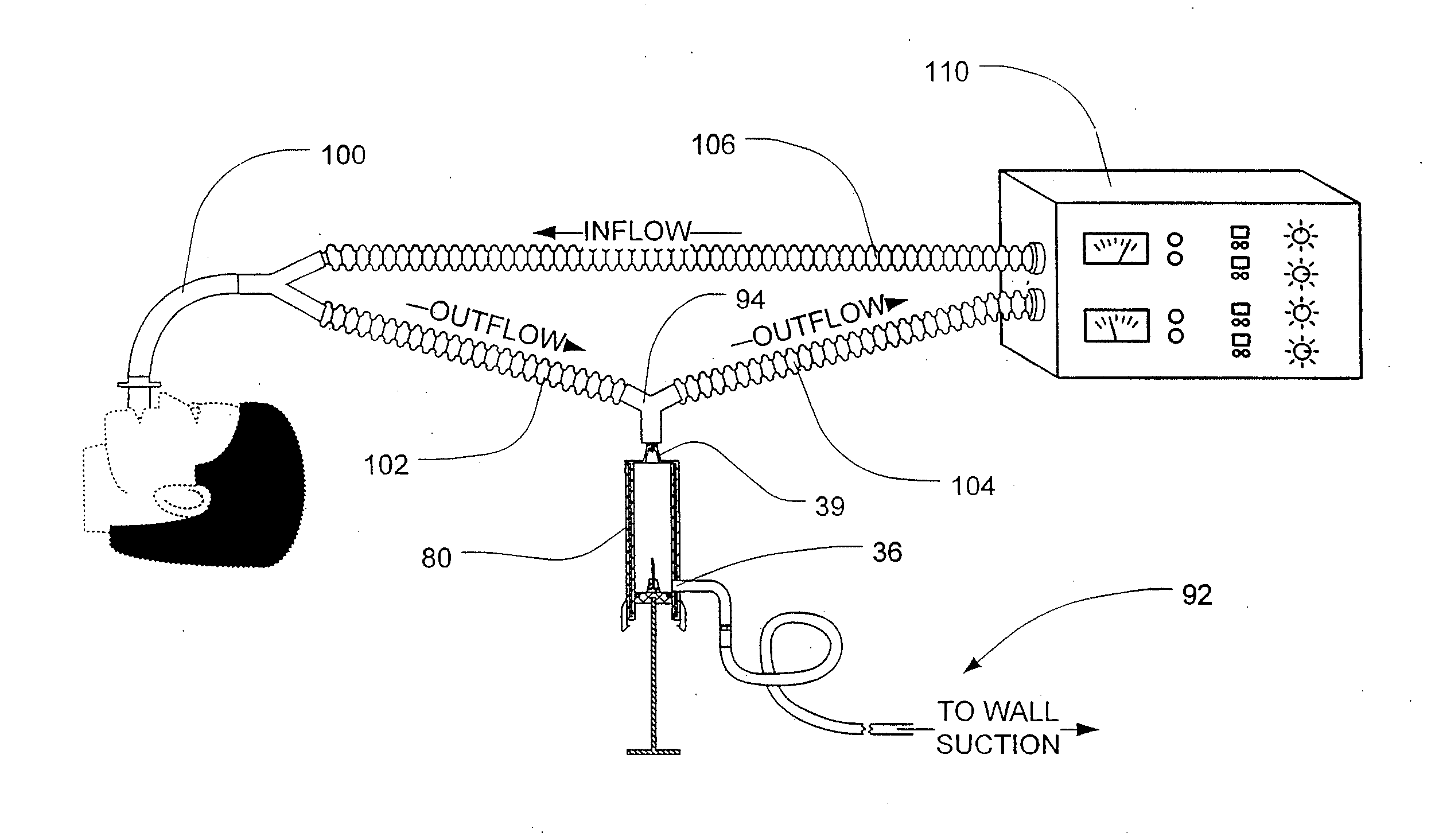
[0132]Breath condensate samples were obtained according to the following procedure. The analyte was obtained from the exhaled breath condensate that accumulated in outflow tubing attached to the endotracheal tube of the ventilation system. Analyte appeared clear and non-turbid upon visual inspection.

[0133]The presence of endotoxin was detected according to the following procedure. An assay was performed on undiluted and diluted condensate using a chromogenic limulus assay ...

example 3

[0135]LPS was detected in exhaled breath condensate according to the following procedure. A commercially available 1-liter volume glass flask was prepared for collecting breath condensate samples according to the following procedure. The glass was heated to 400° C. for 3 hours to render its surfaces LPS-free. A sterilized, flexible polyvinyl tube, 11 mm in internal diameter, was arranged in fluid communication to a side-arm of the flask such that when a patient breathed into the tube, the patient's breath passed through the glass flask and out through an exit port. The flask was partially submerged in a dry ice and ethanol slurry mixture as a coolant to facilitate capture of exhaled breath condensate in the flask. The tube and flask were arranged in a fashion to keep the condensing flask above the level of the patient to prevent any capture of aerosolized saliva.

[0136]Exhaled breath was collected according to the following procedure. Eight subjects of varying health status breathed ...


PUM

PropertyMeasurementUnit
temperatureaaaaaaaaaa

Abstract

A device for collecting exhaled breath condensate from a subject. The device comprises a plunger assembly and a stopper. The stopper is connected to the plunger disk of the plunger assembly by a plurality of support pins and is configured for sealing engagement. The device is utilized to collect exhaled breath condensate from both spontaneously breathing and mechanically ventilated subjects and the devices utilized to determine whether lipopolysaccharide is present in the collected exhaled breath condensate.

Description

CROSS-REFERENCES TO RELATED APPLICATION[0001]This application is a continuation-in-part of and thus is entitled to the benefit of, and claims priority to U.S. Divisional patent application Ser. No. 12 / 157,133 filed Jun. 6, 2008, which claims the benefit of U.S. patent application Ser. No. 11 / 135,265 filed May 23, 2005, now U.S. Pat. No. 7,828,741, which claims the benefit of provisional U.S. Patent Application Ser. No. 60 / 577,641, filed Jun. 7, 2004. U.S. patent application Ser. No. 11 / 135,265 is also a continuation-in-part of U.S. patent application Ser. No. 10 / 742,721 filed Dec. 19, 2003, which claims the benefit of provisional U.S. Patent Application Ser. No. 60 / 434,916 filed Dec. 20, 2002 and provisional U.S. Patent Application Ser. No. 60 / 447,581 filed Feb. 14, 2003. In addition, U.S. patent application Ser. No. 10 / 742,721 is a continuation-in-part of U.S. patent application Ser. No. 10 / 778,477 filed Feb. 13, 2004, now U.S. Pat. No. 7,547,285, which claims the benefit of provis...

Claims


Application Information

Patent Timeline
Patent Type & Authority Applications(United States)
IPC IPC(8): A61B5/08
CPCA61B5/082A61B5/097A61B5/412G01N2400/50C12Q1/14G01N33/579G01N33/92C12Q1/04
Inventor KLINE, JEFFREY A.HERNANDEZ, JACKELINEWATTS, JR., JOHN ALBERT
Owner CHARLOTTE MECKLENBURG HOSPITAL AUTHORITY
Why Eureka
Try Eureka