Method for treating and diagnosing cancer by using cell-derived microvesicles

a cell-derived micro-vesicle and cancer technology, applied in the direction of bacteria material medical ingredients, immunological disorders, antibody medical ingredients, etc., can solve the problems of serious limitations in the capacity of these production methods, the risk of bacterial proliferation in normal tissues or organs surrounding the cancer mass, and the use of bacteria in cancer therapy. , to achieve the effect of enhancing the defense mechanism, reducing the side effects of conventional drugs, and reducing pain and inconvenience of patients in the course of treatmen

Active Publication Date: 2013-08-01
ROSETTA EXOSOME CO LTD
View PDF3 Cites 86 Cited by
  • Summary
  • Abstract
  • Description
  • Claims
  • Application Information

AI Technical Summary

Benefits of technology

[0041]The bacterial cell-derived microvesicles in accordance with the present invention can be applied to the treatment or diagnosis of cancer, with a decrease in the side effects of conventional drugs, whereby the agony and inconvenience of the patient in the course of treatment can be reduced.
[0042]Conventional anticancer agents act non-specifically on proliferating cells, causing side effects upon exertion of their cytotoxicity on normal cells. In addition, conventional administration methods allow anticancer agents to be delivered to not only cancer cells or tissues, but also non-cancerous organs, thus causing significant side effects. With an increase in the senescent population, cancer in elderly persons is rising as a social problem. Abnormality of immune functions which lose the capacity of controlling the growth of cancer mainly accounts for the onset and progression of cancer, but there are only rare cases in which cancer is treated by enhancing the defense mechanism of effectively suppressing the growth of cancer.
[0043]The bacterial cell-derived microvesicles of the present invention can be used in effectively suppressing the grown of cancer. Once they are loaded with a targeting molecule, microvesicles derived from bacterial cells, whether or not transformed, can be directed towards cancer vessels, cells or tissues, thus minimizing the adverse effects resulting from the erroneous targeting of microvesicles. In addition, when a drug such as an anticancer agent, an anti-inflammatory agent, etc., is loaded thereto or

Problems solved by technology

In spite of these efforts, the use of bacteria in cancer therapy always has the risk of bacterial proliferation in norm

Method used


Image

  • Method for treating and diagnosing cancer by using cell-derived microvesicles
  • Method for treating and diagnosing cancer by using cell-derived microvesicles
  • Method for treating and diagnosing cancer by using cell-derived microvesicles

Examples


example 1

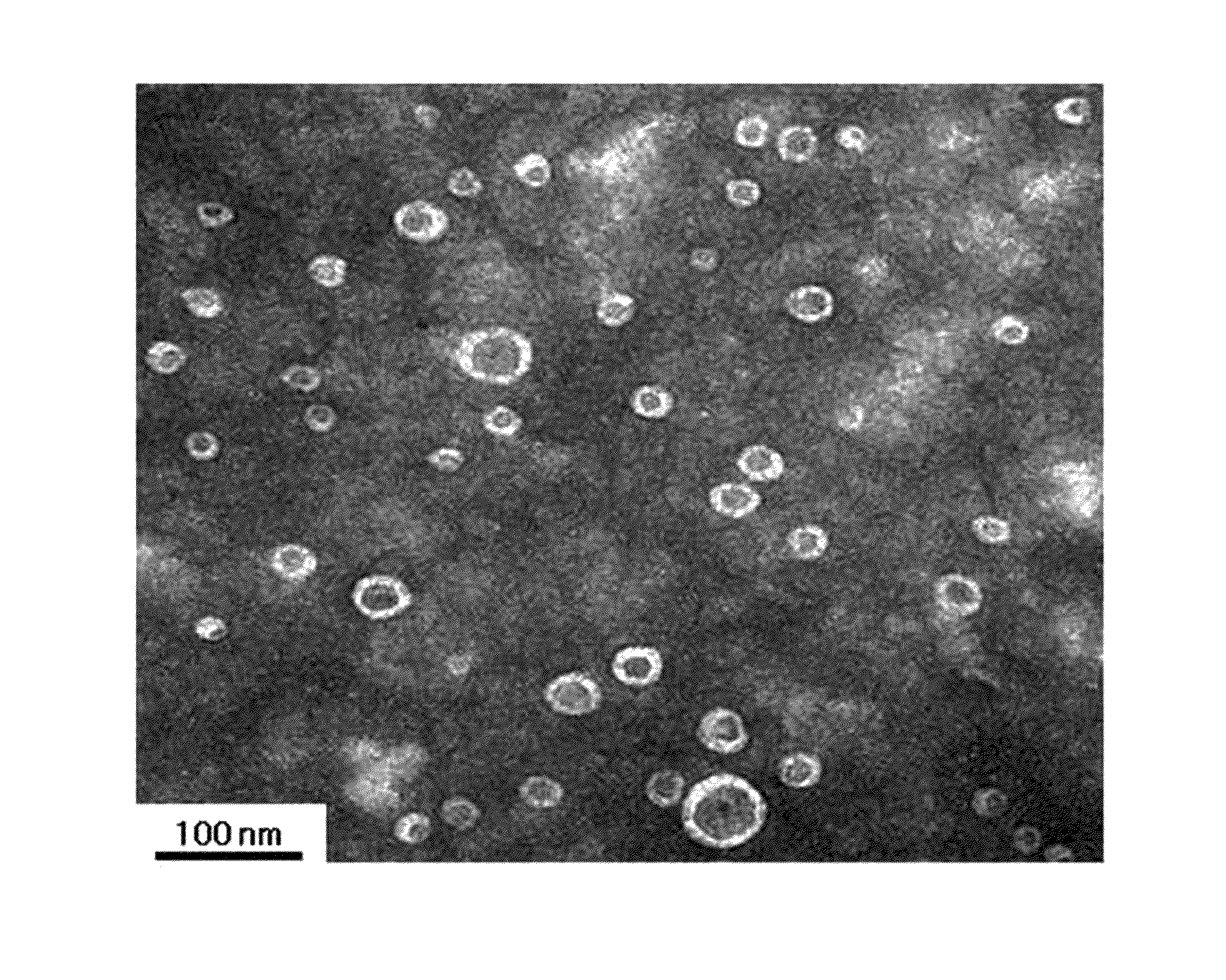
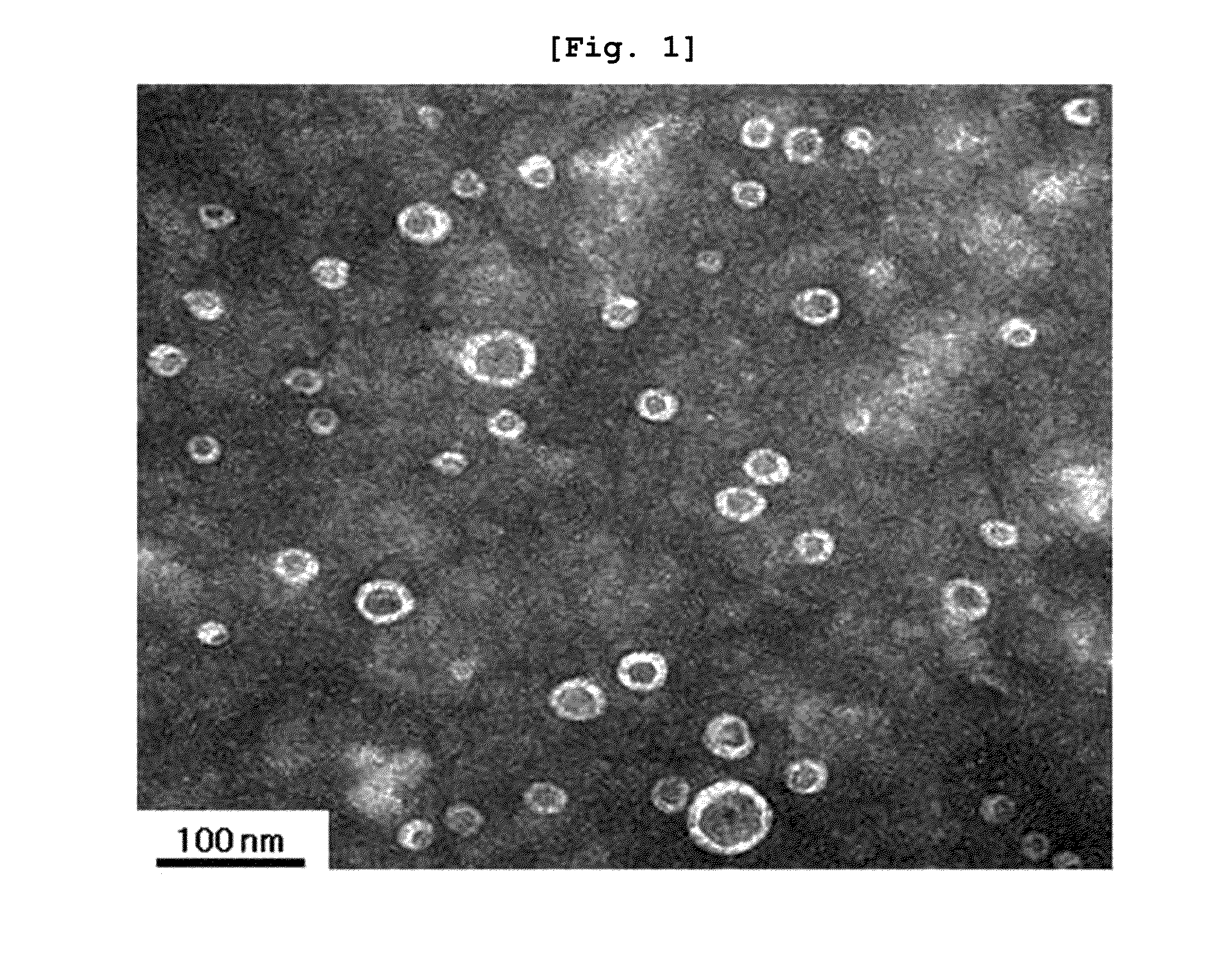
Construction of Artificial Microvesicles Derived from Gram-Negative Bacteria by Extrusion and Properties Thereof

[0208]Artificial microvesicles were constructed from Gram-negative bacteria by extrusion.

[0209]In this experiment, the Gram-negative bacterium E. coli was employed. E. coli was cultured to an optical density of 1.0 (at 600 nm) in 50 mL of LB broth. The bacteria cells were collected as a pellet after centrifugation at 3,500×g for 10 min, and the cell pellet was resuspended in PBS (phosphate buffered saline).

[0210]This cell suspension was passed three times through each of membrane filters with a pore size of 10 μm, 5 μm, and 1 μm, in that order. In a 5 mL ultracentrifuge tube, 1 mL of 50% OptiPrep, 1 mL of 5% OptiPrep, and 3 mL of the cell suspension effluent from the membrane filters were sequentially placed. Ultracentrifugation at 100,000×g for 3 hrs formed a layer of microvesicles between 50% OptiPrep and 5% OptiPrep.

[0211]The artificial microvesicles constructed from th...

example 2

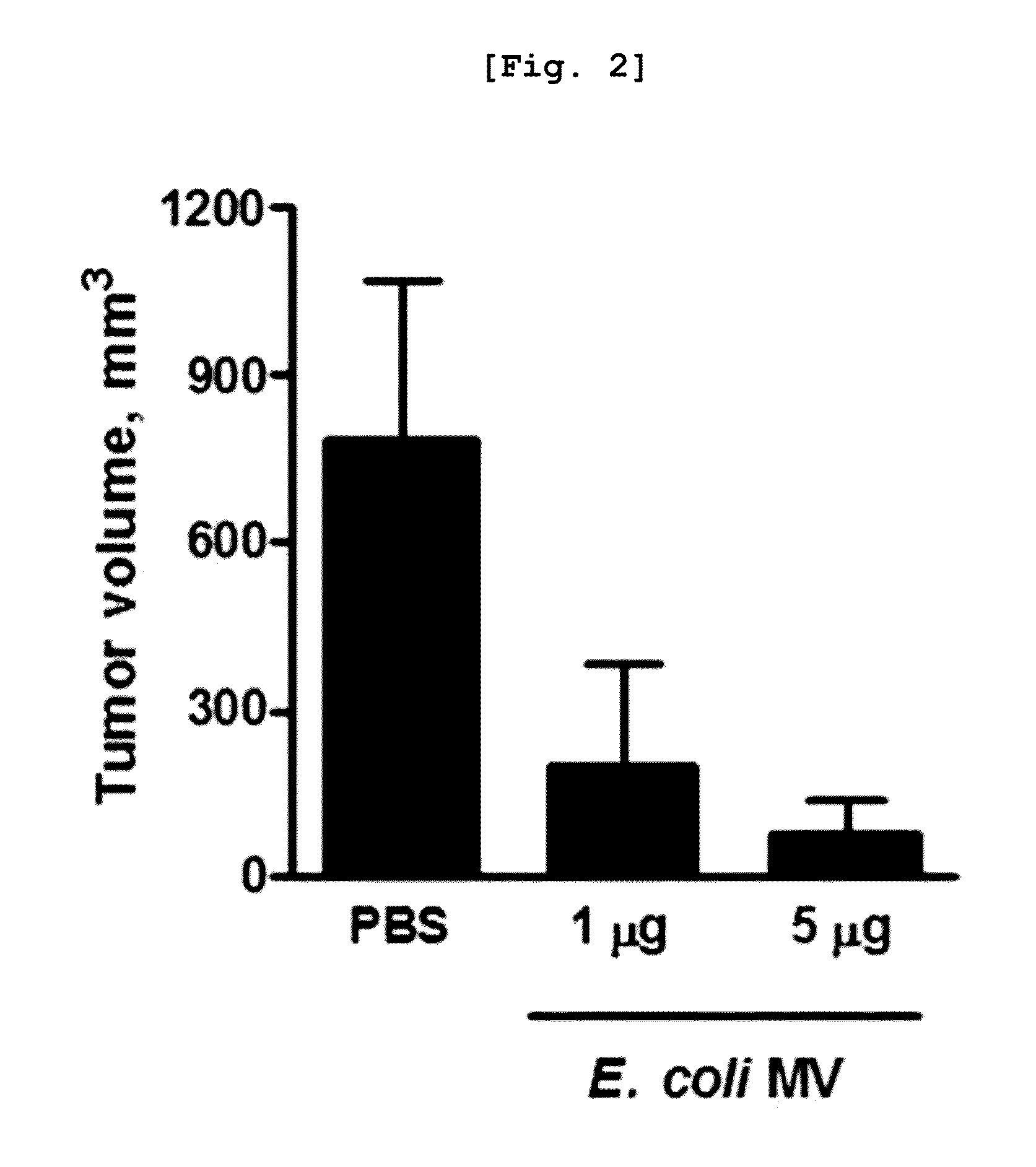
In Vivo Anticancer Activity of Gram-Negative Bacterial Cell-Derived Shedding Microvesicles

[0212]For use in this experiment, microvesicles that were spontaneously shed from Gram-negative bacteria were isolated. The Gram-negative bacteria E. coli, Pseudonomas aeruginosa, and Salmonella enteritidis were used as sources of microvesicles. Bacteria were inoculated into 100 mL of LB in an Erlenmeyer flask and cultured at 37° C. for 6 hrs. Of the culture, 8 mL was transferred into 600 mL of LB broth in a 2 L Erlenmeyer flask and cultured at 37° C. for 5 hrs to an optical density of 1.5 (at 600 nm). The resulting culture was divided into 500 mL high speed centrifuge tubes before centrifugation at 10,000×g and 4° C. for 20 min. The supernatant was forced to pass once through a membrane filter with a pore size of 0.45 μm, and then concentrated 25-fold using a Quixstand system equipped with a membrane having a molecular weight cut-off of 100 kDa. The concentrate was passed once through a membra...

example 3

In Vivo Anticancer Activity of Gram-Positive Bacterial Cell-Derived Shedding Microvesicles

[0215]For use in this experiment, microvesicles that were spontaneously shed from Gram-positive bacteria were isolated. The Gram-positive bacteria Staphylococcus aureus and Lactobacillus acidophilus were used as sources of microvesicles. Bacteria were inoculated into 100 mL of a nutrient broth in an Erlenmeyer flask and cultured at 37° C. for 6 hrs. Of the culture, 8 mL was transferred into 600 mL of a nutrient broth in a 2 L Erlenmeyer flask and cultured at 37° C. for 5 hrs to an optical density of 1.5 (at 600 nm). The resulting culture was divided into 500 mL high speed centrifuge tubes before centrifugation at 10,000×g and 4° C. for 20 min. The supernatant was forced to pass once through a membrane filter with a pore size of 0.45 μm, and then concentrated 25-fold using a Quixstand system equipped with a membrane having a molecular weight cut-off of 100 kDa. The concentrate was passed once th...


PUM

PropertyMeasurementUnit
Densityaaaaaaaaaa
Therapeuticaaaaaaaaaa
Toxicityaaaaaaaaaa

Abstract

Disclosed is a method for the treatment and/or diagnosis of cancer, using bacterial cell-derived microvesicles, which is highly effective in cancer therapy with a significant reduction in side effects. Also, a method is provided for delivering of a drug therapeutic or diagnostic for a disease, using bacterial cell-derived microvesicles loaded with the drug, thereby treating and diagnosing the disease effectively and specifically.

Description

TECHNICAL FIELD[0001]The present invention relates to a method for the treatment and / or diagnosis of cancer, using bacterial cell-derived microvesicles. Particularly, the present invention relates to a method for the therapy of cancer, using components of bacterial cell-derived microvesicles.BACKGROUND ART[0002]In 1813, Vautier reported the apparent cure of cancer in patients who concurrently suffered from gas gangrene caused by the infection of the anaerobic bacterium Clostridium perfringens. Since then, extensive studies have been undertaken on the deliberate injection of various bacteria including Salmonella typhimurium in addition to Clostridium, with the concept of bacteria-mediated cancer therapy. As a cancer grows over a certain size (approximately 1 mm3), it undergoes an undersupply of oxygen from surrounding vessels, with the formation of hypoxia within the cancer tissue. The cancer tissue in a hypoxia state provides an environment in which anaerobic bacteria are readily li...

Claims


Application Information

Patent Timeline
IPC IPC(8): A61K35/74A61K49/00A61K38/12A61K38/16A61K31/616A61K31/704
CPCA61K9/5068A61K47/46A61K49/0017A61K49/0045A61K49/0097A61K47/48776A61K49/005A61K31/616A61K31/704A61K35/74A61K38/12A61K38/164A61K2039/585A61K47/6901A61P35/00A61P37/04A61K39/085
Inventor GHO, YONG SONGKIM, OH YOUNJANG, SU CHULYOON, CHANG MINKIM, YOON KEUN
Owner ROSETTA EXOSOME CO LTD
Why Eureka
Try Eureka