

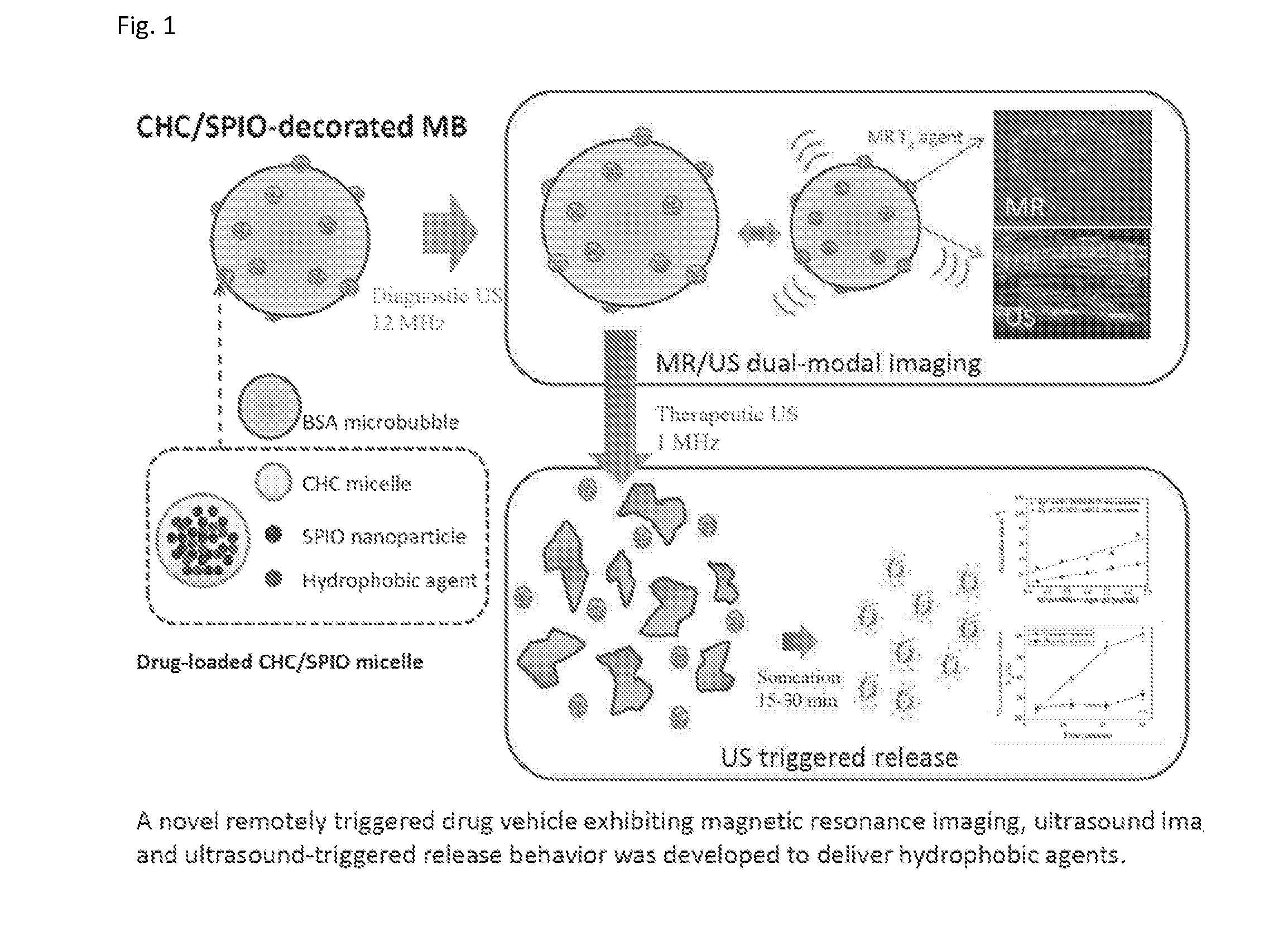
Dual-modal imaging-guided drug vehicle with ultrasound-triggered release function
a drug vehicle and ultrasound technology, applied in the field of dual-modal imagingguided drug vehicles with ultrasound-triggered release functions, can solve the problems of difficult for microbubbles to load enough drugs, the timing and location of drug delivery methods are difficult to control, and the image resolution of ultrasound is not high enough for molecular imaging, so as to avoid the leakage of drugs
- Summary
- Abstract
- Description
- Claims
- Application Information
AI Technical Summary
Benefits of technology
Problems solved by technology
Method used
Image

Examples
Embodiment Construction
[0018]The characteristics and advantages of the present invention will furthermore be illustrated and explained in the following preferred embodiments. The preferred embodiments are for better illustration and not for limiting the scope of the present invention.
[0019]The description and figures below are to disclose the preferred embodiments according to the present invention. Various modifications may be made to this invention for different usages and situations without departing from the scope covered by the appended claims. People familiar with the common senses in the concerned field can make modification of forms, structures or materials based on the present invention.
Preferred Embodiments
[0020]In one of the preferred embodiments, the micelles on the surface of the drug vehicle contain superparamagnetic Fe3O4 (SPIO) nano particles to provide MRI T2 weighted image (as shown in FIG. 7). The structure of the drug vehicle is ultrasonically triggered to destruction and the distance ...
PUM
| Property | Measurement | Unit |
|---|---|---|
| particle diameter | aaaaa | aaaaa |
| particle diameter | aaaaa | aaaaa |
| particle diameter | aaaaa | aaaaa |
Abstract
Description
Claims
Application Information