Methods for screening compounds for capability to inhibit premalignant squamous epithelial cell progression to a malignant state
- Summary
- Abstract
- Description
- Claims
- Application Information
AI Technical Summary
Benefits of technology
Problems solved by technology
Method used
Image
Examples
example 1
General Experimental Procedures
[0061]Cell lines and treatments. MSK-Leuk1 cells were derived from a dysplastic leukoplakia lesion located adjacent to a SCC of the tongue. MSK-Leuk1 cells were cultured in KGM medium (Lonza, Walkersville, Md.). MSK-Leuk1 cells (passage 33) were determined to be identical to the early passage MSK-Leuk1 cells (Identity Mapping Kit, Coriell Institute for Medical Research, Camden, N.J.). All HNSCC cell lines were derived from patients with SCC of the tongue. SCC9 (male) and SCC15 (male) cells were cultured in S-MEM medium, supplemented with 2 mM L-glutamine, 100 units / ml penicillin, 100 μg / ml streptomycin and 10% FBS. UPCI:SCC56 (male), UPCI:SCC103 (female) and UPCI:SCC122 (male) cells were cultured in MEM medium, supplemented with 2 mM L-glutamine, 100 μM non-essential amino acids, 50 μg / ml gentamycin (Gibco) and 10% FBS.
[0062]For all the experiments that involved estradiol (E2) exposure, MSK-Leuk1 cells were cultured in phenol red-free and serum-free De...
example 2
Experimental Results
[0074]A. Estrogen metabolism genes and ERβ are expressed in cells derived from premalignant and malignant head and neck lesions.
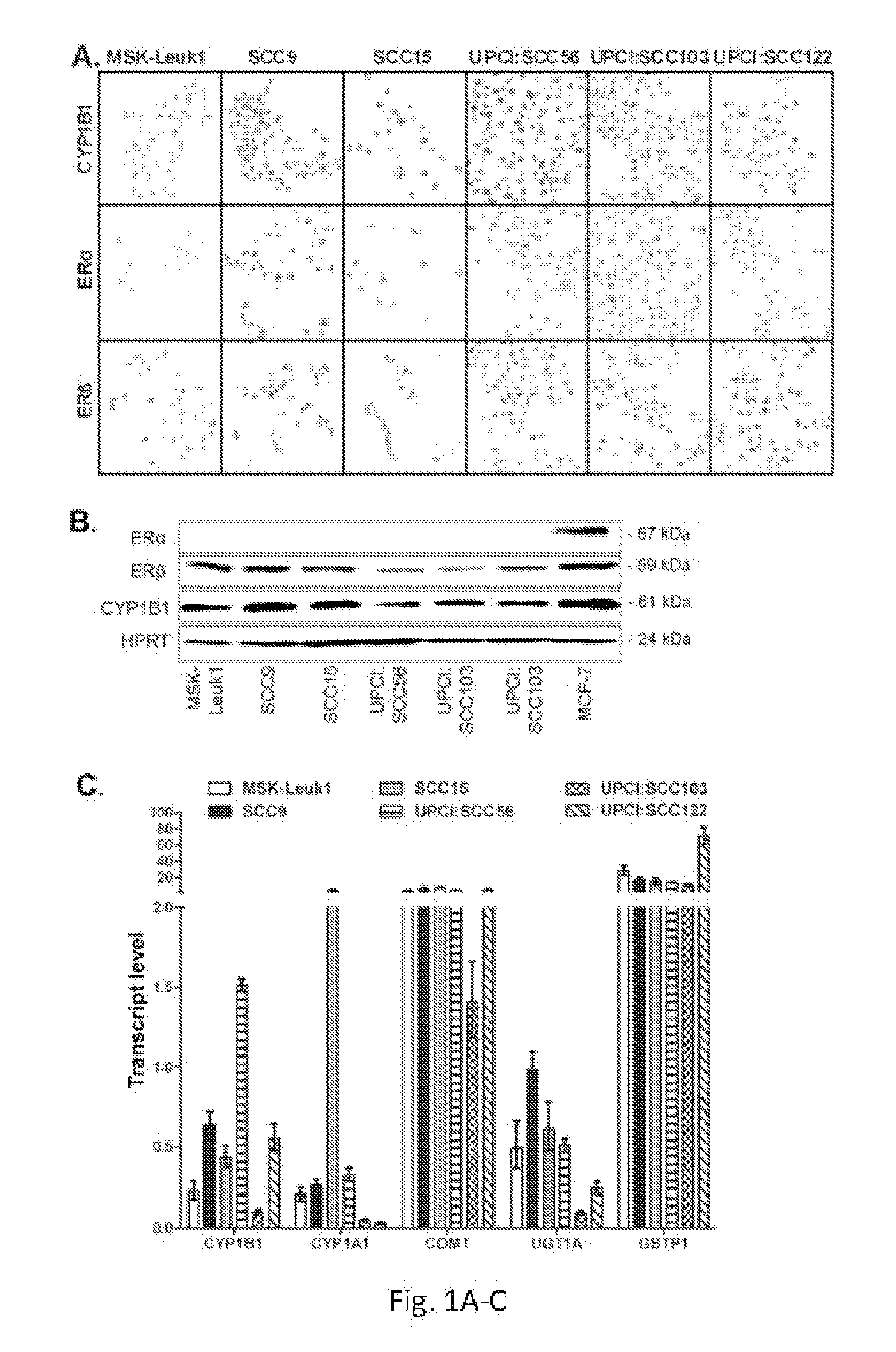
[0075]Immunohistochemical staining of sections from formalin-fixed, paraffin embedded pellets of MSK-Leuk1 cells and five HNSCC cell lines was performed using antibodies specific for ERα, ERβ and CYP1B1. ERβ and CYP1B1 were detected in MSK-Leuk1 cells and all HNSCC cell lines at comparable levels, with staining for both proteins localized to the nucleus. ERα was not detected in any of the cell lines evaluated (FIG. 1A). Consistent with immunohistochemical staining data, ERβ and CYP1B1 were detected by Western blot in all head and neck lines. While ERα was detected in MCF-7 cells (positive control), it was not detectable in any of the head and neck cell lines (FIG. 1B).
[0076]The finding that ERβ and CYP1B1 are present in cultured head and neck cells was extended by examining the expression profile of CYP19 (aromatase), which encodes the r...
example 3
Summary
[0088]The results demonstrate that a panel of estrogen metabolism genes is expressed in cultured human head and neck cells. Without intending to be limited to any particular theory or mechanism of action, it is believed that detection of transcripts for these genes in both premalignant lesions and HNSCCs suggests that these enzymes contribute to cellular metabolism throughout tumorigenesis. It is believed that to date, the contribution of the estrogen pathway to the premalignant stage of head and neck tumorigenesis has not been evaluated.
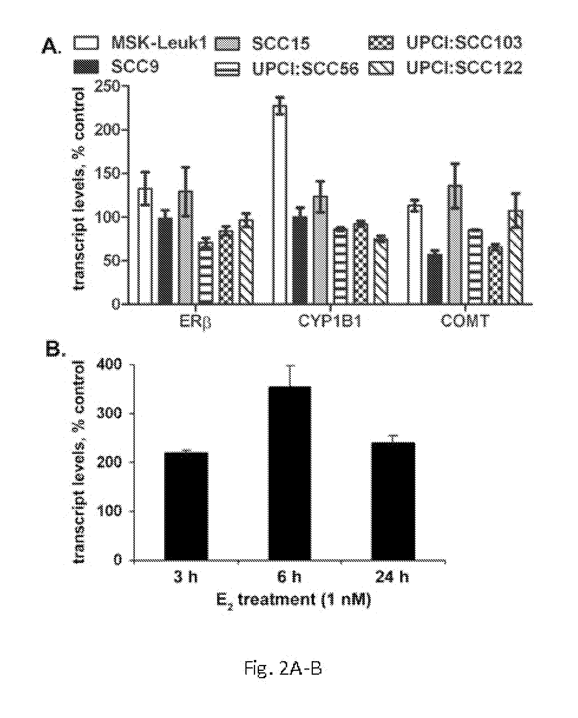
[0089]The results show that CYP1B1 is upregulated in MSK-Leuk1 but not in HNSCC cells following E2 exposure. The mechanistic basis for this differential upregulation of CYP1B1 remains unclear. However, it has been shown for lung cancer that the timing of hormone exposure relative to a diagnosis of lung cancer may make a difference with respect to whether the hormonal effect is protective or adverse (Siegfried J M (2010) Cancer Prev. Res. 3:69...
PUM
 Abstract
Description
Claims
Application Information
 - R&D
- Intellectual Property
- Life Sciences
- Materials
- Tech Scout
- Unparalleled Data Quality
- Higher Quality Content
- 60% Fewer Hallucinations
Browse by: Latest US Patents, China's latest patents, Technical Efficacy Thesaurus, Application Domain, Technology Topic, Popular Technical Reports.
© 2025 PatSnap. All rights reserved.Legal|Privacy policy|Modern Slavery Act Transparency Statement|Sitemap|About US| Contact US: help@patsnap.com


