
Revision Systems, Tools and Methods for Revising Joint Arthroplasty Implants

a joint arthroplasty and joint technology, applied in the field of orthopaedic methods, systems and prosthetic devices, can solve the problems of reducing the chance for a surgeon to correct the failed implant, reducing the chance for surgeons to use high-resource patient and/or implant related information in the planning and execution of implant revision procedures, and reducing the chance of surgery

Active Publication Date: 2014-07-31
CONFORMIS
View PDF1 Cites 107 Cited by
  • Summary
  • Abstract
  • Description
  • Claims
  • Application Information

AI Technical Summary

Benefits of technology

This patent describes devices and methods for replacing a portion of a patient's joint (e.g., cartilage and bone) with a non-pliable, non-liquid implant material that achieves a near anatomic fit with the surrounding structures and tissues of the joint. The devices and methods also include preparing the implantation site with bone cuts and other modifications such as burring or reaming of the articular surface or underlying bone. Overall, this patent provides tools that increase the accuracy of positioning and cutting in joint replacement surgery.

Problems solved by technology

Current surgical revision procedures typically fail to utilize many sources of highly relevant patient and / or implant related information in planning and executing implant revision procedures.
Moreover, the information available to a surgeon (and / or information that is presented to the surgeon) is generally not properly assessed for accuracy and / or cross-referenced / compared to other available relevant information to ensure accurate reflection of the condition of the patient's anatomy and / or the failed implant.
The failure to assess multiple sources of information accurately, the failure to cross-reference and / or evaluate such information to create a more comprehensive picture of the failed implant and the patient's disease state, and the failure to provide a treating surgeon with an accurate pre-operative evaluation of the failed implant, the patient's anatomy, the disease state and / or the information regarding the potential surgical site significantly reduces the opportunity for a surgeon to correct the failed implant in a “least-invasive” and / or “most effective” manner.
Moreover, the failure to have such information available during the planning and implant design phase may result in designing or choosing an incorrect / unsuitable implant and / or an implant that requires removal of healthy bone stock which could have been preserved for future use if the patient's anatomy were appropriately assessed.
Because much of the supporting anatomical structure may have been removed and / or modified (during the original surgery) to accommodate the original failed implant, and because addition anatomical degradation, anatomical structure removal and / or remodeling may have occurred prior to the revision surgery, a significant amount of time and effort, and a significant number of surgical tools, are utilized by the surgeon to locate desired anatomical alignment and / or positioning relative to any “virgin” anatomical reference points (i.e., intramedullary canal reference points, etc.).

Method used


Image

  • Revision Systems, Tools and Methods for Revising Joint Arthroplasty Implants
  • Revision Systems, Tools and Methods for Revising Joint Arthroplasty Implants
  • Revision Systems, Tools and Methods for Revising Joint Arthroplasty Implants

Examples


example 1

Design and Construction of a Three-Dimensional Articular Repair System

[0607]Areas of cartilage are imaged as described herein to detect areas of cartilage loss and / or diseased cartilage. The margins and shape of the cartilage and subchondral bone adjacent to the diseased areas are determined. The thickness of the cartilage is determined. The size of the articular repair system is determined based on the above measurements. In particular, the repair system is either selected (based on best fit) from a catalogue of existing, pre-made implants with a range of different sizes and curvatures or custom-designed using CAD / CAM technology. The library of existing shapes is typically on the order of about 30 sizes.

[0608]The implant is a chromium cobalt implant. The articular surface is polished and the external dimensions slightly greater than the area of diseased cartilage. The shape is adapted to achieve perfect or near perfect joint congruity utilizing shape information of surrounding cart...

example 2

Minimally Invasive, Arthroscopically Assisted Surgical Technique

[0609]The articular repair systems are inserted using arthroscopic assistance. The device does not require the 15 to 30 cm incision utilized in unicompartmental and total knee arthroplasties. The procedure is performed under regional anesthesia, typically epidural anesthesia. The surgeon can apply a tourniquet on the upper thigh of the patient to restrict the blood flow to the knee during the procedure. The leg is prepped and draped in sterile technique. A stylette is used to create two small 2 mm ports at the anteromedial and the anterolateral aspect of the joint using classical arthroscopic technique. The arthroscope is inserted via the lateral port. The arthroscopic instruments are inserted via the medial port. The cartilage defect is visualized using the arthroscope. A cartilage defect locator device is placed inside the diseased cartilage. The probe has a U-shape, with the first arm touching the center of the area ...

example 3

“Failed Implant” Assisted Knee Technique

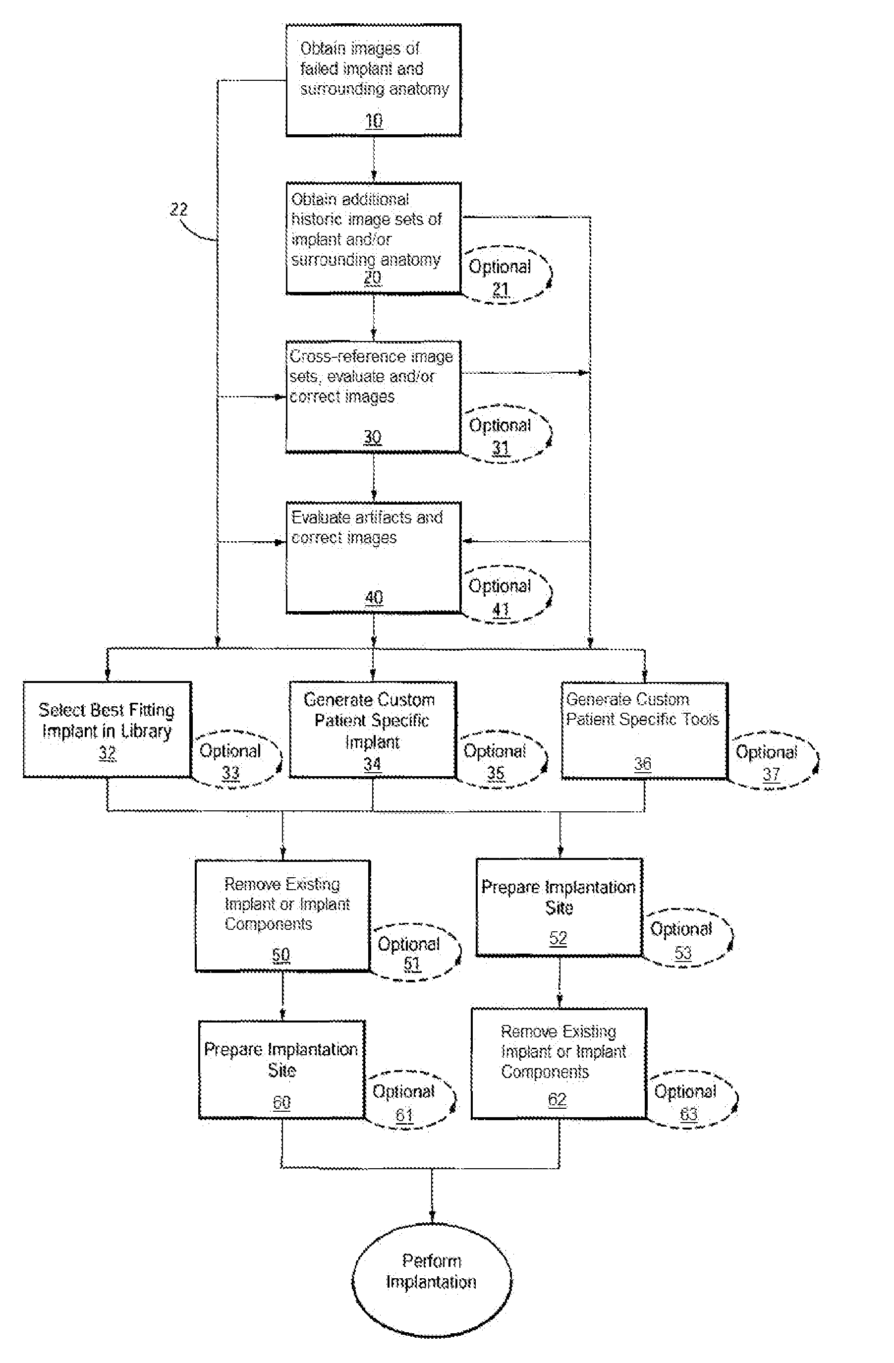
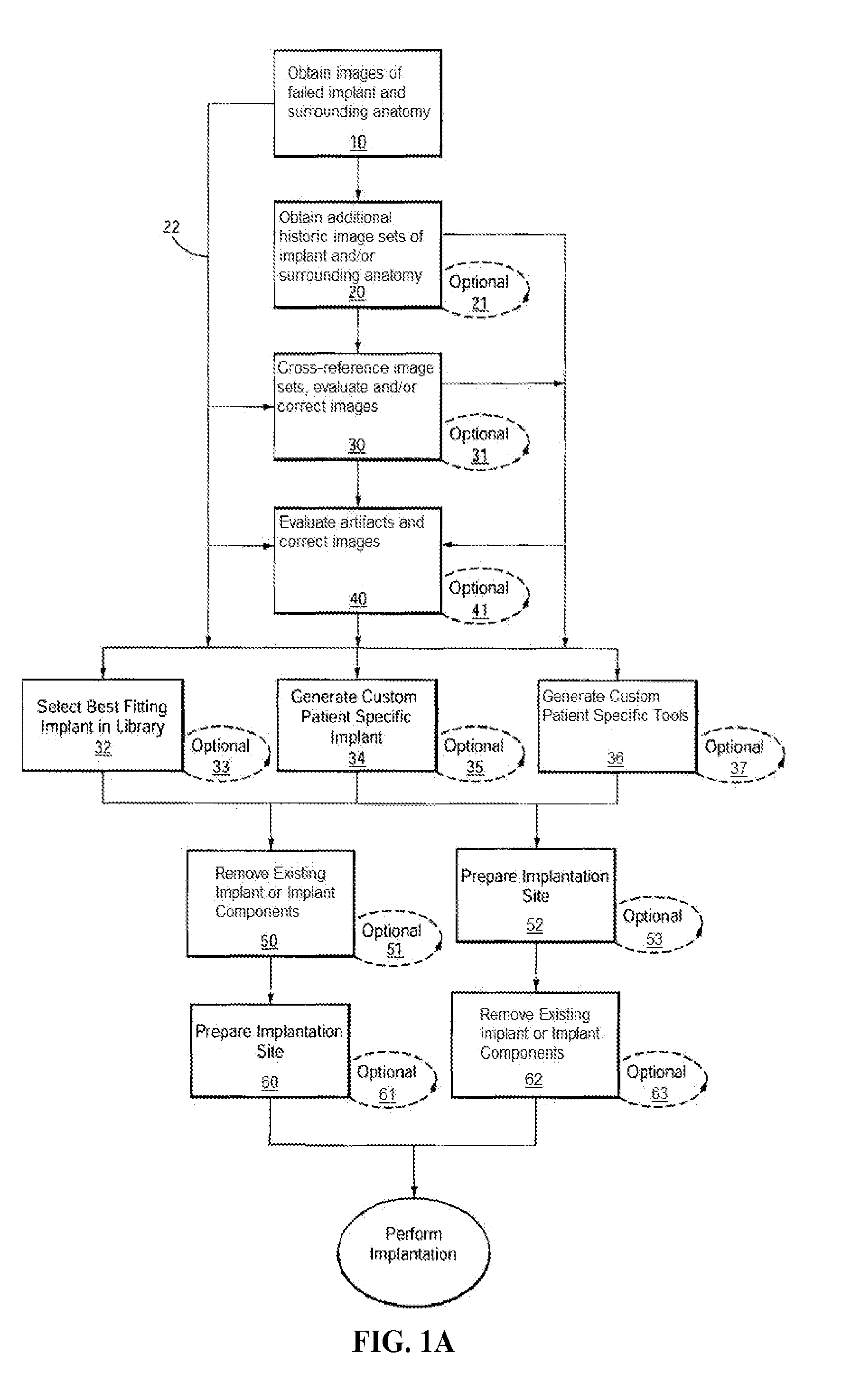
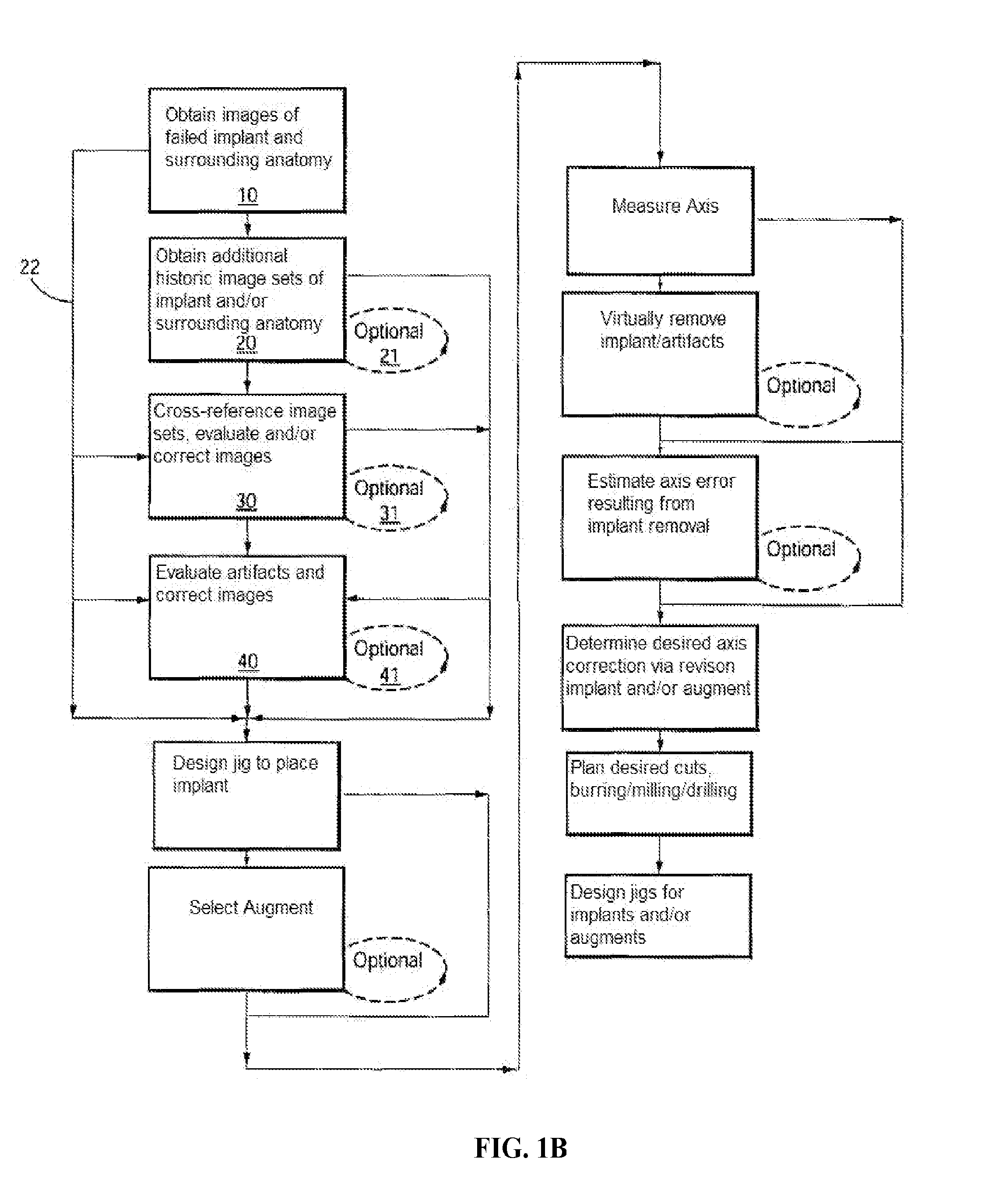
[0614]Example 3 depicts one embodiment of a revision system, method and devices contemplated by the present invention. In this embodiment, a total knee implant is experiencing failure or impending failure for any number of reasons, and requires surgical removal and revision to a replacement total knee implant. While the current embodiment contemplates removal and replacement of all implant components, it should be understood that a partial component replacement and / or implantation, either of one side (i.e., all of the tibial components) of an implant, as well as replacement and / or implantation of individual failed or failing components of the implant, are contemplated by the present invention.

[0615]Initially, the “failed implant” will be assessed and diagnosed. This process typically includes non-invasive imaging of the implant and the patient's anatomy, usually in an attempt to determine the condition of the implant and / or joint as well as to...


PUM

PropertyMeasurementUnit
Surfaceaaaaaaaaaa

Abstract

Disclosed herein are methods, compositions and tools for repairing an articular joint having a failed implant. The articular repair systems are customizable or highly selectable by or adaptable to individual patients and geared toward providing optimal fit and function. The surgical tools are designed to be customizable or highly selectable by or adaptable to individual patients to increase the speed, accuracy and simplicity of performing total or partial arthroplasty including revision surgeries.

Description

RELATED APPLICATIONS[0001]This application claims the benefit of: U.S. Ser. No. 61 / 523,756, entitled “Revision Systems, Tools and Methods for Revising Joint Arthroplasty Implants,” filed Aug. 15, 2011, the disclosure of which is incorporated herein by reference in its entirety.FIELD OF THE INVENTION[0002]This disclosure relates to orthopedic methods, systems and prosthetic devices and more particularly relates to methods, systems and devices for articular resurfacing and for correcting failed surgical resurfacing and / or replacement implants. The disclosure also includes surgical tools, molds and / or jigs designed to achieve optimal cut planes in a joint in preparation for installation of a joint revision implant.BACKGROUND OF THE INVENTION[0003]Joint arthroplasties can be highly invasive and often require surgical resection of the entirety, or a majority of the, articular surface of one or more bones involved in the repair. In various procedures, the marrow space can be fairly extens...

Claims


Application Information

Patent Timeline
IPC IPC(8): A61F2/30B23P15/00
CPCB23P15/00A61F2/30A61F2/38A61F2/30756A61B17/155A61B17/157A61B17/158A61B17/1714A61B17/1746A61B17/1764A61B2017/568A61F2002/3069A61F2002/3096A61F2002/30963Y10T29/49
Inventor LINDERMAN, JUSTINSLAMIN, JOHNLANG, PHILIPPSTEINES, DANIEL
Owner CONFORMIS
Learn More